Mucosal Barrier Injury and Stem Cell Transplant … Barrier Injury and Stem Cell Transplant...
186
Mucosal Barrier Injury and Stem Cell Transplant Recipients Nicole Blijlevens
Transcript of Mucosal Barrier Injury and Stem Cell Transplant … Barrier Injury and Stem Cell Transplant...
Mucosal Barrier Injury and Stem Cell Transplant Recipients
Nicole Blijlevens

Mucosal Barrier Injury and Stem Cell Transplant Recipients
een wetenschappelijke proeve op het gebiedvan de Medische Wetenschappen
Proefschrift
ter verkrijging van de graad van doctor aan de Radboud Universiteit Nijmegen, op gezag van de Rector Magnificus, prof. dr. C.W.P.M. Blom volgens besluit van het College van
Decanen in het openbaar te verdedigen op maandag 27 juni 2005, des namiddags om 3.30 uur precies
door
Nicolina Maria Anna Blijlevens
geboren op 16 februari 1965te Maastricht

Promotor:Prof. Dr. B.E. de Pauw
Co-promotor:Dr. J.P. Donnelly Dr. A.V.M.B. Schattenberg
Manuscript commissie:Prof. Dr. J.B.M.J. JanssenProf. Dr. S. McCann (Trinity College, Dublin)Prof. Dr. B.J. Kullberg
ISBN: 90-9019506-8© 2005 N.M.A. Blijlevens, Nijmegen
Cover: Ans Wilders, NijmegenLayout: Scriptura, NijmegenProduction: Quickprint, Nijmegen

There are some patients whom we cannot helpThere are none whom we cannot harm
Arthur L. Bloomfield

Table of contents

8
_____ Table of contents _______________________________________________________________________________________________________ ________________________________________________________________________________________________________ Table of contents _____
9
8
_____ Table of contents _______________________________________________________________________________________________________ ________________________________________________________________________________________________________ Table of contents _____
9
Table of contents
Chapter 1 Aim and Outline of the Thesis 11
Chapter 2 Mucosal Barrier Injury: biology, pathology, clinical 17 counterparts and consequences of intensive treatment for haematological malignancy: an overview
Bone Marrow Transplant. 2000;25(12):1269-1278
Chapter 3 Role of curcumin and the inhibition of NF-kappaB in the onset of chemotherapy-induced mucosal barrier injury 37
Leukemia. 2004;18(2):276-284
Chapter 4 Measuring mucosal damage induced by cytotoxic therapy 59Support Care Cancer. 2004;12(4):227-233
Chapter 5 Prospective evaluation of gut mucosal barrier injury 73 following various myeloablative regimens for haematopoietic stem cell transplant
Bone Marrow Transplant. 2005;35(7):707-11
Chapter 6 Citrulline: a potentially simple quantitative marker of 85 intestinal epithelial damage following myeloablative therapy
Bone Marrow Transplant. 2004;34(3):193-196
Chapter 7 Monitoring myelo-ablative therapy induced small bowel 95 toxicity by serum citrulline concentration: a comparison with sugar permeability tests
Cancer 2005: 103:191-199
Chapter 8 A randomised, double-blinded, placebo-controlled pilot 111 study of parenteral glutamine for allogeneic stem cell transplant patients
Support Care Cancer 2005 Mar 15; [Epub ahead of print]
10
_____ Table of contents _______________________________________________________________________________________________________
Chapter 9 Inflammatory response to mucosal barrier injury after 125 myeloablative therapy in allogeneic stem cell transplant recipients
Submitted to Bone Marrow Transplant
Chapter 10 Mucosal Barrier Injury: A paradigm for dysregulation 137 of the mucosal barrier and microbial flora
Submitted
Chapter 11 Summarising conclusions and recommendations 151
Chapter 12 Nederlandse samenvatting voor niet-ingewijden 161
Chapter 13 List of abbreviations 169
Chapter 14 List of publications 173
Chapter 15 Curriculum vitae 177
Chapter 16 Dankwoord 181
Aim and Outline of the Thesis
Chapter 1

12
_____ Chapter 1 _______________________________________________________________________________________________________________ ____________________________________________________________________________________ Aim and Outline of the Thesis _____
13
12
_____ Chapter 1 _______________________________________________________________________________________________________________ ____________________________________________________________________________________ Aim and Outline of the Thesis _____
13
Aim and Outline of the Thesis
Oral mucositis is a frequent complication of cytoreductive chemotherapy and radiotherapy and, until recently, was viewed solely as non-specific toxic effect treatment on dividing epithelial cells. Many patients suffer from pain are unable to eat or drink normally and consequently suffer a poorer quality of life. Moreover, mucositis is often dose-limiting especially after myeloablative therapy for haematopoietic stem cell transplantation (HSCT). In the last few years it has become increasingly clear that mucositis is not restricted to the mouth but extends to the rest of the alimentary tract involving the oesophagus, stomach, and the small and large intestines. The explanation for this lies in the fact that the mucosa of the alimentary tract is composed of a lining of epithelial cells that are coated with mucous on the outside and are adjacent to submucosal tissue on the inside. This layer functions as an anatomical and immunological barrier and so the term “mucosal barrier injury” (MBI) is more appropriate to describe the complex biological events that occur along the entire digestive tract following intensive cytotoxic treatment.
The aim of this thesis is to define and diagnose MBI of the alimentary tract of HSCT recipients, to explore the relationship of MBI to other transplant related complications particularly microbial infections and to determine whether MBI of the alimentary tract could be treated with glutamine-dipeptide.
Chapter 2 Mucosal Barrier Injury in haematopoietic stem cell transplant recipients In this chapter we described the biology, pathology and clinical counterparts of mucosal damage after intensive chemotherapy with or without radiotherapy. We adopted the term “Mucosal Barrier Injury” as this encompassed the biological nature as well as the clinical manifestation known as mucositis and emphasised the fact that MBI induced by myeloablative therapy affected the entire alimentary tract of HSCT recipients
Chapter 3 Mucosal Barrier Injury; the initiation phase The activation of the transcription factor, nuclear factor-kappa B (NF-κB) by chemotherapy mediates the release of proinflammatory cytokines. This is probably the first step in initiating MBI. In this chapter we explored whether inhibition of NF-κB by curcumin or caffeic acid phenethyl esther (CAPE) is able to prevent MBI of the small intestines after the administration of methotrexate or cytarabine. These studies were performed in-vitro using an intestinal cell culture model and in vivo in rats.
Chapter 4 Mucosal Barrier Injury to patients after cytotoxic therapyA number of scales have been advocated and several are used in practice to score oral mucositis but little has been done to assess the gastrointestinal counterpart
14
_____ Chapter 1 _______________________________________________________________________________________________________________ ____________________________________________________________________________________ Aim and Outline of the Thesis _____
15
of MBI. Accurate diagnosis of MBI of the intestines is virtually impossible since it requires biopsies to obtain histology and is too hazardous to patients who have received cytotoxic therapy principally because of the risk of bleeding. Pain and diarrhoea are universal symptoms of MBI but cannot be traced easily to a specific section of the alimentary tract. Hence we explored the possibility of using sugar permeability test to measure gut integrity and function using a solution of 4 different sugars; lactulose, L-rhamnose, D-xylose and 3-O-methylglucose. These tests were conducted alongside the scoring of oral mucositis and gut toxicity of patients given chemotherapy for acute leukaemia and of allogeneic HSCT recipients after intense myeloablative therapy.
Chapter 5 Mucosal Barrier Injury following various myeloablative regimens for stem cell transplantIn this chapter we determined the MBI occurring among 129 recipients of an autologous or allogeneic HSCT as measured by oral mucositis, gut toxicity and sugar permeability testing in order to determine whether sugar permeability testing was able to discriminate between various myeloablative regimens.
Chapter 6 Mucosal Barrier Injury measured by citrullineSerum citrulline is a reliable biochemical marker of the enterocyte mass of the small intestine of patients with villous-atrophy. Therefore we investigated the use of citrulline as marker of gut MBI of 32 allogeneic HSCT recipients who had all received the same myeloablative therapy with idarubicin, cyclophosphamide and total body irradiation
Chapter 7 Mucosal Barrier Injury: comparison of sugar permeability testing and citrullineThe sensitivity and specificity of serum citrulline compared with sugar permeability tests was analysed in 10 additional allogeneic HSCT recipients receiving the same conditioning regimen of idarubicin, cyclophosphamide and total body irradiation.
Chapter 8 Mucosal Barrier Injury: a randomised study of parenteral given glutamine-dipeptide Here we describe the results of a prospective, randomised, double-blinded, placebo-controlled pilot study of parenteral nutrition supplemented with 0.57 g/kg glutamine-dipeptide in a homogeneous group of 32 allogeneic HSCT recipients to determine its effect on MBI.
14
_____ Chapter 1 _______________________________________________________________________________________________________________ ____________________________________________________________________________________ Aim and Outline of the Thesis _____
15
Chapter 9 Mucosal Barrier Injury and inflammatory response in relation to bacteraemiaWe prospectively collected plasma of recipients participating in the randomised study of glutamine-dipeptide to investigate the relationship of MBI measured by using oral mucositis scoring, citrulline and sugar permeability testing, the host inflammatory response measured by C-reactive protein, interleukin-8 and LPS-binding protein and the onset of bacteraemia during MBI.
Chapter 10 Mucosal Barrier Injury: paradigm for dysregulation of mucosal barrier and microbial flora Here we postulate that MBI is far more than simply a risk factor for severe infectious complications after anti-cancer treatment. Rather, MBI is a manifestation of the breakdown in the relationship between host (patient) and endogenous microflora. The delicate balance between microbes and a damaged host is known as the “damage-response” framework in which the outcome of that interaction determines microbial pathogenesis and not the particular microbial species.

Mucosal Barrier Injury: biology, pathology, clinical counterparts and consequences of intensive treatment for haematological malignancy:
an overview
Chapter 2
N.M.A. Blijlevens, J.P. Donnelly and B.E. De Pauw
Department of Hematology, University Medical Center St. Radboud, Nijmegen
The Netherlands
Bone Marrow Transplant. 2000;25(12):1269-1278

18
_____ Chapter 2 _______________________________________________________________________________________________________________ ______________________________________________________________________________________________ Mucosal Barrier Injury _____
19
18
_____ Chapter 2 _______________________________________________________________________________________________________________ ______________________________________________________________________________________________ Mucosal Barrier Injury _____
19
Summary
Mucositis is an inevitable side effect of the conditioning regimens used for haematopoietic stem cell transplantation. The condition is better referred to as mucosal barrier injury (MBI) since it is primarily the result of toxicity and is a complex and dynamic pathobiological process manifested not only in the mouth but also throughout the entire digestive tract. A model has been proposed for oral MBI and consists of four phases, an inflammatory, epithelial, ulcerative and a healing phase. A variety of factors are involved in causing and modulating MBI including the nature of the conditioning regimen, the elaboration of pro-inflammatory and other cytokines, translocation of the resident microflora and their products for example endotoxins across the mucosal barrier, exposure to antimicrobial agents and whether or not the haematopoietic stem cell graft is from a donor. Neutropenic typhlitis is the most severe gastrointestinal manifestation of MBI, but it also influences the occurrence of other major transplant-related complications including acute GVHD, veno-occlusive disease and systemic infections. The pathobiology, clinical counterparts and the means of measuring MBI are discussed together with potential approaches for prevention, amelioration and, perhaps, even cure.
Introduction
Mucositis is an inevitable side effect of the intensive conditioning therapy used for haematopoietic stem cell transplantation [1] and usually refers to the mucosal ulceration of mouth and throat. However, it is generally accepted that oral mucositis is in reality the most obvious manifestation of damage or injury elsewhere particularly that of the gut. Hence, mucosal barrier injury (MBI) may be a more appropriate term for this biological process. There exists no clear definition of MBI which is defined by a constellation of signs and symptoms that vary in their clinical expression. Oral MBI is reported to affect 60% to 100% of transplant recipients [2,3] and is characterised by pain, oedema, erythema, lesions, pseudomembrane formation, excessive mucous production, reduced saliva and bleeding all of which reduces the patient’s ability to eat and drink. By contrast, there are no reliable data on the incidence of gut MBI although intestinal symptoms affect almost every transplant recipient to some extent and include nausea, vomiting, abdominal cramping and watery diarrhoea occasionally accompanied by macroscopic blood loss. The exact course and severity of bowel symptoms of MBI is also difficult to ascertain because many patients are in such pain due to oral MBI that they only gain relief from narcotic analgesia which induces constipation as a result of reduced gut motility. There are also a number of scoring systems for oral MBI [4] although none is universally accepted and all lack standardisation but, as yet, there is no system for registering gut MBI although there are published definitions for grading toxicity of individual signs and symptoms. Consequently much more is known about the course of oral MBI than its intestinal counterpart. Oral MBI is known to begin around the time conditioning therapy is completed, and has been shown to worsen until a peak is reached after which it declines
20
_____ Chapter 2 _______________________________________________________________________________________________________________ ______________________________________________________________________________________________ Mucosal Barrier Injury _____
21
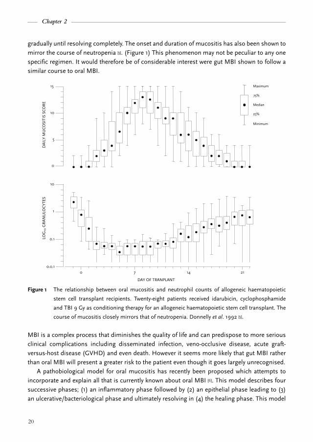
gradually until resolving completely. The onset and duration of mucositis has also been shown to mirror the course of neutropenia [5]. (Figure 1) This phenomenon may not be peculiar to any one specific regimen. It would therefore be of considerable interest were gut MBI shown to follow a similar course to oral MBI.
Figure 1 The relationship between oral mucositis and neutrophil counts of allogeneic haematopoietic
stem cell transplant recipients. Twenty-eight patients received idarubicin, cyclophosphamide
and TBI 9 Gy as conditioning therapy for an allogeneic haematopoietic stem cell transplant. The
course of mucositis closely mirrors that of neutropenia. Donnelly et al. 1992 [5].
MBI is a complex process that diminishes the quality of life and can predispose to more serious clinical complications including disseminated infection, veno-occlusive disease, acute graft-versus-host disease (GVHD) and even death. However it seems more likely that gut MBI rather than oral MBI will present a greater risk to the patient even though it goes largely unrecognised.
A pathobiological model for oral mucositis has recently been proposed which attempts to incorporate and explain all that is currently known about oral MBI [6]. This model describes four successive phases; (1) an inflammatory phase followed by (2) an epithelial phase leading to (3) an ulcerative/bacteriological phase and ultimately resolving in (4) the healing phase. This model
20
_____ Chapter 2 _______________________________________________________________________________________________________________ ______________________________________________________________________________________________ Mucosal Barrier Injury _____
21
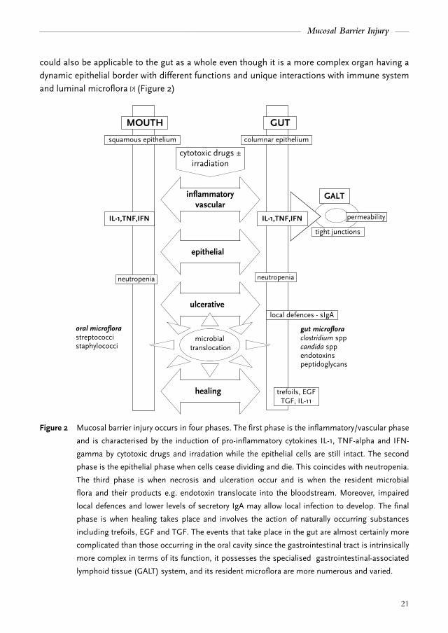
could also be applicable to the gut as a whole even though it is a more complex organ having a dynamic epithelial border with different functions and unique interactions with immune system and luminal microflora [7] (Figure 2)
Figure 2 Mucosal barrier injury occurs in four phases. The first phase is the inflammatory/vascular phase
and is characterised by the induction of pro-inflammatory cytokines IL-1, TNF-alpha and IFN-
gamma by cytotoxic drugs and irradation while the epithelial cells are still intact. The second
phase is the epithelial phase when cells cease dividing and die. This coincides with neutropenia.
The third phase is when necrosis and ulceration occur and is when the resident microbial
flora and their products e.g. endotoxin translocate into the bloodstream. Moreover, impaired
local defences and lower levels of secretory IgA may allow local infection to develop. The final
phase is when healing takes place and involves the action of naturally occurring substances
including trefoils, EGF and TGF. The events that take place in the gut are almost certainly more
complicated than those occurring in the oral cavity since the gastrointestinal tract is intrinsically
more complex in terms of its function, it possesses the specialised gastrointestinal-associated
lymphoid tissue (GALT) system, and its resident microflora are more numerous and varied.
22
_____ Chapter 2 _______________________________________________________________________________________________________________ ______________________________________________________________________________________________ Mucosal Barrier Injury _____
23
Pathobiology
The inflammatory phaseRadiation and cytotoxic drugs induce the systemic release of pro-inflammatory cytokines, interleukin-1 (IL-1) and tumour necrosis factor-alpha (TNF-α) from activated macrophages and monocytes. Ionising radiation also induces cytokine gene expression directly [8]. TNF-α and its receptors are activated and suggest that resident tissue macrophages and monocytes rather than circulating polymorphonuclear (PMN) cells are the main target in vivo [9]. Tissue macrophages are not eliminated by conditioning therapy and may persist up to 4 months after transplantation. In the gut, macrophages reside in the gastrointestinal-associated lymphoid tissue (GALT) which houses the vast majority of total circulating lymphocytes as well as other members of the lymphoreticular system such as monocytes and intraepithelial lymphocytes. Once released into the circulation, the cytokines increase expression of HLA histocompatibility antigens and critical adhesion molecules that amplify local tissue injury by inviting PMN cells and activated lymphocytes to invade [10]. This results in increased vascularity and most likely higher local levels of cytotoxic agents. In an animal model, exposure to bleomycin or 5-fluorouracil (5-FU) resulted in increased cellularity of subepithelial oral tissue, vascular dilation and leukocyte margination within 24 hours [11]. The generation of cytokines is self-limited during autologous transplantation and resolves within 7 –10 days [10].
The inflammatory response may be specific to different classes of chemotherapeutic agents and to the particular sequence of preparative regimens used in haematopoietic stem cell transplantation since a variety of different profiles of cytokines release have been reported [12,13,14]. For instance, elevated TNF-α levels were found in 13 (24%) of 56 patients given either cyclophosphamide and total body irradiation or cyclophosphamide and busulphan and these levels were predictive for transplant related complications within the first 6 months post BMT [14]. Serum levels of TNF-α and IL-1β have also been shown to be markedly higher with higher doses of TBI 1 week after transplant [13]. In contrast, busulphan, VP-16 and cyclophosphamide induced interferon-gamma production directly [12, 15]. Moreover, intestinal damage manifest by villous blunting, apoptosis and brush border loss (the surface area of villous cells amplified by numerous finger-like microvilli ) correlated well with cytokine levels [13].
In a clinical phase I/II study, use of the monoclonal antibody MAK 195F diminished the release of TNF-α but it was also observed that the kinetics of TNF-anti-TNF complexes were different after conditioning therapy with cyclophosphamide and TBI than with cyclophosphamide and busulphan which actually induced less TNF-α release [16]. Elevated cytokine levels detected as early as one week after transplantation might be related to engraftment in the absence of complications or to infectious disease, non-infectious events or GVHD rather than MBI [17] Moreover, other investigators have failed to find elevated levels of cytokines during or shortly after conditioning therapy [12,17,18].
Before total cell destruction, TNF-α, IFN-γ and IL-1 induce major changes in the functionality, permeability, brush border transport, glutamine utilisation (glutamine is the main source of
22
_____ Chapter 2 _______________________________________________________________________________________________________________ ______________________________________________________________________________________________ Mucosal Barrier Injury _____
23
energy source for intestinal cells) and mucosal cell integrity [19,20,21,22]. IFN-γ and TNF-α induce dose-related cellular exfoliation, leading to the formation of a mucoid cap in an vain attempt to protect the mucosa [23].
Epithelial cells are also capable of producing and secreting TNF-α and IL-1α [24]. In an H-2-incompatible transplanted SCID mice model, colonic TNF-α, IL-1α and IL-6 appeared 4 hours after TBI and peaked by 24 hours. If no transplantation followed, TNF-α and IL-1α levels decreased rapidly 3-5 days later [17,18,25]. Epithelial cells are also capable of mounting an immune host response, and of taking up, processing and presenting soluble antigens as well as expressing MHC class II molecules [26]. Thus, taken together, these data support the view that the primary step in MBI is an inflammatory response.
The epithelial phaseCytotoxic drugs and radiation interfere with rapidly dividing cells and the kinetics of proliferating mucosal cells influences their sensitivity to these agents. Normally, cell renewal takes place continuously in crypts from a proliferating pool of clonal undifferentiated stem cells and cell division is completed in about 24 hours [27]. Younger cells migrate up to the villous tips and slough off into the lumen at the extrusion zone. Anti-metabolites, for example cytarabine, are cell cycle dependent and interfere with the synthesis of DNA in dividing cells whereas the intercalating agents such as the anthracyclines are more effective during the G2 phase after mitosis is complete when the cell has time to restore errors. In contrast, alkylating agents such as cyclophosphamide generate lethal DNA lesions even in resting cells by forming cross-links between DNA strands while ionising radiation exerts its main effect during mitosis. Various chemotherapeutic drugs such as adriamycin, bleomycin and 5-FU increase cellular sensitivity to radiation in a synergistic manner [28]. This has also been observed clinically when idarubicin was given at the same time as cyclophosphamide and TBI [29].
Normally, the entire epithelium is renewed in 4-6 days, but decreased cell renewal is thought to lead to mucosal atrophy, thinning and necrosis although in rats, sublethal doses of alkylating agents mainly induced lower absorption rather than villous atrophy [30].
The ulcerative-bacteriological phaseIncreased redness and swelling of the mucosa and underlying tissue are usually the first signs of oral MBI, mainly due to increased vascularity and vascular permeability (inflammatory phase) and thinning of epithelium (epithelial phase). This process usually culminates in the ulcerative phase within about 14 days of starting chemotherapy [1]. It is also during this phase that the resident microflora are assumed to play a role. Normally, these microorganisms contribute to maintaining the integrity of the integument and prevent pathogenic microbes from gaining a foothold. The ecological system tries to maintain its balance but once the mucosa is damaged, microbes may infect the submucosal tissue. Non-pathogenic streptococci specifically bind to, and use, the glycoproteins in the dental plaque that develops in the absence of normal food intake and saliva production. The non-cellular defence depends on amount and quality of mucus and saliva
24
_____ Chapter 2 _______________________________________________________________________________________________________________ ______________________________________________________________________________________________ Mucosal Barrier Injury _____
25
produced which contain diverse host defence peptides (defensins), lactoferrin, lysozyme and immunoglobulins [31]. Secretory IgA inhibits bacterial adherence, specifically of oral streptococci, neutralises toxin and virus, prevents antigen uptake and possesses anti-inflammatory activity [32]. Several classes of host defence peptides can be found in saliva and on surfaces each possessing rapid lytic activity against the membranes of Gram-positive and Gram-negative bacteria as well as yeasts [33]. During conditioning therapy and after transplantation, the salivary immunoglobulins (IgA, IgG, IgM) have been shown to be lower than normal and the elimination of T-cells from engrafted bone marrow results in less initial capacity for immunoglobulin production and secretion [34]. Thus, the local immune defences of the oral cavity are impaired. It is common practice to administer antimicrobial agents, particularly the fluoroquinolones and local antiseptics such as chlorhexidine, to haematopoietic stem cell transplant recipients, leading inevitably to marked shifts in the resident oral flora towards the more resistant species particularly the viridans (alpha-haemolytic) streptococci. This shift is more profound in-patients with overt oral mucositis [35]. There has also been a corresponding increase in bacteraemia due to these streptococci with oral mucositis being an important risk factor in autologous haematopoietic stem cell transplant recipients [36]. Similarly, Donnelly et al. [37] reported a higher incidence of viridans streptococcal bacteraemia due to the marked mucositis associated with treatment intensification. One particular species, Streptococcus mitis, is apparently associated with sepsis and adult respiratory distress syndrome (ARDS), mainly after high-dose cytarabine [38,39]. This syndrome could be provoked by changes in the pulmonary endothelium and lung macrophages induced by cytotoxic chemotherapy which, in turn, induces cytokine production perhaps triggered by infection with Streptococcus mitis [40]. The stomach or small intestine could be also be a portal of entry if colonisation with these streptococci occurs as a result of the achlorhydria induced by H
2-histamine antagonists and proton pump inhibitors since the use of these agents
has been noted as a risk factor for the so-called “alpha-strep syndrome” [38]. Obviously, MBI is itself a risk factor for viridans streptococcal bacteraemia but it might not always indicate systemic infection since transient bacteraemia also occurs in healthy persons after dental manipulation [41]. Moreover, these bacteria do not elaborate exotoxins nor are they professional pathogens. Thus, viridans streptococcal bacteraemia might simply signal the presence of mucosal barrier injury rather than infection.
Although, to some extent, similar to the oral cavity, the gut harbours a much more complex ecosystem comprising a greater variety of aerobic and anaerobic bacteria that share a symbiotic relationship with the host. This relationship plays an important role in maintaining the gut’s histological structure and also provides so-called ‘colonisation resistance’ i.e. the ability of the gut to repel foreign bacteria. The intestinal microflora depends on prebiotics, the fibrous nutrients [42] that enhance probiotic bacteria, like bifidobacteria, lactobacilli, and Clostridium species. These are the species that are thought to provide the colonisation resistance by elaborating antibacterial compounds and competing for nutrients so preventing overgrowth by potentially pathogenic bacteria [43]. These probiotic bacteria also produce nutrients for mucosal cells. Certain antimicrobial agents, particularly those that affect cell-wall synthesis, exert a major
24
_____ Chapter 2 _______________________________________________________________________________________________________________ ______________________________________________________________________________________________ Mucosal Barrier Injury _____
25
impact on the gut’s ecosystem by destroying the ‘protective’ anaerobic flora particularly the probiotic bacteria. When the gut epithelium is disrupted, bacterial translocation occurs and pro-inflammatory bacterial oligopeptides, especially endotoxin (lipopolysaccharide or LPS) readily gain access [44]. In the normal host (whether animal or human) pathogenic bacteria such as Escherichia coli and Pseudomonas aeruginosa penetrate the mucosa and migrate to extra-intestinal sites such as the mesenteric lymph nodes, spleen and liver. The GALT system, together with the Kupffer cells of liver and spleen serve as a backup to trap endotoxins and kill bacteria. The rate of translocation of enterobacteria like E. coli and other gram-negative bacilli such as P. aeruginosa is strongly associated with the degree of neutropenia [45]. Microbial translocation is exacerbated by irradiation [46] and chemotherapy [47] as micro-organisms can be cultured in extra-intestinal sites as well as in blood [48]. Different modes of translocation exist and occur even before any histological damage is apparent. Anaerobic non-pathogenic bacteria rarely translocate but yeasts such as Candida albicans can do so more easily when disruption has occurred [49]. Endotoxin can be transported through the lymphatic channels, bypass the liver or enter the peritoneal cavity directly and can cause systemic endotoxaemia [50]. Endotoxin can also increase intestinal permeability directly [51] or by stimulating primed macrophages to release an excessive amount of cytokines, mostly TNF-α, thereby inducing mucosal inflammation and increasing permeability [52]. Higher levels of circulating endotoxin are obtained after giving intensive TBI containing regimens [13] suggesting that persistent low-grade endotoxaemia or the inflammation associated with MBI induce fever of unknown origin since endotoxaemia and gut mucosal damage occurred in 44 (70%) of 63 HSC transplant recipients (both allogeneic and autologous) all of whom developed fever that could not be explained by infection [53].
Peptidoglycan (the major component of the cell wall of Gram-positive bacteria) may play a similar role as endotoxin as it is also biologically active in tissues and may induce a pro-inflammatory response [54]. Although much less potent than endotoxin gram-for-gram, large amounts of peptidoglycan may well be released into the circulation when gut MBI is present simply because there are many more Gram-positive than Gram-negative bacteria in the gut. Exposure to antibiotics that cause lysis will also liberate cell wall fragments.
Neutropenic typhlitis, a paradigm for gut MBITyphlitis, also called neutropenic enterocolitis, necrotising enterocolitis or ileocaecal syndrome, is a caecitis often extending to both the proximal and distal caecum that may be primarily a severe manifestation of gut MBI. Indeed, all factors that contribute to the development of MBI are present clinically. First, typhlitis occurs after the administration of cytotoxic drugs, particularly high-dose cytarabine, etoposide and anthracyclines at the nadir of neutropenia and thrombocytopenia. Secondly, prolonged exposure to antibiotics results in a marked shift in the gut microflora towards toxin producing bacteria [55] such as Staphylococcus aureus, Pseudomonas aeruginosa and Clostridium septicum [56,57]. In fact, Clostridium species are now more likely to predominate for reasons which are poorly understood such that bacteraemia due to C. tertium or C. septicum is almost pathognomonic for typhlitis. Antimicrobial pressure also predisposes to intestinal overgrowth by
26
_____ Chapter 2 _______________________________________________________________________________________________________________ ______________________________________________________________________________________________ Mucosal Barrier Injury _____
27
Clostridium difficile in transplant recipients [58]. Necrosis of the mucosal surface of the ileocaecal region probably provides a favourable environment for the spores of Clostridium species to germinate and may be their portal of entry into the bloodstream. The pathogenesis of typhlitis would therefore seem to require various elements to be present simultaneously, namely, gut MBI, a perturbed resident microflora and profound neutropenia. Typhlitis is not only a paradigm for MBI but, because of the high mortality rate, it is also the most severe clinical form of MBI and deserves more attention both in terms of developing techniques for early diagnosis as well as in evolving strategies for prevention and treatment. Consequently we can expect to encounter more cases of typhlitis as chemotherapeutic regimens become more intense.
The healing phase In general, the repair of oral MBI parallels haematological reconstitution as peripheral blood counts return to normal [3,5,59] with complete resolution occurring within 2-3 weeks [60]. In contrast, gut function does not return to normal for several more weeks, since malabsorption and diminished enzyme activity still persist even after structural repair. The healing of mucosal damage probably occurs in two phases commencing with the restitution of mucosal integrity and then remodelling of the mucosal architecture. The mucosal repair process depends on the severity of damage since superficial injury can be repaired rapidly by epithelial migration without mitosis [61]. However, proliferation in conjunction with angiogenesis is necessary for deep lesions involving large areas of necrosis to recover [62]. Trefoil peptides (mucin-associated peptides) are secreted by epithelial cells, with each region of the gut probably having its own variant [62]. These peptides act as rapid response molecules to injury by promoting cell migration, cell differentiation and wound healing [63]. Epidermal growth factor, transforming growth factor alpha, interleukin-11 and fibroblast growth factor also appear to promote epithelium repair and regeneration [64]. In contrast, these agents appear to play only a limited role in the healing of the oral mucosa.
Acute Graft-versus-Host DiseaseGut MBI may evolve into acute GVHD since tissue damage caused by conditioning regimens plays a role in both conditions. Any mature donor T lymphocytes within the allograft that recognise host-antigens are activated by endotoxin and pro-inflammatory cytokines [10, 13]. Animal studies [25] and some studies in humans [13,14] suggest that high levels of pro-inflammatory cytokines predispose to acute GVHD, although others could not confirm this observation [12,15,17,65]. The early administration of the anti-TNFα monoclonal antibody MAK 195F changes the nature of the inflammatory response, reduces the number of febrile episodes and delays the onset and severity of acute GVHD [16]. The microflora might also play a role in triggering acute GVHD because less disease was observed in decontaminated mice chimeras [66,67]. Intestinal decontamination with metronidazole also significantly reduced the severity of acute GVHD in HLA-identical sibling transplants [68]. Taken together, these data suggest a role for MBI in triggering of acute GVHD because of either the release of cytokines induced by conditioning regimens or the translocation of microbial toxins.
26
_____ Chapter 2 _______________________________________________________________________________________________________________ ______________________________________________________________________________________________ Mucosal Barrier Injury _____
27
Diagnostic tools
Intestinal permeabilityPermeability refers to the property possessed by a epithelium that enables passage of a solute by unmediated diffusion [69]. Permeability can be measured in vivo by means of the urinary excretion of test substances or by detecting their presence in blood. Lactulose, various polymers of polyethylene glycol (PEG), 51Cr-labeled ethylenediaminetetraacetic acid (51Cr-EDTA) have all been used, but the results are markedly influenced by extraneous factors such as bowel transit time, gastric emptying and renal function. Nonetheless, permeability to 51Cr-EDTA is already increased two days after starting conditioning therapy and continues to increase until shortly after BMT, about 12 days later [70]. Others have shown that the intestinal toxicity induced by melphalan can be monitored using 51Cr-EDTA thus allowing the effectiveness of various treatments for reducing the intestinal toxicity to be assessed [71]. Unfortunately, 51Cr-EDTA is radioactive and not suitable for routine use.
The uptake of antibiotics such as gentamicin and tobramycin may provide a safer means of determining increased permeability since such drugs are normally excluded by the intact gut but can be detected in plasma during mucositis when given by mouth [72]. Studies of epithelial cell handling of cytotoxic drugs, radiation and antimicrobial agents could offer new possibilities for documenting MBI [73,74].
Sugar absorption testsThe principal features of gut MBI are a loss of epithelial surface and a change in the permeability [69]. This can also be measured if at least two different probes are used at once since the extraneous factors equally affect the pre- and post-mucosal determinants and the urinary excretion ratio becomes an index of intestinal permeability. For example, monosaccharides such as mannitol and rhamnose are absorbed through aqueous pores in the cell membrane whilst disaccharides like lactulose gain access through the tight junctions located at the upper end of adjacent epithelial cells. Tight junctions are dynamic structures exerting physiologic control over the flow of solutes through paracellular spaces and play an important role in gut permeability. Reduction of urinary monosaccharide excretion represents a loss of epithelial cell surface area, while increased urinary disaccharide excretion indicates damage to the tight junctions. Sugar absorption tests (SAT) have proven their value in intestinal diseases but they lack diagnostic specificity. SATs offer an easy, reliable means of assessing the onset, duration and severity of gut MBI in patients treated with cytotoxic agents. Absorption is already increased after only two days treatment with chemotherapy [76] suggesting that cytokines might be interfering with the tight junctions (see inflammatory phase) rather than any direct inhibition of cell proliferation, which tends to occur later [77]. Altered permeability continues to progress until reaching a peak about 7 days after conditioning therapy has been completed [78] and returns to normal about 4 weeks later [79]. This mirrors the oral MBI and neutropenia. (Figure 1)
It should be possible to discriminate patients at risk of developing serious toxicity to therapy

28
_____ Chapter 2 _______________________________________________________________________________________________________________ ______________________________________________________________________________________________ Mucosal Barrier Injury _____
29
from those not at risk by using these SATs since a positive correlation was found between progressive non-oral clinical toxicity and increased permeability in transplant recipients [70,71]. Bow et al. also found that the absorption of D-xylose was at its lowest 2 to 3 weeks after remission induction treatment had been started in patients who developed systemic candidosis [80]. Malabsorption of D-xylose was also found to be an independent predictor of neutropenic enterocolitis and hepatosplenic candidosis and also correlated well with bacteraemia [80,81]. As yet, there are no objective means of determining gut MBI in use routinely and none has been validated for use in clinical trials to assess gut toxicity although the data available suggests SATs may be useful for helping adapt supportive care regimens for selected patients in order to reduce morbidity and perhaps mortality.
A consensus in the way MBI is measured in clinical practice analogous to the validated scoring system of oral mucositis of the Mucositis Study Group [82] is a prerequisite for such studies. There is also a pressing need for much simpler tests of each phase of MBI. Data from chemotherapy induced cytokine expression of epithelial cells in single-cell testing or cell lines, quantitation of cytokine profiles in saliva or stools [83] and the results of basal cell kinetic studies like grading of epithelial cells viability by trypan blue dye exclusion obtained by oral washings [84] should be incorporated in clinical research and care. It should be possible to demonstrate overt mucositis using radionuclide imaging techniques, such as indium-labelled leukocytes [85] and technetium-labelled diphytanoylphosphatidylcholine liposomes but there are only a few anecdotal reports and their clinical feasibility is expected to be minimal.
Intervention and treatment of mucosal barrier injury
NutritionEnteral nutrition stimulates gut-responsive hormones, prevents mucosal atrophy, improves mucosal blood flow and gastrointestinal motility, stimulates mucus formation and secretion of sIgA and reduces bacterial translocation [86]. In children without severe MBI, enteral and parenteral nutrition were equally effective in maintaining the nutritional status [87] and a diet containing lactose and bovine milk protein appeared to be well tolerated [88]. Oral administration of short-chain fatty acids (SCFAs) typically produced by the anaerobic flora of the gut reduces the inflammation and necrosis induced by cytarabine in mice [89]. These SCFAs are normally produced by the fermentation of dietary fibre and unabsorbed starch by the same gut microflora and are the preferred fuel of enterocytes.
Total parenteral nutrition (TPN) is advocated for those patients who are either malnourished or who are expected to have inadequate oral intake for a prolonged period (usually 7-10 days) to restore the negative nitrogen and caloric balance. These patients are typically those with MBI that is sufficiently severe that it impedes adequate enteral nutrition leading to malnutrition, weight loss, malabsorption and micronutrient deficiencies. TPN does help to reduce the morbidity of malnourished patients completing a course of myeloablative therapy [1], but at the same time
28
_____ Chapter 2 _______________________________________________________________________________________________________________ ______________________________________________________________________________________________ Mucosal Barrier Injury _____
29
it promotes villous atrophy, increases intestinal permeability, reduces luminal sIgA content and enhances bacterial translocation [90]. Nevertheless, the long-term outcome for allogeneic HSC transplant recipients is better with TPN, even when they are well-nourished [91] whereas autologous HSC transplant recipients gain little or no benefit [92].
GlutamineGlutamine has attracted a lot of attention because it is the primary fuel for intestinal epithelia and the cornerstone of protein and nucleic acid synthesis but mucosal cells cannot synthesise enough themselves making glutamine conditionally essential during stress [93]. Administering glutamine to animals after irradiation and chemotherapy prevented mucosal atrophy, reduced bacterial translocation, endotoxaemia and infections [94-96]. It is much less clear whether glutamine given orally prevents human oral MBI, [97,98] although patients treated with high-dose chemotherapy experienced less diarrhoea [99]. Glutamine supplement given to HSC transplant recipients parenterally helps to preserve hepatic function, lowers the length of stay in hospital, improves the nitrogen balance and lowers the infection rate [100,101] but has no influence on the occurrence of mucositis or fever [102,103]. However, nothing is known about the effect of glutamine on gut integrity or function since permeability tests were not performed.
CytoprotectantsDirect cytoprotectants such as sucralfate and diphenhydramine do not ameliorate oral MBI [1, 104-
106]. Whereas indirect cytoprotectants, like transforming growth factor-β3 and epidermal growth factor, interfere with epithelial cell replication in animals and are being tested in clinical trials for their efficacy and safety in modulating oral MBI [107,108]. Recombinant-human GM-CSF given as a mouthwash shortened the duration of severe oral MBI [109,110] but the mechanism of action remains unclear. GM-CSF might have a direct pleiotropic effect on epithelial cell kinetics. Alternatively, the effect may be indirect as a result of the first neutrophils produced by haematopoietic progenitor cells migrating to the oral mucosa and thereby reducing local infection [111]. Other clinical trials exploring the effects of recombinant growth factors as transforming growth factor-β1 or TGF-β3 and others on MBI are coming [112-114]. A clearly different approach consists of delivering monoclonal antibodies that bind and inactivate doxorubicin (MAD11) in intestinal cells [115].
Antimicrobial agentsIt is common practice to try to reduce the bioburden of Gram-negative bacilli in the oral cavity by giving antimicrobial agents and maintaining good oral hygiene and also to provide remedial dental treatment when necessary to reduce oral complications [116]. Antibiotic lozenges containing tobramycin, polymyxin and amphotericin B reduce oral MBI [117] but the effect of chlorhexidine is unclear [118-121].
There have been no formal studies of the effect of antimicrobial agents whether given for prophylaxis or treatment on MBI although it is usually assumed that they are beneficial. If MBI is not primarily the result of infection as seems to be the case, treatment with antimicrobial
30
_____ Chapter 2 _______________________________________________________________________________________________________________ ______________________________________________________________________________________________ Mucosal Barrier Injury _____
31
agents is unlikely to be of benefit and may even prove harmful in exerting selective pressure on the resident flora. Probiotics may help restore the balance of gut flora in cancer patients [122] but trials of sufficient size are lacking. IgM-enriched immunoglobulin has been shown to reduce endotoxaemia and febrile episodes in transplant recipients [53].
Future directions
Mucosal barrier injury is far more than simply a toxicological side effect of cytotoxic regimens. Enough evidence exists to indicate that MBI is a complex and dynamic pathological process but it is essential to understand its nature more fully.. A model for oral MBI already exists and shows that it is the net result of an almost complete breakdown of the epithelium initiated by the release of pro-inflammatory cytokines induced by the cytotoxic drugs followed by an arrest of the mucosal cell cycle and inhibition of repair leading to apoptosis. Infection, if it plays any role at all, is largely secondary. This model may go some way to explain the corresponding phenomenona in the gut although gut MBI is likely to be much more complex and more difficult to unravel mainly because the damage cannot be seen and the signs and symptoms are too imprecise.
Since gut permeability increases very soon after exposure to chemotherapy and irradiation it seems logical to pursue tests such as the SATs further and to look for other chemical probes. Certainly, a means of objectively monitoring MBI is necessary before drug products can be formally tested for their effects on MBI.
At that moment there are several products ranging from cytokines and defensins to nutrients and probiotics which look promising. For example the growth factor interleukin-11 (IL-11) by reducing pro-inflammatory cytokine expression and secretion by macrophages (phase I), it prevents apoptosis of intestinal crypt cells partially by inhibiting proliferation (phase II) and promotes recovery of these crypt cells while remodelling connective tissue (phase IV). Defensins, trefoil peptides and even sIgA-antibodies could offer additional tools to tackle hostile microbes for example IgA-IgG administered orally has been shown to reduce gut MBI in patients undergoing intensive cytotoxic therapy [123].
With the means of reliably detecting and monitoring gut MBI at our disposal, the process will graduate from being an expected though unpleasant side effect with little therapeutic options to a condition that might actually be preventable.
30
_____ Chapter 2 _______________________________________________________________________________________________________________ ______________________________________________________________________________________________ Mucosal Barrier Injury _____
31
References
1. Berger AM, Kilroy TJ. Adverse effects of treatment; oral complications. In DeVita VT, Hellman S, Rosenberg SA (eds.) Cancer; principles and practice of oncology. Lippincott - Raven Publishers: Philadelphia,1997; 5th edition, pp 2714-25.
2. Schubert MM, Williams BE, Lloid ME et al. Clinical assessment scale for the rating of oral mucosal changes associated with bone marrow transplantation. Development of an oral mucositis index. Cancer 1992; 69 (10):2469-77.
3. Woo SB, Sonis ST, Monopoli MM, Sonis AL. A longitudinal study of oral ulcerative mucositis in bone marrow transplant recipients. Cancer 1993; 72 (5):1612-7.
4. Parulekar W, Mackenzie R, Bjarnason G, Jordan RC. Scoring oral mucositis. Oral Oncol. 1998; 34 (1):63-71.5. Donnelly JP, Muus P, Schattenberg A et al. A scheme for daily monitoring of oral mucositis in allogeneic BMT
recipients. Bone Marrow Transplant. 1992; 9 (6):409-13.6. Sonis ST. Mucositis as a biological process: a new hypothesis for the development of chemotherapy-induced
stomatotoxicity. Oral Oncol. 1998; 34 (1):39-43.7. Sleisenger M.H.; Fordtran J.S. Gastrointestinal disease; pathofysiology, diagnosis, management. W. B. Saunders
Company: Philadelphia, 4th edition, 1989.8. Sherman ML, Datta R, Hallahan DE et al. Regulation of tumor necrosis factor gene expression by ionizing radiation
in human myeloid leukemia cells and peripheral blood monocytes. J.Clin.Invest. 1991; 87 (5):1794-7.9. Hoffmann B., Hintermeier-Knabe R., Holler E. et al. Evidence for induction of TNFa by irradiation and cytotoxic
therapy preceding bone marrow transplantation - in vivo and in vitro studies. European Cytokine Network 1992; 3:256
10. Ferrara JL. Cytokine dysregulation as a mechanism of graft versus host disease. Curr.Opin.Immunol. 1993; 5 (5):794-9.
11. Sonis ST, Tracey C, Shklar G et al. An animal model for mucositis induced by cancer chemotherapy. Oral Surg.Oral Med.Oral Pathol. 1990; 69 (4):437-43.
12. Schwaighofer H, Kernan NA, O’Reilly RJ et al. Serum levels of cytokines and secondary messages after T-cell-depleted and non-T-cell-depleted bone marrow transplantation: influence of conditioning and hematopoietic reconstitution. Transplantation 1996; 62 (7):947-53.
13. Hill GR, Crawford JM, Cooke KR et al. Total body irradiation and acute graft-versus-host disease: the role of gastrointestinal damage and inflammatory cytokines. Blood 1997; 90 (8):3204-13.
14. Holler E, Kolb HJ, Moller A et al. Increased serum levels of tumor necrosis factor alpha precede major complications of bone marrow transplantation. Blood 1990; 75 (4):1011-6.
15. Niederwieser D, Herold M, Woloszczuk W et al. Endogenous IFN-gamma during human bone marrow transplantation. Analysis of serum levels of interferon and interferon-dependent secondary messages. Transplantation 1990; 50 (4):620-5.
16. Holler E, Kolb HJ, Mittermuller J et al. Modulation of acute graft-versus-host-disease after allogeneic bone marrow transplantation by tumor necrosis factor alpha (TNF alpha) release in the course of pretransplant conditioning: role of conditioning regimens and prophylactic application of a monoclonal antibody neutralizing human TNF alpha (MAK 195F). Blood 1995; 86 (3):890-9.
17. Chasty RC, Lamb WR, Gallati H et al. Serum cytokine levels in patients undergoing bone marrow transplantation. Bone Marrow Transplant. 1993; 12 (4):331-6.
18. Schwaighofer H, Herold M, Schwarz T et al. Serum levels of interleukin 6, interleukin 8, and C-reactive protein after human allogeneic bone marrow transplantation. Transplantation 1994; 58 (4):430-6.
19. Adams RB, Planchon SM, Roche JK. IFN-gamma modulation of epithelial barrier function. Time course, reversibility, and site of cytokine binding. J.Immunol. 1993; 150 (6):2356-63.
20. Austgen TR, Chen MK, Dudrick PS et al. Cytokine regulation of intestinal glutamine utilization. Am.J.Surg. 1992; 163 (1):174-9.
21. Marano CW, Lewis SA, Garulacan LA et al. Tumor necrosis factor-alpha increases sodium and chloride conductance across the tight junction of CACO-2 BBE, a human intestinal epithelial cell line. J.Membr.Biol. 1998; 161 (3):263-74.
32
_____ Chapter 2 _______________________________________________________________________________________________________________ ______________________________________________________________________________________________ Mucosal Barrier Injury _____
33
22. Souba WW, Copeland EM. Cytokine modulation of Na(+)-dependent glutamine transport across the brush border membrane of monolayers of human intestinal Caco-2 cells. Ann.Surg. 1992; 215 (5):536-44.
23. Jarry A, Muzeau F, Laboisse C. Cytokine effects in a human colonic goblet cell line. Cellular damage and its partial prevention by 5 aminosalicylic acid. Dig.Dis.Sci. 1992; 37 (8):1170-8.
24. Fox PC, Grisius MM, Bermudez DK, Sun D. Cytokine mRNA expression in labial salivary glands and cytokine secretion in parotid saliva in Sjogren’s syndrome. Adv.Exp.Med.Biol. 1998; 438 :909-15.
25. Xun CQ, Thompson JS, Jennings CD et al. Effect of total body irradiation, busulfan-cyclophosphamide, or cyclophosphamide conditioning on inflammatory cytokine release and development of acute and chronic graft-versus-host disease in H-2-incompatible transplanted SCID mice. Blood 1994; 83 (8):2360-7.
26. Goke M, Podolsky DK. Regulation of the mucosal epithelial barrier. Baillieres.Clin.Gastroenterol. 1996; 10 (3):393-405.
27. Wright NA. Aspects of the biology of regeneration and repair in the human gastrointestinal tract. Philos.Trans.R.Soc.Lond.B.Biol.Sci. 1998; 353 (1370):925-33.
28. Steel G. Combination of radiotherapy and chemotherapy. In Steel G (ed).Basic clinical radiobiology; for radiation oncologists. Edward Arnold; London: 1993, pp 151-62.
29. Muus P, Donnelly P, Schattenberg A et al. Idarubicin-related side effects in recipients of T-cell-depleted allogeneic bone marrow transplants are schedule dependent. Semin.Oncol. 1993; 20 (6 Suppl 8):47-52.
30. Shaw MT, Spector MH, Ladman AJ. Effects of cancer, radiotherapy and cytotoxic drugs on intestinal structure and function. Cancer Treat.Rev. 1979; 6 (3):141-51.
31. McNabb PC, Tomasi TB. Host defense mechanisms at mucosal surfaces. Annu.Rev.Microbiol. 1981; 35 :477-9632. Mestecky J., Russell M.W., Elson C.O. intestinal IgA: novel views on its function in the defence of the largest
mucosal surface. Gut 1999; 44 :2-5.33. Ganz T, Lehrer RI. Defensins. Pharmacol.Ther. 1995; 66 (2):191-205.34. Garfunkel AA, Tager N, Chausu S et al. Oral complications in bone marrow transplantation patients: recent
advances. Isr.J.Med.Sci. 1994; 30 (1):120-4.35. Meurman JH, Pyrhonen S, Teerenhovi L, Lindqvist C. Oral sources of septicaemia in patients with malignancies.
Oral Oncol. 1997; 33 (6):389-97.36. Ruescher TJ, Sodeifi A, Scrivani SJ et al. The impact of mucositis on alpha-hemolytic streptococcal infection in
patients undergoing autologous bone marrow transplantation for hematologic malignancies. Cancer 1998; 82 (11):2275-81.
37. Donnelly JP, Dompeling EC, Meis JF, de-Pauw BE. Bacteremia due to oral viridans streptococci in neutropenic patients with cancer: cytostatics are a more important risk factor than antibacterial prophylaxis. Clin.Infect.Dis. 1995; 20 (2):469-70.
38. Elting LS, Bodey GP, Keefe BH. Septicemia and shock syndrome due to viridans streptococci: a case-control study of predisposing factors. Clin.Infect.Dis. 1992; 14 (6):1201-7.
39. Lucas VS, Beighton D, Roberts GJ, Challacombe SJ. Changes in the oral streptococcal flora of children undergoing allogeneic bone marrow transplantation. J.Infect. 1997; 35 (2):135-41.
40. Dompeling EC, Donnelly JP, Raemaekers JM, de-Pauw BE. Pre-emptive administration of corticosteroids prevents the development of ARDS associated with Streptococcus mitis bacteremia following chemotherapy with high-dose cytarabine. Ann.Hematol. 1994; 69 (2):69-71.
41. Hall G, Heimdahl A, Nord CE. Bacteremia after oral surgery and antibiotic prophylaxis for endocarditis. Clin.Infect.Dis. 1999; 29 (1):1-8.
42. Berg RD. Probiotics, prebiotics or ‘conbiotics’? Trends.Microbiol. 1998; 6 (3):89-92.43. Gibson GR, Roberfroid MB. Dietary modulation of the human colonic microbiota: introducing the concept of
prebiotics. J.Nutr. 1995; 125 (6):1401-12.44. Ferry DM, Butt TJ, Broom MF et al. Bacterial chemotactic oligopeptides and the intestinal mucosal barrier.
Gastroenterology 1989; 97 (1):61-7.45. Tancrede CH, Andremont AO. Bacterial translocation and gram-negative bacteremia in patients with hematological
malignancies. J.Infect.Dis. 1985; 152 (1):99-103.46. Guzman SG, Bonsack M, Liberty J, Delaney JP. Abdominal radiation causes bacterial translocation. J.Surg.Res. 1989;
46 (2):104-7.
32
_____ Chapter 2 _______________________________________________________________________________________________________________ ______________________________________________________________________________________________ Mucosal Barrier Injury _____
33
47. Berg R.D. Bacterial Translocation from the Gastrointestinal Tracts of Mice Receiving Immunosuppressive Chemotherapeutic Agents. Current Microbiology 1983; 8 :285-92.
48. Wells CL, Maddaus MA, Simmons RL. Proposed mechanisms for the translocation of intestinal bacteria. Rev.Infect.Dis. 1988; 10 (5):958-79.
49. Alexander JW, Boyce ST, Babcock GF et al. The process of microbial translocation. Ann.Surg. 1990; 212 (4):496-510.
50. van-Leeuwen PA, Boermeester MA, Houdijk AP et al. Clinical significance of translocation. Gut 1994; 35 (1 Suppl):S28-S34
51. O’Dwyer ST, Michie HR, Ziegler TR et al. A single dose of endotoxin increases intestinal permeability in healthy humans. Arch.Surg. 1988; 123 (12):1459-64.
52. Nestel FP, Price KS, Seemayer TA, Lapp WS. Macrophage priming and lipopolysaccharide-triggered release of tumor necrosis factor alpha during graft-versus-host disease. J.Exp.Med. 1992; 175 (2):405-13.
53. Jackson SK, Parton J, Barnes RA et al. Effect of IgM-enriched intravenous immunoglobulin (Pentaglobin) on endotoxaemia and anti-endotoxin antibodies in bone marrow transplantation. Eur.J.Clin.Invest. 1993; 23 (9):540-5.
54. Schrijver IA, Melief MJ, Eulderink F et al. Bacterial peptidoglycan polysaccharides in sterile human spleen induce proinflammatory cytokine production by human blood cells. J.Infect.Dis. 1999; 179 (6):1459-68.
55. Gomez L, Martino R, Rolston KV. Neutropenic Enterocolitis: Spectrum of the Disease and Comparison of Definite and Possible Cases. Clin.Infect.Dis. 1998; 27 :695-9.
56. Gorbach SL. Editorial Response: Neutropenic Enterocolitis. Clin.Infect.Dis. 1998; 27 :700-1.57. Pouwels MJ, Donnelly JP, Raemaekers JM et al. Clostridium septicum sepsis and neutropenic enterocolitis in a
patient treated with intensive chemotherapy for acute myeloid leukemia. Ann.Hematol. 1997; 74 (3):143-7.58. Yuen KY, Woo PCY, Liang RHS et al. Clinical significance of alimentary tract microbes in bone marrow transplant
recipients. Diagn Microbiol Infect Dis. 1998; 30 (2):75-81.59. Berkowitz RJ, Strandjord S, Jones P et al. Stomatologic complications of bone marrow transplantation in a pediatric
population. Pediatr.Dent. 1987; 9 (2):105-10.60. McDonald GB, Shulman HM, Sullivan KM, Spencer GD. Intestinal and hepatic complications of human bone
marrow transplantation. Part II. Gastroenterology 1986; 90 (3):770-84.61. Lacy ER. Epithelial restitution in the gastrointestinal tract. J.Clin.Gastroenterol. 1988; 10 Suppl 1:S72-S7762. Moss SF, Wright NA. Molecular aspects of mucosal repair: a summary [comment]. Yale J.Biol.Med. 1996; 69 (2):
155-8.63. Modlin IM, Poulsom R. Trefoil peptides: mitogens, motogens, or mirages? J.Clin.Gastroenterol. 1997; 25 Suppl 1:
S94-100.64. Podolsky DK. Healing the epithelium: solving the problem from two sides. J.Gastroenterol. 1997; 32 (1):122-6.65. Imamura M, Hashino S, Kobayashi H et al. Serum cytokine levels in bone marrow transplantation: synergistic
interaction of interleukin-6, interferon-gamma, and tumor necrosis factor-alpha in graft-versus-host disease. Bone Marrow Transplant. 1994; 13 (6):745-51.
66. Pollard M, Chang CF, Srivastava KK. The role of microflora in development of graft-versus-host disease. Transplant.Proc. 1976; 8 (4):533-6.
67. van-Bekkum DW, Knaan S. Role of bacterial microflora in development of intestinal lesions from graft-versus-host reaction. J.Natl.Cancer Inst. 1977; 58 (3):787-90.
68. Beelen D.W., Elmaagacil A., Müller K-D et al. Influence of intestinal bacterial decontamination using metronidazole and ciprofloxacin or ciprofloxacin alone on the development of acute Graft-Versus-Host Disease after marrow transplantation in patients with hematologic malignancies: final results and long-term follow-up of an open-label prospective randomised trial. Blood 1999; 93 (10):3267-75.
69. Travis S, Menzies I. Intestinal permeability: functional assessment and significance. Clin.Sci.Colch. 1992; 82 (5):471-88.
70. Johansson JE, Ekman T. Gastro-intestinal toxicity related to bone marrow transplantation: disruption of the intestinal barrier precedes clinical findings. Bone Marrow Transplant. 1997; 19 (9):921-5.
71. Selby P, McElwain TJ, Crofts M et al. 51Cr-EDTA test for intestinal permeability. Lancet 1984; 2 (8393):38-9.72. Rohrbaugh TM, Anolik R, August CS et al. Absorption of oral aminoglycosides following bone marrow
transplantation. Cancer 1984; 53 (7):1502-6.

34
_____ Chapter 2 _______________________________________________________________________________________________________________ ______________________________________________________________________________________________ Mucosal Barrier Injury _____
35
73. Ranaldi G, Islam K, Sambuy Y. Epithelial cells in culture as a model for the intestinal transport of antimicrobial agents. Antimicrob.Agents Chemother. 1992; 36 (7):1374-81.
74. Meunier V, Bourrie M, Berger Y, Fabre G. The human intestinal epithelial cell line Caco-2; pharmacological and pharmacokinetic applications. Cell Biol.Toxicol. 1995; 11 (3-4):187-94.
75. Uil JJ, van-Elburg RM, van-Overbeek FM et al. Clinical implications of the sugar absorption test: intestinal permeability test to assess mucosal barrier function. Scand.J.Gastroenterol. 1997; 223 Suppl:20-8
76. Parrilli G, Iaffaioli RV, Capuano G et al. Changes in intestinal permeability to lactulose induced by cytotoxic chemotherapy. Cancer Treat.Rep. 1982; 66 (6):1435-6.
77. Parrilli G, Iaffaioli RV, Martorano M et al. Effects of anthracycline therapy on intestinal absorption in patients with advanced breast cancer. Cancer Res. 1989; 49 (13):3689-91.
78. Keefe DM, Cummins AG, Dale BM et al. Effect of high-dose chemotherapy on intestinal permeability in humans. Clin.Sci.Colch. 1997; 92 (4):385-9.
79. Fegan C, Poynton CH, Whittaker JA. The gut mucosal barrier in bone marrow transplantation. Bone Marrow Transplant. 1990; 5 (6):373-7.
80. Bow EJ, Loewen R, Cheang MS, Schacter B. Invasive fungal disease in adults undergoing remission-induction therapy for acute myeloid leukemia: the pathogenetic role of the antileukemic regimen. Clin.Infect.Dis. 1995; 21 (2):361-9.
81. Bow EJ, Loewen R, Cheang MS, Shore TB et al. Cytotoxic therapy-induced D-xylose malabsorption and invasive infection during remission-induction therapy for acute myeloid leukemia in adults. J.Clin.Oncol. 1997; 15 (6):2254-61.
82. Sonis ST, Eilers JP, Epstein JB et al. Validation of a new scoring system for the assessment of clinical trial research of oral mucositis induced by radiation or chemotherapy. Cancer 1999; 85 (10):2103-13.
83. Saiki T, Mitsuyama K, Toyonaga A et al. Detection of pro- and anti-inflammatory cytokines in stools of patients with inflammatory bowel disease. Scand.J.Gastroenterol. 1998; 33 (6):616-22.
84. Wymenga AN, van-der-Graaf WT, Spijkervet FL et al. A new in vitro assay for quantitation of chemotherapy-induced mucositis. Br.J.Cancer 1997; 76 (8):1062-6.
85. Adkins D, Goodgold H, Hendershott L et al. Indium-labeled white blood cells apheresed from donors receiving G-CSF localize to sites of inflammation when infused into allogeneic bone marrow transplant recipients. Bone Marrow Transplant. 1997; 19 (8):809-12.
86. Deitch EA. Bacterial translocation: the influence of dietary variables. Gut 1994; 35 (1 Suppl):S23-S2787. Papadopoulou A, Williams MD, Darbyshire PJ, Booth IW. Nutritional support in children undergoing bone marrow
transplantation. Clinical Nutrition 1998; 17 (2):57-63.88. Papadopoulou A, MacDonald A, Williams MD et al. Enteral nutrition after bone marrow transplantation. Arch Dis
Child. 1997; 77 (2):131-6.89. Ramos MG, Bambirra EA, Cara DC et al. Oral administration of short-chain fatty acids reduces the intestinal
mucositis caused by treatment with Ara-C in mice fed commercial or elemental diets. Nutr.Cancer 1997; 28 (2):212-7.
90. Illig KA, Ryan CK, Hardy DJ et al. Total parenteral nutrition-induced changes in gut mucosal function: atrophy alone is not the issue. Surgery 1992; 112 (4):631-7.
91. Weisdorf SA, Lysne J, Wind D et al. Positive effect of prophylactic total parenteral nutrition on long-term outcome of bone marrow transplantation. Transplantation 1987; 43 (6):833-8.
92. Iestra J.A., Fibbe W.E., Zwinderman A.H et al. Parenteral nutrition following intensive cytotoxic therapy: an exploratory study on the need for parenteral nutrition after various treatment approaches for haematological malignancies. Bone Marrow Transplant. 1999; 23 :933-9.
93. Lacey JM, Wilmore DW. Is glutamine a conditionally essential amino acid? Nutr.Rev. 1990; 48 (8):297-309.94. Karatzas T, Scopa S, Tsoni I et al. Effect of glutamine on intestinal mucosal integrity and bacterial translocation after
abdominal radiation. Clinical Nutrition 1991; 10 :199-205.95. Klimberg VS, Souba WW, Dolson DJ et al. Prophylactic glutamine protects the intestinal mucosa from radiation
injury. Cancer 1990; 66 (1):62-8.96. Klimberg VS, Nwokedi E, Hutchins LF et al. Glutamine facilitates chemotherapy while reducing toxicity. JPEN. 1992;
16 (6 Suppl):83S-7S.
34
_____ Chapter 2 _______________________________________________________________________________________________________________ ______________________________________________________________________________________________ Mucosal Barrier Injury _____
35
97. Anderson PM, Ramsay NKC, Shu XO et al. Effect of low-dose oral glutamine on painful stomatitis during bone marrow transplantation. Bone Marrow Transplant 1998; 22 (4):339-44.
98. Anderson PM, Schroeder G, Skubitz KM. Oral glutamine reduces the duration and severity of stomatitis after cytotoxic cancer chemotherapy. Cancer 1998; 83 (7):1433-9.
99. Muscaritoli M, Micozzi A, Conversano L et al. Oral glutamine in the prevention of chemotherapy-induced gastrointestinal toxicity. Eur.J.Cancer 1997; 33 (2):319-20
100. Brown SA, Goringe A, Fegan C et al. Parenteral glutamine protects hepatic function during bone marrow transplantation. Bone Marrow Transplant. 1998; 22 (3):281-4.
101. Ziegler TR, Young LS, Benfell K et al. Clinical and metabolic efficacy of glutamine-supplemented parenteral nutrition after bone marrow transplantation. A randomized, double-blind, controlled study. Ann.Intern.Med. 1992; 116 (10):821-8.
102. Schloerb PR, Skikne BS. Oral and parenteral glutamine in bone marrow transplantation: A randomized, double-blind study. JPEN 1999; 23 (3):117-22.
103. van-Zaanen HC, van-der-Lelie H, Timmer JG et al. Parenteral glutamine dipeptide supplementation does not ameliorate chemotherapy-induced toxicity. Cancer 1994; 74 (10):2879-84.
104. Franzen L, Henriksson R, Littbrand B, Zackrisson B. Effects of sucralfate on mucositis during and following radiotherapy of malignancies in the head and neck region. A double-blind placebo-controlled study. Acta Oncol. 1995; 34 (2):219-23.
105. Makkonen TA, Bostrom P, Vilja P, Joensuu H. Sucralfate mouth washing in the prevention of radiation-induced mucositis: a placebo-controlled double-blind randomized study. Int.J.Radiat.Oncol.Biol.Phys. 1994; 30 (1):177-82.
106. Barker G, Loftus L, Cuddy P, Barker B. The effects of sucralfate suspension and diphenhydramine syrup plus kaolin-pectin on radiotherapy-induced mucositis. Oral Surg.Oral Med.Oral Pathol. 1991; 71 (3):288-93.
107. Sonis ST, Van-Vugt AG, Brien JP et al. Transforming growth factor-beta 3 mediated modulation of cell cycling and attenuation of 5-fluorouracil induced oral mucositis. Oral Oncol. 1997; 33 (1):47-54.
108. Sonis ST, Costa-JW J, Evitts SM et al. Effect of epidermal growth factor on ulcerative mucositis in hamsters that receive cancer chemotherapy. Oral Surg.Oral Med.Oral Pathol. 1992; 74 (6):749-55.
109. Gordon B, Spadinger A, Hodges E et al. Effect of granulocyte-macrophage colony-stimulating factor on oral mucositis after hematopoietic stem-cell transplantation. J.Clin.Oncol. 1994; 12 (9):1917-22.
110. Ibrahim EM, al-Mulhim FA. Effect of granulocyte-macrophage colony-stimulating factor on chemotherapy-induced oral mucositis in non-neutropenic cancer patients. Med.Oncol. 1997; 14 (1):47-51.
111. Lieschke GJ, Ramenghi U, O’Connor MP et al. Studies of oral neutrophil levels in patients receiving G-CSF after autologous marrow transplantation. Br.J.Haematol. 1992; 82 (3):589-95.
112. Keith-JC J, Albert L, Sonis ST et al. IL-11, a pleiotropic cytokine: exciting new effects of IL-11 on gastrointestinal mucosal biology. Stem.Cells Dayt. 1994; 12 Suppl 1:79-89.
113. Sonis ST, Van-Vugt AG, McDonald J et al. Mitigating effects of interleukin 11 on consecutive courses of 5-fluorouracil-induced ulcerative mucositis in hamsters. Cytokine. 1997; 9 (8):605-12.
114. Danilenko DM. Preclinical and early clinical development of keratinocyte growth factor, an epithelial-specific tissue growth factor. Toxicol.Pathol. 1999; 27 (1):64-71.
115. Reilly RM, Domingo R, Sandhu J. Oral delivery of antibodies. Future pharmacokinetic trends. Clin.Pharmacokinet. 1997; 32 (4):313-23.
116. Barker GJ. Current practices in the oral management of the patient undergoing chemotherapy or bone marrow transplantation. Support Care Cancer 1999; 7 (1):17-20.
117. Bondi E, Baroni C, Prete A et al. Local antimicrobial therapy of oral mucositis in paediatric patients undergoing bone marrow transplantation. Oral Oncol. 1997; 33 (5):322-6.
118. Ferretti GA, Ash RC, Brown AT et al. Chlorhexidine for prophylaxis against oral infections and associated complications in patients receiving bone marrow transplants. J.Am.Dent.Assoc. 1987; 114 (4):461-7.
119. Foote RL, Loprinzi CL, Frank AR et al. Randomized trial of a chlorhexidine mouthwash for alleviation of radiation-induced mucositis. J.Clin.Oncol. 1994; 12 (12):2630-3.
120. Weisdorf DJ, Bostrom B, Raether D et al. Oropharyngeal mucositis complicating bone marrow transplantation: prognostic factors and the effect of chlorhexidine mouth rinse. Bone Marrow Transplant. 1989; 4 (1):89-95.
121. Spijkervet FK, van-Saene HK, van-Saene JJ et al. Mucositis prevention by selective elimination of oral flora in irradiated head and neck cancer patients. J.Oral Pathol.Med. 1990; 19 (10):486-9.
36
_____ Chapter 2 _______________________________________________________________________________________________________________
122. Salminen S, Isolauri E, Salminen E. Clinical uses of probiotics for stabilizing the gut mucosal barrier: successful strains and future challenges. Antonie Van Leeuwenhoek 1996; 70 (2-4):347-58.
123. Johansson JE, Ekman T. Gut mucosa barrier preservation by orally administered IgA-IgG to patients undergoing bone marrow transplantation: a randomised pilot study. Bone Marrow Transplant. 1999; 24 (1):35-9.
Role of curcumin and the inhibition of NF-kappaB in the onset of chemotherapy-induced mucosal barrier injury
Chapter 3
B. van’t Land 1, N.M.A. Blijlevens2, J. Marteijn1, S. Timal1, J P. Donnelly2, T.J.M. de Witte2 and L. M’Rabet1
1Numico-Research BV, Department of Condition and Disease Specific Research, Wageningen2Department of Hematology, University Medical Center St. Radboud, Nijmegen
The Netherlands
Leukemia. 2004;18(2):276-284

38
_____ Chapter 3 _______________________________________________________________________________________________________________ _____________________________________________________ Role of NF-kappaB inhibition in mucosal barrier injury _____
39
38
_____ Chapter 3 _______________________________________________________________________________________________________________ _____________________________________________________ Role of NF-kappaB inhibition in mucosal barrier injury _____
39
Summary
The inhibition of nuclear factor-kappa B (NF-κB) by, for instance, curcumin is becoming an important new approach in combination with chemotherapy or irradiation for the treatment of a variety of cancers including haematological malignancies. A dose-limiting side effect of anti-cancer therapy in the gastrointestinal tract, is mucosal barrier injury. It is hypothesised that mucosal barrier injury is initiated and amplified by pro-inflammatory- and NF-κB-regulated mediators. Therefore, the effect of NF-κB inhibition was studied in the onset of mucosal barrier injury. In response to cytostatic drug treatment (arabinoside cytosine (Ara-C) and methotrexate (MTX)), NF-κB was activated in intestinal epithelial cells (IEC-6) resulting in a NF-κB related induction of TNF-α and monocyte chemoattractant protein-1 (MCP-1). NF-κB inhibition increased susceptibility of IEC-6 cells to Ara-C as well as MTX induced cell death when obtained by the addition of caffeic acid phenethyl ester (CAPE), but not using curcumin. In an animal model for MTX-induced mucosal barrier injury, the induction of NF-κB-related cytokines and chemokines was detected upon treatment with MTX. Despite increased susceptibility shown in vitro, inhibition of NF-κB resulted in a partial amelioration of villous atrophy normally seen in the small intestine upon MTX treatment. These results show the inhibition of NF-κB does not increase intestinal side effects of the anti-cancer treatment, suggesting a safe use of curcumin and CAPE in combination with anti-cancer treatment.
Introduction
Targeting nuclear factor-kappa B (NF-κB) is a promising new approach to treat a variety of cancers including haematological malignancies, this in combination with chemotherapy or irradiation. NF-κB is an ubiquitous transcription factor that regulates the transcription of many genes involved in immune- and inflammatory responses as well as cell survival [1]. In non-stimulated cells, NF-κB is localised in the cytoplasm associated with inhibitory proteins of the inhibitor κB (IκB) family, rendering NF-κB inactive. The best understood pathway of NF-κB activation is the activation by IκB kinases [2,3]. In response to multiple activating signals, IκB is phosphorylated, and subsequently ubiquitinated. This ubiquitination leads to rapid proteasomal degradation of IκB, unmasking the nuclear localisation signal of NF-κB. NF-κB is subsequently translocated into the nucleus, resulting in activation and transcription of several genes [4,5,6]. Transcription of both pro-inflammatory protein TNF-α as well as chemokine monocyte chemoattractant protein-1 (MCP-1) is regulated by NF-κB [7,8]. In addition, IκBα and p105 are up-regulated by NF-κB which is thought to ensure a successful feedback, terminating the NF-κB signal eventually resulting in an efficient regulation mechanism [9,10].
NF-κB associated mediators, like certain cytokines and chemokines, play an important role in the onset and augmentation of various inflammatory diseases in the gastrointestinal tract. A deregulated function of NF-κB is associated with Crohn’s disease and ulcerative colitis, as well as
40
_____ Chapter 3 _______________________________________________________________________________________________________________ _____________________________________________________ Role of NF-kappaB inhibition in mucosal barrier injury _____
41
other inflammatory diseases like asthma and rheumatoid arthritis [11,12,13,14]. Mucosal barrier injury, an important dose limiting side effect of anti-cancer treatment, is defined and characterised by an increased villous atrophy and subsequently induced intestinal permeability. It is hypothesised that, in the onset of mucosal barrier injury the release of cyto- and chemokines is crucial [15,16]. Additionally, DNA-damage induced by anti-cancer therapy, is known to activate NF-κB [3,17,18]. Therefore, the inhibition of NF-κB could be beneficial in the reduction of mucosal barrier injury.
However, it should be realised that NF-κB activation is essential for cell proliferation and cell survival [11,19,20]. Moreover, it has been postulated that NF-κB inhibition increases susceptibility of various cancers to chemotherapeutic- or radiation-induced apoptosis [21,22]. For instance, the blockade of NF-κB activity in prostate cancer cells inhibits angiogenesis, invasion and metastasis. In human ovarian cancer cells also an inhibition of tumorigenicity could be detected [23,24]. This suggests that inhibition of NF-κB during anti-cancer therapy, may increase the susceptibility of both the transformed cancer cells and the non-transformed cells, that is, the intestinal epithelia, to anti-cancer treatment. Taking all these findings into account, it can be hypothesised that inhibition of NF-κB might have positive as well as negative health effects during anti-cancer therapy.
A well known NF-κB inhibitor is curcumin originating from powered rhizomes of the plant Curcuma longa Linn. Curcumin is known for its wide ranges of beneficial effects, such as anti-inflammatory and cancer chemopreventing activities [25]. These activities include anti-oxidation [26], suppression of c-jun/AP-1 [27], inhibition of prostaglandin biosynthesis [28], and inhibition of the activity and expression of the enzyme cyclooxygenase [29,30]. A second NF-κB inhibitor is caffeic acid phenethyl ester (CAPE). It does not affect other transcription factors like AP-1 or Oct-1 [31]. Furthermore, besides the inhibition of NF-κB, CAPE suppresses lipid peroxidation [32], displays anti-oxidant activity [33], inhibits ornithine decarboxylase, protein tyrosine kinase and lipoxygenase activities [34]. Both inhibitors could effectively be used in vivo to inhibit NF-κB activation during anti-cancer treatment.
The present study addresses, the use of curcumin and its effects on the small intestine during methotrexate (MTX) treatment in rats. In addition, the inhibition of NF-κB by CAPE was investigated in the onset of mucosal barrier injury as a deleterious side effect of anti-cancer treatment, that is, chemotherapy.
Material and methods
CellsThe non-transformed small intestinal rat epithelial cell line (IEC-6, CRL-1592) was obtained from the ATCC (Rockville, MD, USA). Cells were cultured in Dulbecco’s modified Eagle’s medium adjusted to contain 1.5 g/L sodium bicarbonate, 4.5 g/L glucose, 50 IU/ml Penicillin, 50 µg/ml Streptomycin and 10 % foetal calf serum (FCS). Media and supplements were obtained from
40
_____ Chapter 3 _______________________________________________________________________________________________________________ _____________________________________________________ Role of NF-kappaB inhibition in mucosal barrier injury _____
41
Gibco BRL (Breda, The Netherlands). The cell line was kept in a continuing culture in a humidified atmosphere at 37 ºC and 5 % CO
2 and was used in the experiments between passage 18 and 35.
Immuno-fluorescence localisation of NF-κBFor localisation of the NF-κB subunit, that is, p65, an immuno-fluorescence technique was used. For this technique the IEC-6 cells were plated on a chamber slide system (Nunc, Invitrogen, Merelbeke, Belgium). Cells were stimulated for 24 hours with 2.5 µg/ml of the chemotherapeutic agent arabinoside cytosine (Ara-C) (cytarabine; Sigma, Zwijndrecht, The Netherlands), with or without 0.25 µg/ml CAPE (Calbiochem, Darmstadt, Germany) or 2.5 µg/ml curcumin (ICN Biomedical, Zoetermeer, The Netherlands) both inhibitors of NF-κB. After fixation with 4 % paraformaldehyde for 30 minutes, permeability of the cells was induced using 0.1 % Triton X-100 (Sigma) and 0.5 % Bovine Serum Albumin (BSA, Sigma) in phosphate-buffered saline (PBS) for 30 minutes. After washing the cells with PBS containing 0.1 % Triton X-100 and 0.01 % BSA, the cells were incubated with a-p65 (0.1 µg/ml mouse-anti-rat monoclonal antibodies (mAb), Santa Cruz Biotechnology, Heidelberg, Germany) for 1 hour at room temperature, followed by incubation for 1 hour with a secondary anti-mouse-FITC (Sigma, Goat anti-mouse IgG). After extensive washing with PBS, the slides were mounted with a water based immu-mount (Shandon, Pittsburgh USA) and analysed using the KS300 image analysis software (Zeiss, Weesp, The Netherlands).
Quantification of NF-κB activationIEC-6 cells were incubated with 2.5 µg/ml Ara-C or with 2.5 µg/ml MTX in Petri dishes (Costar, Badhoevedorp, The Netherlands) and harvested at 0, 8, 16 and 24 hours with cell lysis buffer containing, 20 mM HEPES (pH 7.5), 350 mM NaCl, 20 % glycerol, 1 % Igepal-CA630, 1mM MgCl
2, 0.5 mM EDTA, 0.1 mM EGTA. In order to prevent protein degradation and additional NF-
κB activation the lysis buffer contained 0.2% PMSF and a protease inhibitor cocktail (Boehringer Mannheim, Germany). Additional plates of cells were co-incubated with either 0.25 µg/ml CAPE or 2.5 µg/ml or curcumin and harvested at 24 hours. A total of 10 µg cell lysate was used for the detection of p65 binding to DNA using a TransAM NF-κB ELISA (Active Motive, Rixensart, Beligium). The method was performed according manufacturer’s protocol. Briefly, 10 µg of total cell lysates were incubated for 1 hour in 50µl of binding buffer (4 mM Hepes (pH 7.5), 120 mM KCl, 8% glycerol, 1% BSA) in a 96 wells plate pre-coated with the NF-κB consensus site (5’-GGGACTTTCC-3’) [35]. Only the activated (nuclear) NF-κB subunit p65 binding will be detected, since cytosolic NF-κB bound to IκB is unable to bind DNA. After washing with supplied buffer, the wells were incubated for 1 hour with 0.1 µg/ml α-p65 monoclonal antibody, followed by an incubation with the supplied horse radish peroxidase-conjugated antibody (0.2 µg/ml). Finally, the presence of activated p65 was visualised using 3,3’,5,5’-tetramethylbenzidine substrate, and stopped with H
2SO4,
followed by detection on at 450 nm.
42
_____ Chapter 3 _______________________________________________________________________________________________________________ _____________________________________________________ Role of NF-kappaB inhibition in mucosal barrier injury _____
43
Cellular viabilityThe metabolic activity of IEC-6 cells plated in 96 wells plate was measured for the time periods as indicated in the figures with 2.5 µg/ml Ara-C or 2.5 µg/ml MTX in the presence or absence of the NF-κB inhibitors CAPE (0.25 µg/ml) or curcumin (2.5 µg/ml). After 4 hours incubation at 37 ºC with 50 µl tetrazolium salt 2,3-bis[2-methoxy-4-nitro-5-[(sulfenylamino)carbonyl]-2H-tetrazolium-hydroxide] (XTT) solution as provided in the kit from Boehringer Mannheim, formation of the formazan dye by metabolically active cells was measured at 490 nm.
FlowcytometryCells were incubated for 48 hours with 2.5 µg/ml Ara-C with or without 0.25 µg/ml CAPE or 2.5 µg/ml or curcumin in Petri dishes (Costar) and prepared for flowcytometry as described previously [36]. Briefly, after fixation using 250 µl 96 % (-20 °C) ethanol for at least 30 minutes, cells were centrifuged at 4 °C for 5 minutes at 2000 rpm, and were subsequently washed twice with 1 ml PBS. For cellular DNA-labelling, cells were incubated for 45 minutes at room temperature in the dark with 500 µl propidium iodide (5 µg/ml, Sigma) DNAse free RNAse (0.05 mg/ml, Boehringer) solution. Finally cells were analysed using a Flowcytometer (Coulter, Mijdrecht, The Netherlands).
RT-PCRTotal RNA was isolated from 2 x 10⁶ IEC-6 cells or 20 mg tissue using the RNeasy kit from Qiagen (Hilden, Germany) according to manufacturer’s protocol. The total RNA fractions were treated with 30 Units DNAse (Qiagen) during isolation for 30 minutes at room temperature, to obtain pure samples of total RNA. Subsequently, mRNA was than converted to cDNA by incubating 1 µg total RNA for 45 minutes at 42 °C with 0.5 µg Oligo(dT)
12-18 primers and 200 Units SuperscriptTMII
RNase H- Reverse Transcriptase, both obtained from Gibco BRL in the conditions described by the manufacturer. Finally, using specific primers for rat TNF-α and MCP-1 obtained from Biosource Inc (Nivelles, Belgium), PCR products were induced. The quantification of the PCR products separated on a 2 % agarose gel was performed using a Lumi-imagerTM (Boehringer Mannheim, Germany).
MTX-animal model The animal experiments were approved by the Animal care and Ethics Committee (DEC Consult, The Netherlands) and were performed according to the UKCCCR guidelines [37]. Female Wag/Rij rats (Charles River, Wiga, Germany), 8 weeks of age at the time of the first MTX injection with an average weight of 111.2±3.7 g were used. The animals were housed in pairs in a conventional environment under controlled humidity and a 12:12-h-reversed light/dark cycle, with free access to chow and water. At 7 days before the first MTX injection, all rats were switched to a semi-synthetic diet (Hope-farms, Woerden, The Netherlands) containing a constant amount of 1 mg/kg folic acid. At the same time they were trained to expect to be given a supplement twice daily at 9:00 and 16:00 using a placebo containing 2.5 % casein, 2.5 % sucrose and strawberry
42
_____ Chapter 3 _______________________________________________________________________________________________________________ _____________________________________________________ Role of NF-kappaB inhibition in mucosal barrier injury _____
43
vanilla flavour dissolved in water, which was added in addition to the normal chow. The placebo and semi-synthetic diet were given to the animals until the end of the experiment. The dosage schedule of MTX treatment leading to intestinal damage and regeneration without causing death was obtained from pilot studies.
To investigate the onset of mucosal barrier injury in this MTX-model, 20 rats were injected i.v. on day 0 with 20 mg/kg body weight MTX (Ledertrexate SP Forte, AHP Pharma Hoofddorp, The Netherlands), followed by a second MTX injection of 20 mg/kg body weight 24 hours later. A control group of four rats received 0.9 % NaCl solution instead of MTX. The body weight of the animals was monitored daily. The animals were killed in groups of 4, that is, directly after the first MTX injection (day 0) and subsequently in 24 hour periods until the last group was killed 96 hour after the first MTX injection.
In order to investigate the effect of NF-κB inhibition, 24 rats (n = 4 per group) were used, undergoing the same treatment as described above, of which 2 groups were supplemented with 2.5 mg/kg/day CAPE (n = 8) or 2.5 mg/kg/day curcumin (n = 8). These NF-κB inhibitors were added to the placebo and given in addition to normal chow starting one day before the first MTX-injection, until the end of the experiment. In this experiment, the animals were killed on day 4 (96 h) for histopathology measurements, and on day 2 (48 h) for the analysis of cytokine and chemokine responses. All animals were killed under sedation using 2 - 3 % Isoflurane (Abbot Laboratories, Kent, Great Britain) and segments of the small intestine were taken from the animals for (immuno-) histochemistry or mRNA-analysis. All segments were carefully rinsed once with ice cold PBS and frozen immediately in liquid nitrogen or were fixed in 4 % paraformaldehyde (pH 7.4) (Sigma) for 16 hours, dehydrated and embedded in paraffin.
Immuno-histologyTo examine histopathological changes in the intestine, 5 µm sections were cut, and rehydrated, before staining with 1 mg/ml haematoxylin (Sigma) and 10 mg/ml eosin (Sigma). Histopathological changes were measured as absorption surface-distance ratios as described by Howart et. al.[38], using an image analyses software (Zeiss, KS300 image analysis software). The effects were studied and analysed at a magnitude of 10x for each rat. At least 30 measurements per rat were performed without knowledge of the treatment of the animal. Directly after rehydration, myelo peroxidase positive (MPO+) cells were detected on similar slices and were visualised using 3,3’-diaminobenzidine staining tablets (Sigma) that were dissolved in water. The cells were counted at a magnitude of 60x in frames of 0.05 mm2. At least 30 measurements per rat were performed without knowledge of the treatment of the animal.
The localisation of TNF-α in the small intestine was performed on frozen sections (7 µm) using an acetone fixation followed by the incubation with mouse anti-rat TNF-α (0.5 µg/ml mouse mAb, Santa Cruz Biotechnology,) for 1 hour. Thereafter, sections were incubated with 0.5 µg/ml anti-mouse IgG (Vector Laboratories, Brunschwig Chemie, Amsterdam, The Netherlands) for 1 hour, followed by thorough washing with PBS. Using ABC-complex and AEC substrate (Vector Laboratories) the final “staining” was performed. After thorough washing, the slides were
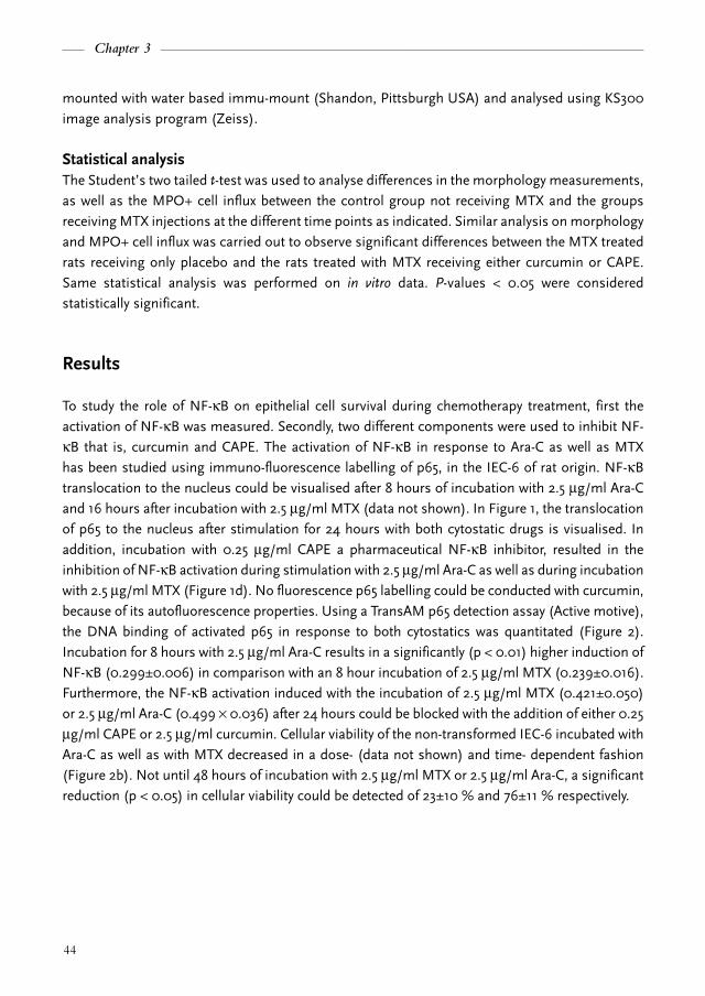
44
_____ Chapter 3 _______________________________________________________________________________________________________________ _____________________________________________________ Role of NF-kappaB inhibition in mucosal barrier injury _____
45
mounted with water based immu-mount (Shandon, Pittsburgh USA) and analysed using KS300 image analysis program (Zeiss).
Statistical analysisThe Student’s two tailed t-test was used to analyse differences in the morphology measurements, as well as the MPO+ cell influx between the control group not receiving MTX and the groups receiving MTX injections at the different time points as indicated. Similar analysis on morphology and MPO+ cell influx was carried out to observe significant differences between the MTX treated rats receiving only placebo and the rats treated with MTX receiving either curcumin or CAPE. Same statistical analysis was performed on in vitro data. P-values < 0.05 were considered statistically significant.
Results
To study the role of NF-κB on epithelial cell survival during chemotherapy treatment, first the activation of NF-κB was measured. Secondly, two different components were used to inhibit NF-κB that is, curcumin and CAPE. The activation of NF-κB in response to Ara-C as well as MTX has been studied using immuno-fluorescence labelling of p65, in the IEC-6 of rat origin. NF-κB translocation to the nucleus could be visualised after 8 hours of incubation with 2.5 µg/ml Ara-C and 16 hours after incubation with 2.5 µg/ml MTX (data not shown). In Figure 1, the translocation of p65 to the nucleus after stimulation for 24 hours with both cytostatic drugs is visualised. In addition, incubation with 0.25 µg/ml CAPE a pharmaceutical NF-κB inhibitor, resulted in the inhibition of NF-κB activation during stimulation with 2.5 µg/ml Ara-C as well as during incubation with 2.5 µg/ml MTX (Figure 1d). No fluorescence p65 labelling could be conducted with curcumin, because of its autofluorescence properties. Using a TransAM p65 detection assay (Active motive), the DNA binding of activated p65 in response to both cytostatics was quantitated (Figure 2). Incubation for 8 hours with 2.5 µg/ml Ara-C results in a significantly (p < 0.01) higher induction of NF-κB (0.299±0.006) in comparison with an 8 hour incubation of 2.5 µg/ml MTX (0.239±0.016). Furthermore, the NF-κB activation induced with the incubation of 2.5 µg/ml MTX (0.421±0.050) or 2.5 µg/ml Ara-C (0.499 × 0.036) after 24 hours could be blocked with the addition of either 0.25 µg/ml CAPE or 2.5 µg/ml curcumin. Cellular viability of the non-transformed IEC-6 incubated with Ara-C as well as with MTX decreased in a dose- (data not shown) and time- dependent fashion (Figure 2b). Not until 48 hours of incubation with 2.5 µg/ml MTX or 2.5 µg/ml Ara-C, a significant reduction (p < 0.05) in cellular viability could be detected of 23±10 % and 76±11 % respectively.
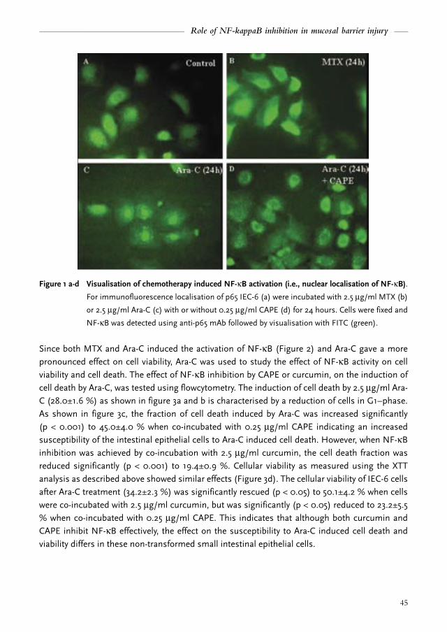
44
_____ Chapter 3 _______________________________________________________________________________________________________________ _____________________________________________________ Role of NF-kappaB inhibition in mucosal barrier injury _____
45
Figure 1 a-d Visualisation of chemotherapy induced NF-κB activation (i.e., nuclear localisation of NF-κB).
For immunofluorescence localisation of p65 IEC-6 (a) were incubated with 2.5 µg/ml MTX (b)
or 2.5 µg/ml Ara-C (c) with or without 0.25 µg/ml CAPE (d) for 24 hours. Cells were fixed and
NF-κB was detected using anti-p65 mAb followed by visualisation with FITC (green).
Since both MTX and Ara-C induced the activation of NF-κB (Figure 2) and Ara-C gave a more pronounced effect on cell viability, Ara-C was used to study the effect of NF-κB activity on cell viability and cell death. The effect of NF-κB inhibition by CAPE or curcumin, on the induction of cell death by Ara-C, was tested using flowcytometry. The induction of cell death by 2.5 µg/ml Ara-C (28.0±1.6 %) as shown in figure 3a and b is characterised by a reduction of cells in G1–phase. As shown in figure 3c, the fraction of cell death induced by Ara-C was increased significantly (p < 0.001) to 45.0±4.0 % when co-incubated with 0.25 µg/ml CAPE indicating an increased susceptibility of the intestinal epithelial cells to Ara-C induced cell death. However, when NF-κB inhibition was achieved by co-incubation with 2.5 µg/ml curcumin, the cell death fraction was reduced significantly (p < 0.001) to 19.4±0.9 %. Cellular viability as measured using the XTT analysis as described above showed similar effects (Figure 3d). The cellular viability of IEC-6 cells after Ara-C treatment (34.2±2.3 %) was significantly rescued (p < 0.05) to 50.1±4.2 % when cells were co-incubated with 2.5 µg/ml curcumin, but was significantly (p < 0.05) reduced to 23.2±5.5 % when co-incubated with 0.25 µg/ml CAPE. This indicates that although both curcumin and CAPE inhibit NF-κB effectively, the effect on the susceptibility to Ara-C induced cell death and viability differs in these non-transformed small intestinal epithelial cells.
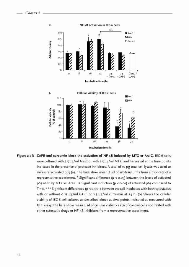
46
_____ Chapter 3 _______________________________________________________________________________________________________________ _____________________________________________________ Role of NF-kappaB inhibition in mucosal barrier injury _____
47
Figure 2 a-b CAPE and curcumin block the activation of NF-κB induced by MTX or Ara-C. IEC-6 cells
were cultured with 2.5 µg/ml Ara-C or with 2.5 µg/ml MTX, and harvested at the time points
indicated in the presence of protease inhibitors. A total of 10 µg total cell lysate was used to
measure activated p65 (a). The bars show mean ± sd of arbitrary units from a triplicate of a
representative experiment. * Significant difference (p < 0.05) between the levels of activated
p65 at 8h by MTX vs. Ara-C. # Significant induction (p < 0.01) of activated p65 compared to
T = 0. *** Significant differences (p < 0.001) between the cell incubated with both cytostatics
with or without 0.25 µg/ml CAPE or 2.5 µg/ml curcumin at 24 h. (b) Shows the cellular
viability of IEC-6 cell cultures as described above at time points indicated as measured with
XTT assay. The bars show mean ± sd of cellular viability as % of control cells not treated with
either cytostatic drugs or NF-κB inhibitors from a representative experiment.

46
_____ Chapter 3 _______________________________________________________________________________________________________________ _____________________________________________________ Role of NF-kappaB inhibition in mucosal barrier injury _____
47
Figure 3 a-d Inhibition of NF-κB by CAPE but not by curcumin increases susceptibility of IEC-6 cells to
Ara-C induced cell death. The percentage of cell death is detected by flowcytometry analysis
of propidium iodide labelled IEC-6 cells incubated with 2.5 µg/ml Ara-C, with or without 0.25
µg/ml CAPE or 2.5 µg/ml curcumin. A total of 10,000 cells were counted and divided into
four sections representative for their DNA content, as indicated in (a) (control cells) and (b)
(Ara-C treated cells). The bars (c) show the % of cell death as mean ± sd of three independent
experiments. *** Significant differences in the apoptotic fraction (p < 0.001). Cellular viability
was measured using XTT assay (d). IEC-6 cells were cultured with 2.5 µg/ml Ara-C with or
without 0.25 µg/ml CAPE or 2.5 µg/ml curcumin at 72 h. The bars show mean ± sd of cellular
viability as % of control cells not treated with either cytostatic drugs or NF-κB inhibitors from
a representative experiment.

48
_____ Chapter 3 _______________________________________________________________________________________________________________ _____________________________________________________ Role of NF-kappaB inhibition in mucosal barrier injury _____
49
In addition to the NF-κB activation by Ara-C, an induction varying from 18- to 129-fold mRNA specific for TNF-α and MCP-1 respectively, was observed after 16 hours of incubation with 2.5 µg/ml Ara-C in a time dependent fashion (Figure 4a). Treatment with MTX resulted in a comparable induction of both TNF-α and MCP-1 (data not shown). The production of TNF-α and MCP-1 seen in response to Ara-C as well as MTX (data not shown) was reduced, although to a different extent, when IEC-6 cells were incubated with 2.5 µg/ml curcumin, indicating a functional inhibition of Ara-C induced NF-κB activation by curcumin (Figure 4b).
Figure 4 a,b Inhibition of NF-κB by curcumin functionally reduces Ara-C induced cytokine and chemokine
response. IEC-6 cells were cultured in the presence of 2.5 µg/ml Ara-C and mRNA was isolated
at time points indicated for RT-PCR (a). (b) Represents IEC-6 cells, which were incubated with
2.5 µg/ml Ara-C with or without 2.5 µg/ml curcumin for 24 hours. PCR products specific for TNF-
α and MCP-1 were normalised to β-actin expressions. The bars represent mean ± sd of three
independent experiments as fold induction compared to cells cultured in the absence of Ara-C.
To determine the effect of curcumin supplementation on chemotherapy mucosal barrier injury, a well-described rat model for MTX-induced intestinal damage was used [39]. First of all NF-κB activation and its mediators were determined in the onset of mucosal barrier injury. CAPE was used in these studies as a second NF-κB inhibitor known to increase the susceptibility to chemotherapy induced cell death. In accordance with in vitro data as depicted in figure 4 on IEC-6 cells, an induction of 17- to 30-fold of rat specific mRNA for TNF-α and MCP-1 could be detected in vivo in the duodenum as soon as 24 hours after the first MTX injection (Figure 5a). Also, an MTX induced cytokine and chemokine response could be detected in the jejunum and to a lesser extent in the ileum (data not shown). No data was obtained in vivo of earlier time points to establish a dose and time dependent relation of the cytokine and chemokine mRNA induction. To locate the cells responsible for the MTX induced TNF-α production, an immuno-histological analysis on frozen sections was performed. As shown in Figure 5b, a specific increase in TNF-α was found in the crypt cell area in response to the MTX treatment, indicating the epithelial cells in this region are responsible for the induction of TNF-α.
48
_____ Chapter 3 _______________________________________________________________________________________________________________ _____________________________________________________ Role of NF-kappaB inhibition in mucosal barrier injury _____
49
Figure 5 a-d Methotrexate induced damage of the small intestine is associated with cytokine and
chemokine responses, localised in the crypt cell compartment of rats. Small intestinal
damage was induced using two injections of 20 mg/kg methotrexate. The induction of
PCR-products from mRNA specific for TNF-α and MCP-1 in the duodenum normalised for
β-actin are shown as fold induction compared to duodenum values of rats at day 0 (a). Bars
represent mean ± sd of three independent experiments. (b) Shows Cross-sections of the
duodenum of rats non-treated or treated with MTX respectively. The sections were stained
for the presence of TNF-α using specific monoclonal antibodies and AEC substrate, which
gives a red colouring. The sections were counter-stained with haematoxylin, and pictures of
representative area were made at magnification of 40x using KS 300 (Zeiss). At time points.
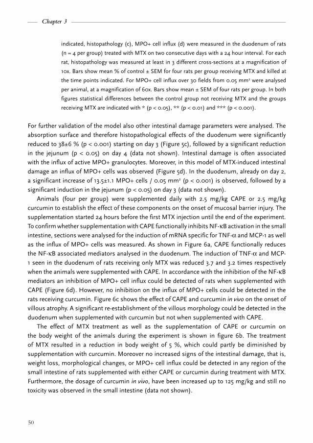
50
_____ Chapter 3 _______________________________________________________________________________________________________________ _____________________________________________________ Role of NF-kappaB inhibition in mucosal barrier injury _____
51
For further validation of the model also other intestinal damage parameters were analysed. The absorption surface and therefore histopathological effects of the duodenum were significantly reduced to 38±6 % (p < 0.001) starting on day 3 (Figure 5c), followed by a significant reduction in the jejunum (p < 0.05) on day 4 (data not shown). Intestinal damage is often associated with the influx of active MPO+ granulocytes. Moreover, in this model of MTX-induced intestinal damage an influx of MPO+ cells was observed (Figure 5d). In the duodenum, already on day 2, a significant increase of 13.5±1.1 MPO+ cells / 0.05 mm2 (p < 0.001) is observed, followed by a significant induction in the jejunum (p < 0.05) on day 3 (data not shown).
Animals (four per group) were supplemented daily with 2.5 mg/kg CAPE or 2.5 mg/kg curcumin to establish the effect of these components on the onset of mucosal barrier injury. The supplementation started 24 hours before the first MTX injection until the end of the experiment. To confirm whether supplementation with CAPE functionally inhibits NF-κB activation in the small intestine, sections were analysed for the induction of mRNA specific for TNF-α and MCP-1 as well as the influx of MPO+ cells was measured. As shown in Figure 6a, CAPE functionally reduces the NF-κB associated mediators analysed in the duodenum. The induction of TNF-α and MCP-1 seen in the duodenum of rats receiving only MTX was reduced 3.7 and 3.2 times respectively when the animals were supplemented with CAPE. In accordance with the inhibition of the NF-κB mediators an inhibition of MPO+ cell influx could be detected of rats when supplemented with CAPE (Figure 6d). However, no inhibition on the influx of MPO+ cells could be detected in the rats receiving curcumin. Figure 6c shows the effect of CAPE and curcumin in vivo on the onset of villous atrophy. A significant re-establishment of the villous morphology could be detected in the duodenum when supplemented with curcumin but not when supplemented with CAPE.
The effect of MTX treatment as well as the supplementation of CAPE or curcumin on the body weight of the animals during the experiment is shown in figure 6b. The treatment of MTX resulted in a reduction in body weight of 5 %, which could partly be diminished by supplementation with curcumin. Moreover no increased signs of the intestinal damage, that is, weight loss, morphological changes, or MPO+ cell influx could be detected in any region of the small intestine of rats supplemented with either CAPE or curcumin during treatment with MTX. Furthermore, the dosage of curcumin in vivo, have been increased up to 125 mg/kg and still no toxicity was observed in the small intestine (data not shown).
indicated, histopathology (c), MPO+ cell influx (d) were measured in the duodenum of rats
(n = 4 per group) treated with MTX on two consecutive days with a 24 hour interval. For each
rat, histopathology was measured at least in 3 different cross-sections at a magnification of
10x. Bars show mean % of control ± SEM for four rats per group receiving MTX and killed at
the time points indicated. For MPO+ cell influx over 30 fields from 0.05 mm2 were analysed
per animal, at a magnification of 60x. Bars show mean ± SEM of four rats per group. In both
figures statistical differences between the control group not receiving MTX and the groups
receiving MTX are indicated with * (p < 0.05), ** (p < 0.01) and *** (p < 0.001).
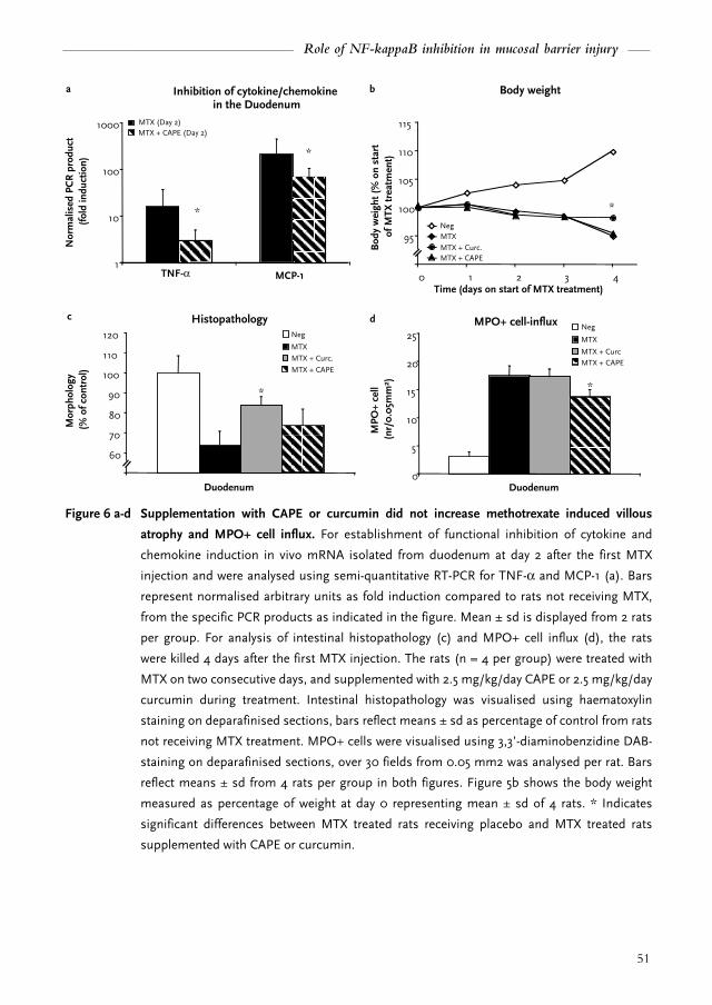
50
_____ Chapter 3 _______________________________________________________________________________________________________________ _____________________________________________________ Role of NF-kappaB inhibition in mucosal barrier injury _____
51
Figure 6 a-d Supplementation with CAPE or curcumin did not increase methotrexate induced villous
atrophy and MPO+ cell influx. For establishment of functional inhibition of cytokine and
chemokine induction in vivo mRNA isolated from duodenum at day 2 after the first MTX
injection and were analysed using semi-quantitative RT-PCR for TNF-α and MCP-1 (a). Bars
represent normalised arbitrary units as fold induction compared to rats not receiving MTX,
from the specific PCR products as indicated in the figure. Mean ± sd is displayed from 2 rats
per group. For analysis of intestinal histopathology (c) and MPO+ cell influx (d), the rats
were killed 4 days after the first MTX injection. The rats (n = 4 per group) were treated with
MTX on two consecutive days, and supplemented with 2.5 mg/kg/day CAPE or 2.5 mg/kg/day
curcumin during treatment. Intestinal histopathology was visualised using haematoxylin
staining on deparafinised sections, bars reflect means ± sd as percentage of control from rats
not receiving MTX treatment. MPO+ cells were visualised using 3,3’-diaminobenzidine DAB-
staining on deparafinised sections, over 30 fields from 0.05 mm2 was analysed per rat. Bars
reflect means ± sd from 4 rats per group in both figures. Figure 5b shows the body weight
measured as percentage of weight at day 0 representing mean ± sd of 4 rats. * Indicates
significant differences between MTX treated rats receiving placebo and MTX treated rats
supplemented with CAPE or curcumin.

52
_____ Chapter 3 _______________________________________________________________________________________________________________ _____________________________________________________ Role of NF-kappaB inhibition in mucosal barrier injury _____
53
Discussion
The activation of NF-κB is not only associated with the induction of inflammatory reactions but also plays an important role in the survival of cells under various cellular stresses [1,40]. It has been hypothesised that through the activation of NF-κB both these processes might modulate the onset of mucosal barrier injury. In more detail, inhibition of the inflammatory responses during chemotherapy might reduce the onset of mucosal barrier injury, whereas inhibition of cell survival regulated by NF-κB might increase villous atrophy the dose limiting side effects of chemotherapy treatment. Therefore, the inhibition of NF-κB by, for instance, curcumin, might have positive as well as negative health effects during anti-cancer therapy. In addition, CAPE was used as a second NF-κB inhibitor to study the possible role for NF-κB in the onset of intestinal atrophy during chemotherapy.
As shown in this study, both the cytostatic drugs MTX and Ara-C induced NF-κB activation in IEC-6 cells, which was followed by the production of pro-inflammatory cytokines and chemokines as well as the induction of cell death. Also in vivo the production of pro-inflammatory cytokines and chemokines after MTX injection could be measured on both mRNA and protein level. The NF-κB related TNF-α production could be especially detected in the crypt cells, suggesting that cytokines and chemokines are produced in particularly by the epithelial stem cells in response to MTX treatment. These results demonstrate the production of pro-inflammatory cytokines and the activation of NF-κB after chemotherapy in vitro and indirectly in vivo. This is in accordance with the hypothesis that chemotherapy induces a pro-inflammatory response in the intestine as previously suggested [15,16]. Besides a direct effect of MTX induced NF-κB activation on the induction of an inflammatory response in the epithelium as shown in Figures 5a,b and 6a, the activation of NF-κB might also be related in a later stage to barrier destruction. This might subsequently lead to the influx of bacteria, resulting in an indirect cytokine response. However, this will not be related to the early onset of mucosal barrier injury.
A significant activation of NF-κB and subsequently the NF-κB regulated cytokine production could be measured after 16 hours of incubation with either MTX or Ara-C,which is in contrast with the finding by others. These previous results indicate that MTX inhibits TNF-α induced NF-κB activation [41]. This discrepancy in findings is probably due to differences in incubation periods used. The TNF-α induced NF-κB activation could only effectively be inhibited when pre-incubation with MTX, and thus pyrimidine pool depletion was established before TNF-α incubation. This indeed might occur before DNA-damage induced NF-κB activation is achieved. Furthermore, TNF-α induced NF-κB activation is receptor mediated, whereas early DNA-damage induced NF-κB activation is not. This indicates different routes of activation. The activation of NF-κB might be related to this induction of cell death. This is, however, probably not the case, since cellular viability did not decrease significantly until 48 hours of incubation with either Ara-C or MTX.
With respect to the role of NF-κB and cytokines on cell survival of epithelial cells in the intestine after chemotherapy treatment, differences between the inhibitors used were obtained
52
_____ Chapter 3 _______________________________________________________________________________________________________________ _____________________________________________________ Role of NF-kappaB inhibition in mucosal barrier injury _____
53
in both IEC-6 cells and MTX treated rats. The inhibition of NF-κB in IEC-6 cells by CAPE resulted in an increased susceptibility of IEC-6 cell to Ara-C induced cell death, whereas inhibition of NF-κB by curcumin resulted in decreased susceptibility to Ara-C induced cell death (Figure 3). Both agents did inhibit NF-κB activation in IEC-6 cells after incubation with Ara-C and MTX (Figure 2). The difference between both agents may in part be due to the fact that inhibition of NF-κB by curcumin and CAPE are achieved at two different levels. Curcumin inhibit NF-κB through the suppression of phoshorylation and degradation of IκBα [42,43,44]. CAPE modifies the NF-κB protein so that it can no longer bind to DNA without any effect on IκBα phosphorylation or IκBα degradation [31,45]. Perhaps, by inhibiting the phosphorylation and degradation of IκBα, degradation of other proteins are also inhibited, whereas the inhibition of NF-κB binding to DNA could induce apoptosis. The difference in the NF-κB inhibitor pathway and response to chemotherapy induced cell death is currently under investigation. The differences between CAPE and curcumin cannot be explained by differences in cytokine production inhibition. Both agents inhibited the production of cytokines (Figure 4). In addition, cytokines it self did not influence Ara-C induced cell death or viability in IEC-6 cells, because addition of cytokines or cytokine specific antibodies to IEC-6 cells during incubation with Ara-C did not have any effect on the induction of cell death (data not shown). Irrespective of its mechanism, in the in vitro study with IEC-6 cells, it was shown that curcumin inhibits the Ara-C induced cell death (Figure 3). This is in contrast with previously described results. Prostate cancer cells become more susceptible to cytostatic drug induced apoptosis when incubated with curcumin [21]. Perhaps, differences between transformed and non-transformed cells account for these different findings.
To study the effect of curcumin supplementation and the inhibition of NF-κB in vivo on chemotherapy-induced mucosal barrier injury, both CAPE and curcumin were tested in an animal model, wherein rats were treated with MTX. The inhibition NF-κB activation using curcumin as shown by Sugimoto et al., [46] in addition to our data, is effective throughout the entire gastrointestinal tract. Although inhibition of NF-κB by CAPE, did increase susceptibility of IEC-6 cells to Ara-C induced cell death, supplementation with CAPE did not increase the small intestinal damage induced by MTX in vivo. Supplementation of CAPE, however, did show an inhibition on the MPO+ cell influx in the duodenum, which correlates with the reduced MCP-1 response (Figure 6). This again suggests an effective NF-κB inhibition by the supplementation of CAPE, since both TNF-α and MCP-1 are NF-κB regulated. The latter result might suggest that the influx of MPO+ cells has only a minor effect on the onset on mucosal barrier injury. Despite the fact, that curcumin reduced in vitro the cytokine response, in vivo supplementation with curcumin did not have any effect on the influx of MPO+ cells (Figure 6). Since supplementation of curcumin shows a significant protection on villous atrophy, this result is in contrast with the in vitro data, where curcumin inhibited chemotherapy induced cell death, at the same concentration at which the activation of NF-κB and subsequently the cytokine and chemokine production was inhibited. In summary a reduction of villous atrophy was achieved without inhibition of the inflammatory response as measured by MPO+ cell influx. This again suggests a minor role for the inflammatory response in the onset of mucosal barrier injury. The results also directly indicate
54
_____ Chapter 3 _______________________________________________________________________________________________________________ _____________________________________________________ Role of NF-kappaB inhibition in mucosal barrier injury _____
55
that the use of CAPE and curcumin does not stimulate the onset of mucosal barrier injury. Since both compounds do not worsen villous atrophy, this also implies that the use of NF-κB inhibitors in order to increase the susceptibility of cancer cells to cytostatic drug induced cell death, does not have a negative effect on the onset of mucosal barrier injury. However, since the effects in vitro using curcumin and CAPE does have a detrimental effect on chemotherapy induced cell death, caution should be exercised when extrapolating these data to the clinic, assuming that the use of NF-κB inhibitors in the clinic to sensitise cancer cells to the chemotherapy treatment moves forward [47,48]. However, due to the differences in pathways to inhibit NF-κB, like with curcumin and CAPE, and also using proteasome inhibitors like PS-341 [47,48], the effect on the deleterious side effects normally seen during anti-cancer treatment, will need extra attention during the trials.
It should be realised that it is not known so far as to whether the NF-κB inhibition obtained by CAPE and curcumin in the doses tested in this study, will increase the susceptibility of various cancers in vivo to cytostatic drug induced cell death. In mice a dosage of 0.2 % w/v curcumin in diet inhibited the development or promotion/progression to disease stages of azoxymethane-induced colonic adenocarcinomas in mice [25,49]. Moreover, a 64 % reduction in adenoma formation in the intestine of Min-mice, which harbour genetic mutation similar to the predisposition to development of adenomatous polyposis in humans was seen using 0.1 % curcumin [50]. Taking into account that rats eat approximately 5 g /day which suggests that with 0.2 % curcumin in the diet an effective concentration of 10 mg/day is required for the chemopreventive properties of curcumin. The highest concentration of curcumin tested in vivo in our studies was 12.5 mg/day without signs of intestinal toxicity. An effective dosage of curcumin to inhibiting NF-κB systemically however remains to be elucidated in humans.
In conclusion, MTX induced NF-κB activation in epithelial cells resulted in production of pro-inflammatory cytokines and chemokines both in vitro and in vivo, which subsequently could be reduced by curcumin and CAPE. Moreover, in vivo supplementation of both curcumin and CAPE did not increase susceptibility to MTX induced small intestinal damage.
Acknowledgement
We thank all biotechnicians of the small animal facility in Wageningen for housing, and assistance with the performance of the animal experiments.
54
_____ Chapter 3 _______________________________________________________________________________________________________________ _____________________________________________________ Role of NF-kappaB inhibition in mucosal barrier injury _____
55
References
1. Chen, F., Castranova, V. & Shi, X. (2001). New insights into the role of nuclear factor-kappaB in cell growth regulation. Am J Pathol, 159, 387-97.
2. Li, J.D., Feng, W., Gallup, M., Kim, J.H., Gum, J., Kim, Y. & Basbaum, C. (1998). Activation of NF-kappaB via a Src-dependent Ras-MAPK-pp90rsk pathway is required for Pseudomonas aeruginosa-induced mucin overproduction in epithelial cells. Proc Natl Acad Sci U S A, 95, 5718-23.
3. Bender, K., Gottlicher, M., Whiteside, S., Rahmsdorf, H.J. & Herrlich, P. (1998). Sequential DNA damage-independent and -dependent activation of NF-kappaB by UV. Embo J, 17, 5170-81.
4. Ghosh, S., May, M.J. & Kopp, E.B. (1998). NF-kappa B and Rel proteins: evolutionarily conserved mediators of immune responses. Annu Rev Immunol, 16, 225-60.
5. Ghosh, S. & Karin, M. (2002). Missing pieces in the NF-kappaB puzzle. Cell, 109, S81-96.6. Finco, T.S. & Baldwin, A.S. (1995). Mechanistic aspects of NF-kappa B regulation: the emerging role of
phosphorylation and proteolysis. Immunity, 3, 263-72.7. Ueda, A., Ishigatsubo, Y., Okubo, T. & Yoshimura, T. (1997). Transcriptional regulation of the human monocyte
chemoattractant protein-1 gene. Cooperation of two NF-kappaB sites and NF-kappaB/Rel subunit specificity. J Biol Chem, 272, 31092-9.
8. Collart, M.A., Baeuerle, P. & Vassalli, P. (1990). Regulation of tumor necrosis factor alpha transcription in macrophages: involvement of four kappa B-like motifs and of constitutive and inducible forms of NF-kappa B. Mol Cell Biol, 10, 1498-506.
9. Baldassarre, F., Mallardo, M., Mezza, E., Scala, G. & Quinto, I. (1995). Regulation of NF-kappa B through the nuclear processing of p105 (NF-kappa B1) in Epstein-Barr virus-immortalized B cell lines. J Biol Chem, 270, 31244-8.
10. Read, M.A., Whitley, M.Z., Williams, A.J. & Collins, T. (1994). NF-kappa B and I kappa B alpha: an inducible regulatory system in endothelial activation. J Exp Med, 179, 503-12.
11. Schmid, R.M. & Adler, G. (2000). NF-kappaB/rel/IkappaB: implications in gastrointestinal diseases. Gastroenterology, 118, 1208-28.
12. Chen, F., Castranova, V., Shi, X. & Demers, L.M. (1999). New insights into the role of nuclear factor-kappaB, a ubiquitous transcription factor in the initiation of diseases. Clin Chem, 45, 7-17.
13. Barnes, P.J. & Karin, M. (1997). Nuclear factor-kappaB: a pivotal transcription factor in chronic inflammatory diseases. N Engl J Med, 336, 1066-71.
14. Wulczyn, F.G., Krappmann, D. & Scheidereit, C. (1996). The NF-kappa B/Rel and I kappa B gene families: mediators of immune response and inflammation. J Mol Med, 74, 749-69.
15. Sonis, S.T. (1998). Mucositis as a biological process: a new hypothesis for the development of chemotherapy-induced stomatotoxicity. Oral Oncol, 34, 39-43.
16. Blijlevens, N.M.A., Donnelly, J.P. & De Pauw, B.E. (2000). Mucosal barrier injury: biology, pathology, clinical counterparts and consequences of intensive treatment for haematological malignancy: an overview. Bone marrow transplantation, 25, 1269-1278.
17. Li, N. & Karin, M. (1998). Ionizing radiation and short wavelength UV activate NF-kappaB through two distinct mechanisms. Proc Natl Acad Sci U S A, 95, 13012-7.
18. Granville, D.J., Carthy, C.M., Jiang, H., Levy, J.G., McManus, B.M., Matroule, J.Y., Piette, J. & Hunt, D.W. (2000). Nuclear factor-kappaB activation by the photochemotherapeutic agent verteporfin. Blood, 95, 256-62.
19. Rothwarf, D.M. & Karin, M. (1999). The NF-kappaB Activation Pathway: A Paradigm in Information Transfer from Membrane to Nucleus. Science Signal Transduction, 5, 1-16.
20. Karin, M. & Lin, A. (2002). NF-kappaB at the crossroads of life and death. Nat Immunol, 3, 221-7.21. Hour, T.C., Chen, J., Huang, C.Y., Guan, J.Y., Lu, S.H. & Pu, Y.S. (2002). Curcumin enhances cytotoxicity of
chemotherapeutic agents in prostate cancer cells by inducing p21WAF1/CIP1 and C/EBPbeta expressions and suppressing NF-kappaB activation. Prostate, 51, 211-218.
22. Chuang, S.E., Yeh, P.Y., Lu, Y.S., Lai, G.M., Liao, C.M., Gao, M. & Cheng, A.L. (2002). Basal levels and patterns of anticancer drug-induced activation of nuclear factor-kappaB (NF-kappaB), and its attenuation by tamoxifen, dexamethasone, and curcumin in carcinoma cells. Biochem Pharmacol, 63, 1709-16.
56
_____ Chapter 3 _______________________________________________________________________________________________________________ _____________________________________________________ Role of NF-kappaB inhibition in mucosal barrier injury _____
57
23. Huang, S., Pettaway, C.A., Uehara, H., Bucana, C.D. & Fidler, I.J. (2001). Blockade of NF-kappaB activity in human prostate cancer cells is associated with suppression of angiogenesis, invasion, and metastasis. Oncogene, 20, 4188-97.
24. Huang, S., Robinson, J.B., Deguzman, A., Bucana, C.D. & Fidler, I.J. (2000). Blockade of nuclear factor-kappaB signaling inhibits angiogenesis and tumorigenicity of human ovarian cancer cells by suppressing expression of vascular endothelial growth factor and interleukin 8. Cancer Res, 60, 5334-9.
25. Rao, C.V., Rivenson, A., Simi, B. & Reddy, B.S. (1995). Chemoprevention of colon carcinogenesis by dietary curcumin, a naturally occurring plant phenolic compound. Cancer Res, 55, 259-66.
26. Joe, B. & Lokesh, B.R. (1994). Role of capsaicin, curcumin and dietary n-3 fatty acids in lowering the generation of reactive oxygen species in rat peritoneal macrophages. Biochim Biophys Acta, 1224, 255-63.
27. Huang, T.S., Lee, S.C. & Lin, J.K. (1991b). Suppression of c-Jun/AP-1 activation by an inhibitor of tumor promotion in mouse fibroblast cells. Proc Natl Acad Sci U S A, 88, 5292-6.
28. Conney, A.H., Lysz, T., Ferraro, T., Abidi, T.F., Manchand, P.S., Laskin, J.D. & Huang, M.T. (1991). Inhibitory effect of curcumin and some related dietary compounds on tumor promotion and arachidonic acid metabolism in mouse skin. Adv Enzyme Regul, 31, 385-96.
29. Huang, M.T., Lysz, T., Ferraro, T., Abidi, T.F., Laskin, J.D. & Conney, A.H. (1991a). Inhibitory effects of curcumin on in vitro lipoxygenase and cyclooxygenase activities in mouse epidermis. Cancer Res, 51, 813-9.
30. Plummer, S.M., Holloway, K.A., Manson, M.M., Munks, R.J., Kaptein, A., Farrow, S. & Howells, L. (1999). Inhibition of cyclo-oxygenase 2 expression in colon cells by the chemopreventive agent curcumin involves inhibition of NF-kappaB activation via the NIK/IKK signalling complex. Oncogene, 18, 6013-20.
31. Natarajan, K., Singh, S., Burke, T.R., Jr., Grunberger, D. & Aggarwal, B.B. (1996). Caffeic acid phenethyl ester is a potent and specific inhibitor of activation of nuclear transcription factor NF-kappa B. Proc Natl Acad Sci U S A, 93, 9090-5.
32. Laranjinha, J., Vieira, O., Madeira, V. & Almeida, L. (1995). Two related phenolic antioxidants with opposite effects on vitamin E content in low density lipoproteins oxidized by ferrylmyoglobin: consumption vs regeneration. Arch Biochem Biophys, 323, 373-81.
33. Jaiswal, A.K., Venugopal, R., Mucha, J., Carothers, A.M. & Grunberger, D. (1997). Caffeic acid phenethyl ester stimulates human antioxidant response element-mediated expression of the NAD(P)H:quinone oxidoreductase (NQO1) gene. Cancer Res, 57, 440-6.
34. Zheng, Z.S., Xue, G.Z., Grunberger, D. & Prystowsky, J.H. (1995). Caffeic acid phenethyl ester inhibits proliferation of human keratinocytes and interferes with the EGF regulation of ornithine decarboxylase. Oncol Res, 7, 445-52
35. Verma, I.M., Stevenson, J.K., Schwarz, E.M., Van Antwerp, D. & Miyamoto, S. (1995). Rel/NF-kappa B/I kappa B family: intimate tales of association and dissociation. Genes Dev, 9, 2723-35.
36. Fraker, P.J., King, L.E., Lill-Elghanian, D. & Telford, W.G. (1995). Quantification of apoptotic events in pure and heterogeneous populations of cells using the flow cytometer. Methods Cell Biol, 46, 57-76.
37. United Kingdom Co-ordinating Committee on Cancer Research (1998).(UKCCCR)Guidelines for the Welfare of Animals in Experimental Neoplasia (Second Edition). Br J Cancer, 77, 1-10.
38. Howarth, G.S., Francis, G.L., Cool, J.C., Xu, X., Byard, R.W. & Read, L.C. (1996). Milk growth factors enriched from cheese whey ameliorate intestinal damage by methotrexate when administered orally to rats. J Nutr, 126, 2519-30.
39. Verburg, M., Renes, I.B., Meijer, H.P., Taminiau, J.A., Buller, H.A., Einerhand, A.W. & Dekker, J. (2000). Selective sparing of goblet cells and paneth cells in the intestine of methotrexate-treated rats. Am J Physiol Gastrointest Liver Physiol, 279, G1037-47.
40. Doostzadeh-Cizeron, J., Yin, S. & Goodrich, D.W. (2000). Apoptosis induced by the nuclear death domain protein p84N5 is associated with caspase-6 and NF-kappa B activation. J Biol Chem, 275, 25336-41.
41. Majumdar, S. & Aggarwal, B.B. (2001). Methotrexate suppresses NF-kappaB activation through inhibition of IkappaBalpha phosphorylation and degradation. J Immunol, 167, 2911-20.
42. Singh, S. & Aggarwal, B.B. (1995). Activation of transcription factor NF-kappa B is suppressed by curcumin (diferuloylmethane). J Biol Chem, 270, 24995-5000.
43. Manna, S.K., Sah, N.K., Newman, R.A., Cisneros, A. & Aggarwal, B.B. (2000). Oleandrin suppresses activation of nuclear transcription factor-kappaB, activator protein-1, and c-Jun NH2-terminal kinase. Cancer Res, 60, 3838-47.
44. Manna, S.K., Mukhopadhyay, A., Van, N.T. & Aggarwal, B.B. (1999). Silymarin suppresses TNF-induced activation of NF-kappa B, c-Jun N-terminal kinase, and apoptosis. J Immunol, 163, 6800-9.
56
_____ Chapter 3 _______________________________________________________________________________________________________________ _____________________________________________________ Role of NF-kappaB inhibition in mucosal barrier injury _____
57
45. Egan, L.J., Mays, D.C., Huntoon, C.J., Bell, M.P., Pike, M.G., Sandborn, W.J., Lipsky, J.J. & McKean, D.J. (1999). Inhibition of interleukin-1-stimulated NF-kappaB RelA/p65 phosphorylation by mesalamine is accompanied by decreased transcriptional activity. J Biol Chem, 274, 26448-53.
46. Sugimoto, K., Hanai, H., Tozawa, K., Aoshi, T., Uchijima, M., Nagata, T. & Koide, Y. (2002). Curcumin prevents and ameliorates trinitrobenzene sulfonic acid-induced colitis in mice. Gastroenterology, 123, 1912-22.
47. Ma, M.H., Yang, H.H., Parker, K., Manyak, S., Friedman, J.M., Altamirano, C., Wu, Z.Q., Borad, M.J., Frantzen, M., Roussos, E., Neeser, J., Mikail, A., Adams, J., Sjak-Shie, N., Vescio, R.A. & Berenson, J.R. (2003). The Proteasome Inhibitor PS-341 Markedly Enhances Sensitivity of Multiple Myeloma Tumor Cells to Chemotherapeutic Agents. Clin Cancer Res, 9, 1136-44.
48. Elliott, P.J., Zollner, T.M. & Boehncke, W.H. (2003). Proteasome inhibition: a new anti-inflammatory strategy. J Mol Med, 81, 235-45.
49. Kawamori, T., Lubet, R., Steele, V.E., Kelloff, G.J., Kaskey, R.B., Rao, C.V. & Reddy, B.S. (1999). Chemopreventive effect of curcumin, a naturally occurring anti-inflammatory agent, during the promotion/progression stages of colon cancer. Cancer Res, 59, 597-601.
50. Mahmoud, N.N., Carothers, A.M., Grunberger, D., Bilinski, R.T., Churchill, M.R., Martucci, C., Newmark, H.L. & Bertagnolli, M.M. (2000). Plant phenolics decrease intestinal tumors in an animal model of familial adenomatous polyposis. Carcinogenesis, 21, 921-7.

Measuring mucosal damage induced by cytotoxic therapy
Chapter 4
N.M.A. Blijlevens1, B. van’t Land2, J.P. Donnelly1, L.M’Rabet2 and B.E. De Pauw1
1Department of Hematology, University Medical Center St. Radboud, Nijmegen2Numico-Research BV, Department of Clinical Nutrition and Therapeutic Diets Wageningen,
The Netherlands
Support Care Cancer. 2004;12(4):227-233

60
_____ Chapter 4 _______________________________________________________________________________________________________________ ____________________________________________________ Measuring mucosal damage induced by cytotoxic therapy _____
61
60
_____ Chapter 4 _______________________________________________________________________________________________________________ ____________________________________________________ Measuring mucosal damage induced by cytotoxic therapy _____
61
Summary
We scored oral mucositis, and gut toxicity and measured sugar permeability testing among 56 recipients of a haematopoietic stem cell transplant (HSCT) given myeloablative conditioning with idarubicin, cyclophosphamide and TBI, and a group of 18 patients given cytotoxic chemotherapy for newly diagnosed acute myeloid leukaemia (AML) or myelodysplastic syndrome (MDS). Gut integrity was already disturbed in the AML/MDS group as measured by the lactulose/rhamnose ratio (L/R ratio = 0.09) before therapy and was severely perturbed (L/R ratio > 0.13) for a month after HSCT. Oral mucositis and to a lesser extent gut toxicity was only significantly correlated with disturbed permeability in the transplant group. The data suggest that sugar permeability, oral mucositis and gut toxicity measure different features of mucosal damage after intensive cytotoxic therapy.
Introduction
Oral mucositis is reported to affect 60 to 100 % of haematopoietic stem cell transplant (HSCT) recipients when myeloablative preparative regimens are used [1]. Less is known about the gastrointestinal counterpart, although gut injury results in significant morbidity and even mortality [2]. While the severe pain of oral mucositis is often the most obvious and troublesome symptom, the extent of gut mucosal injury is difficult to determine because the signs and symptoms such as nausea, vomiting, diarrhoea and abdominal cramps affect almost every HSCT recipient. An alternative means of defining gut injury might be to employ sugar permeability tests as these are generally accepted as surrogate markers of gut disease or damage in patients treated for cancer [3]. Sugar permeability tests are also non-invasive and can test changes in the permeability and absorption (loss of epithelial surface) which are the principle features of gut injury. The conditioning regimen used for HSCT is reported to be an independent risk factor of oral mucositis [4]. In our institution, a higher leukaemia-free survival was found for patients with standard risk acute leukaemia who had been prepared for an allogeneic HSCT with an intensified myeloablative regimen comprising idarubicin (4-demethoxydaunorubicin), cyclophosphamide and TBI [5]. However, the patients experienced prolonged and worsened oral mucositis which was ascribed to the addition of idarubicin [6]. It is not known whether this was also true of the gut. Therefore, we employed a sugar permeability test consisting of a solution of the disaccharide lactulose and the monosaccharides 3-O-methylglucose, L-rhamnose and D-xylose in a cohort of HSCT recipients given idarubicin as part of their myeloablative therapy. Lactulose is absorbed by paracellular transport that is, via open tight-junctions and villous tip extrusion zone and any increase of urinary excretion represents loss of integrity. The other three sugars measure absorption: 3-O-methylglucose measures ATP-dependent transcellular transport, D-xylose detects carrier-mediated transcellular transport and L-rhamnose monitors passive transcellular diffusion, a decrease in urinary excretion of each sugar represents loss of epithelial cells and/or
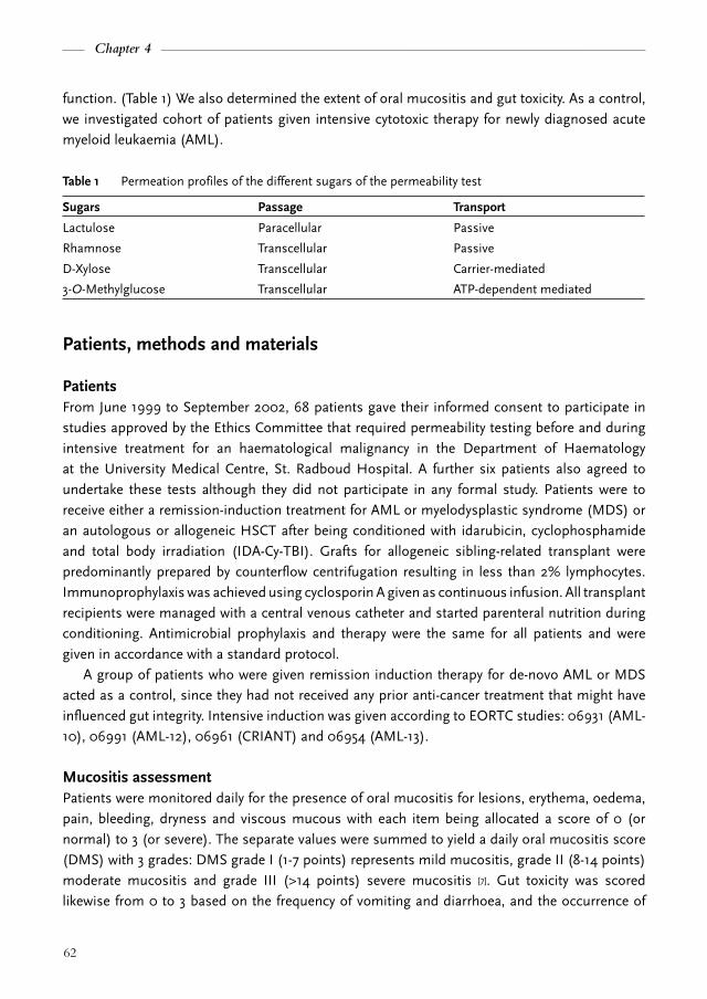
62
_____ Chapter 4 _______________________________________________________________________________________________________________ ____________________________________________________ Measuring mucosal damage induced by cytotoxic therapy _____
63
function. (Table 1) We also determined the extent of oral mucositis and gut toxicity. As a control, we investigated cohort of patients given intensive cytotoxic therapy for newly diagnosed acute myeloid leukaemia (AML).
Table 1 Permeation profiles of the different sugars of the permeability test
Sugars Passage Transport
Lactulose Paracellular Passive
Rhamnose Transcellular Passive
D-Xylose Transcellular Carrier-mediated
3-O-Methylglucose Transcellular ATP-dependent mediated
Patients, methods and materials
PatientsFrom June 1999 to September 2002, 68 patients gave their informed consent to participate in studies approved by the Ethics Committee that required permeability testing before and during intensive treatment for an haematological malignancy in the Department of Haematology at the University Medical Centre, St. Radboud Hospital. A further six patients also agreed to undertake these tests although they did not participate in any formal study. Patients were to receive either a remission-induction treatment for AML or myelodysplastic syndrome (MDS) or an autologous or allogeneic HSCT after being conditioned with idarubicin, cyclophosphamide and total body irradiation (IDA-Cy-TBI). Grafts for allogeneic sibling-related transplant were predominantly prepared by counterflow centrifugation resulting in less than 2% lymphocytes. Immunoprophylaxis was achieved using cyclosporin A given as continuous infusion. All transplant recipients were managed with a central venous catheter and started parenteral nutrition during conditioning. Antimicrobial prophylaxis and therapy were the same for all patients and were given in accordance with a standard protocol.
A group of patients who were given remission induction therapy for de-novo AML or MDS acted as a control, since they had not received any prior anti-cancer treatment that might have influenced gut integrity. Intensive induction was given according to EORTC studies: 06931 (AML-10), 06991 (AML-12), 06961 (CRIANT) and 06954 (AML-13).
Mucositis assessmentPatients were monitored daily for the presence of oral mucositis for lesions, erythema, oedema, pain, bleeding, dryness and viscous mucous with each item being allocated a score of 0 (or normal) to 3 (or severe). The separate values were summed to yield a daily oral mucositis score (DMS) with 3 grades: DMS grade I (1-7 points) represents mild mucositis, grade II (8-14 points) moderate mucositis and grade III (>14 points) severe mucositis [7]. Gut toxicity was scored likewise from 0 to 3 based on the frequency of vomiting and diarrhoea, and the occurrence of
62
_____ Chapter 4 _______________________________________________________________________________________________________________ ____________________________________________________ Measuring mucosal damage induced by cytotoxic therapy _____
63
nausea, abdominal complaints, faecal incontinency and volume of diarrhoea. Summation of the separate score yielded the daily gut score (DGS) grade I (1-6 points) represents mild toxicity, grade II (7-12 points) moderate toxicity and grade III (>12 points) severe toxicity. All gut toxicities developing after chemotherapy had ceased were ascribed to mucositis unless there was a more plausible alternative cause, such as an adverse drug reaction or a proven infectious cause, such as toxigenic enterocolitis.
Sugar permeability testAn isotonic solution was prepared by dissolving 5 g lactulose (LAC), 1 g L-rhamnose (RHA), 0.2 g 3-O-methylglucose (OMG), 0.5 g D-xylose (XYL) in 100 ml demineralised water. The sugar test was produced according to good manufacturing practice by our own hospital pharmacy department. The first sugar permeability test (baseline day) was performed before starting remission-induction or conditioning therapy. After an overnight fast, the subjects emptied their bladders and then drank the solution. A light breakfast with a drink was allowed after 2 hours. Urine was collected for 5 hours, after which the total volume passed was recorded and aliquots of approximately 5 mL were stored in -80°C until analysis was performed. The test was repeated every Wednesday after baseline.
Determination of the sugars in urineThe concentration of each sugar was determined using the method of Jansen et al [8]. Briefly, before analyses, samples were thawed and centrifuged at 3,000g for 30 minutes to remove any precipitates that might have been formed during storage. Next, 100 µL of internal standard sugar (1.0 mmol/L D-glucoheptose (99%), Sigma, Zwijndrecht, The Netherlands) was added to 100 µL aqueous sugar standard or urine before being completely dried using a SpeedVac (Savant Plus, Thermoquest, Breda, The Netherlands). Sugar-rings were made accessible for 100 µl of N-Methyl-N-(trimethylsilyl) trifluoroacetamide (MSTFA), (ICN Biochemicals, Aurora, Ohio) by adding 100 µL ortho-methylhydroxylamine (Fluka, Switserland) solution of 20 g/L pyridine (Fluka) and incubating at 60°C for 1 hour while vortexing (Vibrax). At the end of this time, 100 µl MSTFA was added, and the mixture was again incubated at 60°C while vortexing for 30 minutes. Extraction of the stable derivatives was achieved by adding 1 mL water and 2 mL hexane (Merck, Darmstadt, Germany) and then thoroughly mixing for at least 1 minute. Then the hexane layer containing the derivatives was transferred to a new glass tube, the hexane was evaporated in a SpeedVac to complete dryness and the derivatives were re-dissolved in a mixture of 75 µL MSTFA with 75 µL pyridine and incubated for 30 minutes at 60°C while vortexing. Samples were transferred to airtight vials and 1 µL was injected into a gas chromatograph connected to a Flame Ionisation Detector (Varian 3800 GC, Tempe, Arizona) using a split ratio of 1:25 at 250°C. The separation was carried out on an Alltech DB-5 column (0.25 mm ID, 0.25 µm film thickness, length 30 m (Alltech, Deerfield IL). The helium flow was 1 mL/min, and the split-rate was between carrier gas and the sample was 25:1. The column temperature program started at 125°C for 5 min then increased initially by 3°C/min to 180°C, and by 5°C/min to 230°C which was sustained for
64
_____ Chapter 4 _______________________________________________________________________________________________________________ ____________________________________________________ Measuring mucosal damage induced by cytotoxic therapy _____
65
5 minutes. The sugar-derivatives were detected using the FID detector set at 250°C. Data were collected on-line using Star Chromatography software (Varian, Tempe, Arizona, USA) and peak areas were corrected to the internal standard.
The concentration of each sugar was then estimated in µmol/L using standard calibration curves and urinary recovery of each sugar was calculated as a percentage of intake. The coefficient of variance for LAC, XYL, RHA and OMG were 2.3%, 3.7%, 3.1% and 2.3%. Expressing the urinary excretion of two sugars, for example lactulose and rhamnose, as a ratio minimises the influence of extraneous factors such as gastric emptying and bowel transit time. Therefore the lactulose/rhamnose ratio (L/R) was used as an index of intestinal permeability or integrity and a L/R ratio of 0.02 or less was considered to be normal. The D-xylose/3-O-methylglucose (XYL/OMG) ratio was used as an index of absorptive capacity and a ratio of 0.65 was considered normal [9].
Data analysisThe DMS and DGS that had been noted on the days that the sugar permeability test was performed or, if missing, scores recorded the day before or after, were used for the analysis which was undertaken using SPSS 10.
The Mann-Whitney U test was used for significance calculations of differences in DMS and DGS between baseline and consecutive samples within a treatment group and between treatment groups on the same sampling days. Sugar concentrations were transformed to logarithmic values in order to obtain a normal distribution. Urinary recovery was used to calculate the ratios and expressed as the mean ± the standard deviation and a 2-sided unpaired student’s t-test was used to calculate significant differences with respect to baseline and analysis of variance for differences between groups. A p-value < 0.05 was considered statistically significant. Pearson’s ρ test and Kendall’s τ test were used respectively to estimate correlations between parametric and non-parametric data.
Results
Baseline dataDemographic data of the control group and the transplant group are reported in Table 2. There were slightly more males than females in each group but their median age was similar.
At baseline, DMS and DGS were significantly higher in the control group (p < 0.001). The sugar concentrations and urinary recoveries were not significantly different between each group and the urinary recovery of RHA, XYL, OMG and LAC is depicted in Table 3. The recovery of RHA, XYL and OMG was lower in both groups.
Subsequently, the L/R ratio was significantly (p < 0.001) higher in the control group (0.09±0.05) than in the transplant group (L/R 0.04±0.02), and both baseline L/R ratios were higher than the as normal reported value of less than 0.02. (Table 4) The XYL/OMG ratio was not significantly different between both groups (0.57±0.15 and 0.65±0.15) before starting therapy and was comparable with the reference ratio of 0.65.
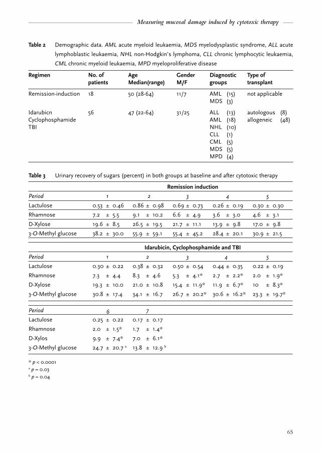
64
_____ Chapter 4 _______________________________________________________________________________________________________________ ____________________________________________________ Measuring mucosal damage induced by cytotoxic therapy _____
65
Table 2 Demographic data. AML acute myeloid leukaemia, MDS myelodysplastic syndrome, ALL acute
lymphoblastic leukaemia, NHL non-Hodgkin’s lymphoma, CLL chronic lymphocytic leukaemia,
CML chronic myeloid leukaemia, MPD myeloproliferative disease
Regimen No. of patients
AgeMedian(range)
GenderM/F
Diagnostic groups
Type of transplant
Remission-induction 18 50 (28-64) 11/7 AML (15)MDS (3)
not applicable
IdarubicnCyclophosphamideTBI
56 47 (22-64) 31/25 ALL (13)AML (18)NHL (10)CLL (1)CML (5)MDS (5)MPD (4)
autologous (8)allogeneic (48)
Table 3 Urinary recovery of sugars (percent) in both groups at baseline and after cytotoxic therapy
Remission induction
Period 1 2 3 4 5
Lactulose 0.53 ± 0.46 0.86 ± 0.98 0.69 ± 0.73 0.26 ± 0.19 0.30 ± 0.30
Rhamnose 7.2 ± 5.5 9.1 ± 10.2 6.6 ± 4.9 3.6 ± 3.0 4.6 ± 3.1
D-Xylose 19.6 ± 8.5 26.5 ± 19.5 21.7 ± 11.1 13.9 ± 9.8 17.0 ± 9.8
3-O-Methyl glucose 38.2 ± 30.0 55.9 ± 59.1 55.4 ± 45.2 28.4 ± 20.1 30.9 ± 21.5
Idarubicin, Cyclophosphamide and TBI
Period 1 2 3 4 5
Lactulose 0.30 ± 0.22 0.38 ± 0.32 0.50 ± 0.54 0.44 ± 0.35 0.22 ± 0.19
Rhamnose 7.3 ± 4.4 8.3 ± 4.6 5.3 ± 4.1* 2.7 ± 2.2* 2.0 ± 1.9*
D-Xylose 19.3 ± 10.0 21.0 ± 10.8 15.4 ± 11.9* 11.9 ± 6.7* 10 ± 8.3*
3-O-Methyl glucose 30.8 ± 17.4 34.1 ± 16.7 26.7 ± 20.2* 30.6 ± 16.2* 23.3 ± 19.7*
Period 6 7
Lactulose 0.25 ± 0.22 0.17 ± 0.17
Rhamnose 2.0 ± 1.5* 1.7 ± 1.4*
D-Xylos 9.9 ± 7.4* 7.0 ± 6.1*
3-O-Methyl glucose 24.7 ± 20.7 a 13.8 ± 12.9 b
* p < 0.0001a p = 0.03b p = 0.04
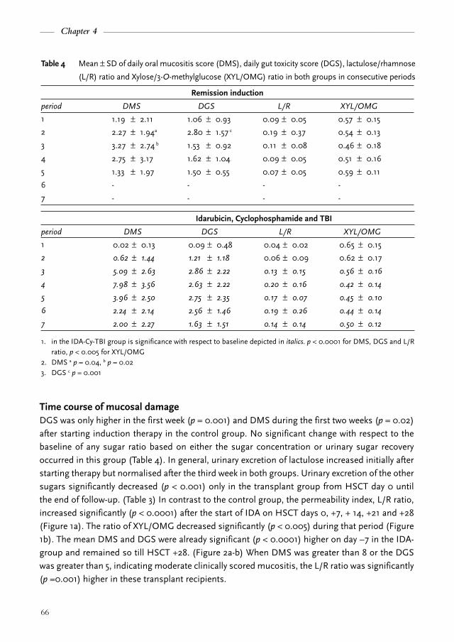
66
_____ Chapter 4 _______________________________________________________________________________________________________________ ____________________________________________________ Measuring mucosal damage induced by cytotoxic therapy _____
67
Time course of mucosal damage DGS was only higher in the first week (p = 0.001) and DMS during the first two weeks (p = 0.02) after starting induction therapy in the control group. No significant change with respect to the baseline of any sugar ratio based on either the sugar concentration or urinary sugar recovery occurred in this group (Table 4). In general, urinary excretion of lactulose increased initially after starting therapy but normalised after the third week in both groups. Urinary excretion of the other sugars significantly decreased (p < 0.001) only in the transplant group from HSCT day 0 until the end of follow-up. (Table 3) In contrast to the control group, the permeability index, L/R ratio, increased significantly (p < 0.0001) after the start of IDA on HSCT days 0, +7, + 14, +21 and +28 (Figure 1a). The ratio of XYL/OMG decreased significantly (p < 0.005) during that period (Figure 1b). The mean DMS and DGS were already significant (p < 0.0001) higher on day –7 in the IDA-group and remained so till HSCT +28. (Figure 2a-b) When DMS was greater than 8 or the DGS was greater than 5, indicating moderate clinically scored mucositis, the L/R ratio was significantly (p =0.001) higher in these transplant recipients.
Table 4 Mean ± SD of daily oral mucositis score (DMS), daily gut toxicity score (DGS), lactulose/rhamnose
(L/R) ratio and Xylose/3-O-methylglucose (XYL/OMG) ratio in both groups in consecutive periods
Remission induction
period DMS DGS L/R XYL/OMG
1 1.19 ± 2.11 1.06 ± 0.93 0.09 ± 0.05 0.57 ± 0.15
2 2.27 ± 1.94a 2.80 ± 1.57 c 0.19 ± 0.37 0.54 ± 0.13
3 3.27 ± 2.74 b 1.53 ± 0.92 0.11 ± 0.08 0.46 ± 0.18
4 2.75 ± 3.17 1.62 ± 1.04 0.09 ± 0.05 0.51 ± 0.16
5 1.33 ± 1.97 1.50 ± 0.55 0.07 ± 0.05 0.59 ± 0.11
6 - - - -
7 - - - -
Idarubicin, Cyclophosphamide and TBI
period DMS DGS L/R XYL/OMG
1 0.02 ± 0.13 0.09 ± 0.48 0.04 ± 0.02 0.65 ± 0.15
2 0.62 ± 1.44 1.21 ± 1.18 0.06 ± 0.09 0.62 ± 0.17
3 5.09 ± 2.63 2.86 ± 2.22 0.13 ± 0.15 0.56 ± 0.16
4 7.98 ± 3.56 2.63 ± 2.22 0.20 ± 0.16 0.42 ± 0.14
5 3.96 ± 2.50 2.75 ± 2.35 0.17 ± 0.07 0.45 ± 0.10
6 2.24 ± 2.14 2.56 ± 1.46 0.19 ± 0.26 0.44 ± 0.14
7 2.00 ± 2.27 1.63 ± 1.51 0.14 ± 0.14 0.50 ± 0.12
1. in the IDA-Cy-TBI group is significance with respect to baseline depicted in italics. p < 0.0001 for DMS, DGS and L/R ratio, p < 0.005 for XYL/OMG2. DMS a p = 0.04, b p = 0.023. DGS c p = 0.001
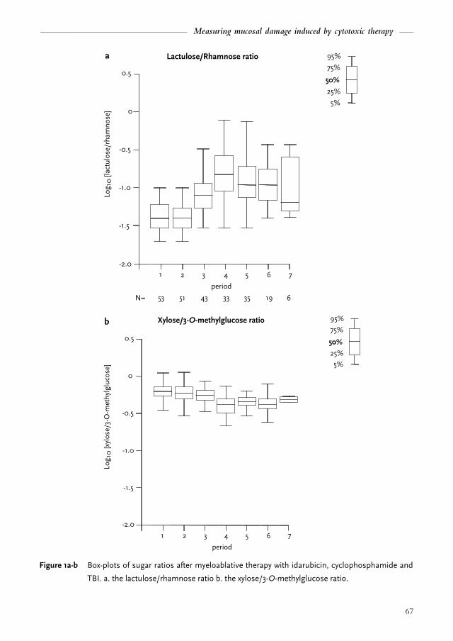
66
_____ Chapter 4 _______________________________________________________________________________________________________________ ____________________________________________________ Measuring mucosal damage induced by cytotoxic therapy _____
67
Figure 1a-b Box-plots of sugar ratios after myeloablative therapy with idarubicin, cyclophosphamide and
TBI. a. the lactulose/rhamnose ratio b. the xylose/3-O-methylglucose ratio.
a
b
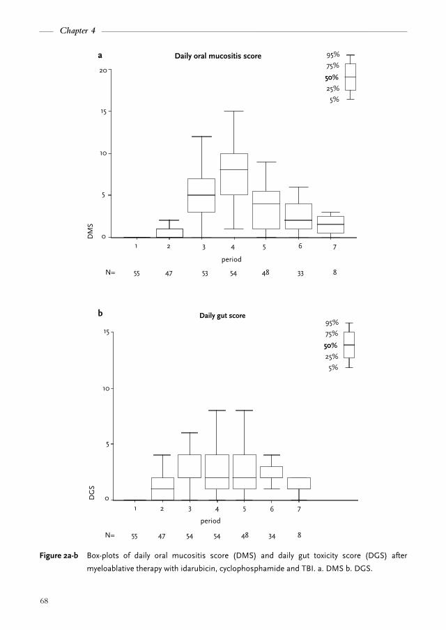
68
_____ Chapter 4 _______________________________________________________________________________________________________________ ____________________________________________________ Measuring mucosal damage induced by cytotoxic therapy _____
69
Figure 2a-b Box-plots of daily oral mucositis score (DMS) and daily gut toxicity score (DGS) after
myeloablative therapy with idarubicin, cyclophosphamide and TBI. a. DMS b. DGS.
a
b
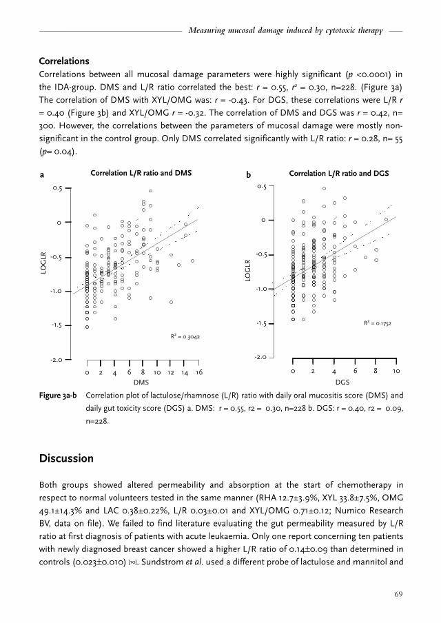
68
_____ Chapter 4 _______________________________________________________________________________________________________________ ____________________________________________________ Measuring mucosal damage induced by cytotoxic therapy _____
69
CorrelationsCorrelations between all mucosal damage parameters were highly significant (p <0.0001) in the IDA-group. DMS and L/R ratio correlated the best: r = 0.55, r2 = 0.30, n=228. (Figure 3a) The correlation of DMS with XYL/OMG was: r = -0.43. For DGS, these correlations were L/R r = 0.40 (Figure 3b) and XYL/OMG r = -0.32. The correlation of DMS and DGS was r = 0.42, n= 300. However, the correlations between the parameters of mucosal damage were mostly non-significant in the control group. Only DMS correlated significantly with L/R ratio: r = 0.28, n= 55 (p= 0.04).
Figure 3a-b Correlation plot of lactulose/rhamnose (L/R) ratio with daily oral mucositis score (DMS) and
daily gut toxicity score (DGS) a. DMS: r = 0.55, r2 = 0.30, n=228 b. DGS: r = 0.40, r2 = 0.09,
n=228.
Discussion
Both groups showed altered permeability and absorption at the start of chemotherapy in respect to normal volunteers tested in the same manner (RHA 12.7±3.9%, XYL 33.8±7.5%, OMG 49.1±14.3% and LAC 0.38±0.22%, L/R 0.03±0.01 and XYL/OMG 0.71±0.12; Numico Research BV, data on file). We failed to find literature evaluating the gut permeability measured by L/R ratio at first diagnosis of patients with acute leukaemia. Only one report concerning ten patients with newly diagnosed breast cancer showed a higher L/R ratio of 0.14±0.09 than determined in controls (0.023±0.010) [10]. Sundstrom et al. used a different probe of lactulose and mannitol and
a b

70
_____ Chapter 4 _______________________________________________________________________________________________________________ ____________________________________________________ Measuring mucosal damage induced by cytotoxic therapy _____
71
also found a higher ratio in 16 patients with AML de novo [11]. We too, found a higher baseline L/R ratio of 0.09±0.05 in patients with newly AML, suggesting that the gut function is already altered in those patients having cancer even before any therapy is given. The L/R ratio reflects both an increase in lactulose permeation, through loss of integrity of the mucosal barrier constituted by tight junctions between epithelial cells and decrease in passive rhamnose absorption indicating a lowered surface area. The alteration in L/R ratio before starting chemotherapy was largely the result of the lower recovery of rhamnose. The mediated absorption of xylose and 3-O-methylglucose was also lower. It is therefore, tempting to speculate that this was caused by the presence of active leukaemia via the production of pro-inflammatory cytokines that depressed gut function [12]. These patients had also a higher DMS and DGS score at baseline that might also reflect active disease. The urinary recoveries were also lower before starting myeloablative conditioning but this might have been the result of previous exposure to cytotoxic therapy rather than active disease since the haematological malignancy was either in remission or inactive. The L/R ratio we found before myeloablative therapy was in the same range (0.04±0.02) as reported for 35 patients undergoing an autologous HSC transplant after high-dose chemotherapy [13].
Permeability increased whilst absorption decreased after conditioning with IDA-Cy-TBI with respect to patients given remission-induction chemotherapy for AML/MDS. Sugar tests measure different aspects of gut toxicity than do clinical measures and there was only a marginal correlation with oral mucositis. Moreover, the L/R ratio remained unchanged throughout the following weeks after induction therapy just as Sunstrom et al found when using their lactulose/mannitol probe [11].
The highest L/R ratio of 0.20±0.16 was seen on HSCT day +7, when Keefe et al also noted a maximum L/R ratio of 0.62 [13]. The L/R ratio has almost returned to within the normal range by HSCT day +28 (that is, 6 weeks after starting chemotherapy) in Keefe’s group, whereas the L/R ratio in our transplant group remained elevated. The protracted declining XYL/OMG ratio after conditioning reflects loss of mediated transport capacity of the gut since there was less uptake of xylose, which takes place predominantly in the duodenum and jejunum. OMG transport was less affected presumably because of the huge reserve capacity of the gut [14]. The prolonged disturbance of the ratios suggest that both increased permeability and decreased absorptive capacity of the gut persists long after morphological restoration of the direct toxic effects on the epithelium [15]. It is conceivable that evolving acute graft-versus host disease of the gut might have played a role but this would have to be investigated by endoscopy which is not done routinely. While these ratios are non-specific they do indicate when disruption of the mucosal barrier can be expected following treatment with a specific myeloablative regimen.
The pattern of increase in DMS and to a lesser extent in DGS resembled that found for permeability and absorptive capacity and the correlation reached statistical significance but was only marginal from a clinical perspective. We found a better correlation between oral mucositis and perturbed permeability (r= 0.55) than with gut toxicity unlike Johansen et al who observed a correlation (r= 0.63) between permeability using 51Cr-EDTA, which measures paracellular transport and gut toxicity using the WHO criteria [16]. The correlation between permeability and
70
_____ Chapter 4 _______________________________________________________________________________________________________________ ____________________________________________________ Measuring mucosal damage induced by cytotoxic therapy _____
71
gut toxicity for our HSCT recipients was much lower suggesting that permeability tests cannot predict the onset or severity of either oral mucositis or gut toxicity or vice versa. Similarly there was virtually no correlation between the DMS and DGS amongst HSCT recipients suggesting that each instrument measure different aspects of the mucosal damage that follows intensive cytotoxic therapy. The instruments are also non-specific but can be used to assess the onset of mucosal barrier injury for specific chemotherapy regimens. Each instrument also indicated that mucosal barrier injury developed concurrently with bone marrow aplasia essentially placing the patient in double jeopardy since there is neither a natural obstacle to opportunistic pathogens translocating from the gut to the tissues nor are there effective phagocytes to tackle them. It seems prudent to employ the sugar test because it provides an objective measure for estimating gut toxicity together with an established instrument for measuring oral mucositis to explore the relationship between mucosal barrier injury and the complications that occur following myeloablative therapy.
Acknowledgements
The authors would like to thank M Balvers for technical assistance.
References
1. Parulekar, W., Mackenzie, R., Bjarnason, G. & Jordan, R.C. (1998) Scoring oral mucositis. Oral Oncol., 34, 63-71.2. Rapoport AP, Miller Watelet LF, Linder T, Rberly S, Raubertas RF, Lipp J, Duerst R, Abboud CN, Constine L, Andrews
J, Etter MA, Spear L, Powley E, Packman CH, Rowe JM, Schwertschlag U, Bedrosian C & Liesveld JI (1999) Analysis of factors that correlate with mucositis in recipients of autologous and allogeneic stem-cell transplants. J Clin Oncol, 17, 2446-2453.
3. Bjarnason, I., MacPherson A & Hollander D (1995) Intestinal permeability: an overview. Gastroenterology, 108, 1566-1581.
4. Wardley, A.M., Jayson GC, Swindell R, Morgenstern GR, Chang J, Bloor R, Fraser CJ & Scarffe JH (2000) Prospective evaluation of oral mucositis in patients receiving myeloablative conditioning regimens and haematopoietic progenitor rescue. Br.J.Haematol., 110:292-299.
5. Schaap, N., Schattenberg, A., Bar B, Preijers F, Geurts van Kessel A, van der Maazen R, De Boo T & De Witte T (1997) Outcome of transplantation for standard-risk leukaemia with grafts depleted of lymphocytes after conditioning with an intensified regimen. British Journal of Haematology, 98, 750-759.
6. Muus, P., Donnelly, P., Schattenberg, A., Linssen, P., Minderman, H., Dompeling, E. & de-Witte, T. (1993) Idarubicin-related side effects in recipients of T-cell-depleted allogeneic bone marrow transplants are schedule dependent. Semin.Oncol., 20, 47-52.
7. Donnelly, J.P., Muus, P., Schattenberg, A., de-Witte, T., Horrevorts, A. & DePauw, B.E. (1992) A scheme for daily monitoring of oral mucositis in allogeneic BMT recipients. Bone Marrow Transplant., 9, 409-413.
8. Jansen G, Muskiet F.A.J., Schierbeek H., Berger R. & Van melle G (1986) Capillary gas chromatographic profiling of urinary, plasma and erythrocyte sugars and polyols as their trimethylsilyl derivates, preceded by a simple and rapid prepurification method. Clinica Chimica Acta, 157, 277-294.
9. Lim, S.G., Menzies, I.S., Lee, C.A., Johnson, M.A. & Pounder, R.E. (1993) Intestinal permeability and function in patients infected with human immunodeficiency virus: a comparison with celiac disease. Scand.J.Gastroenterol, 28, 573-580.
72
_____ Chapter 4 _______________________________________________________________________________________________________________
10. Parrilli, G., Iaffaioli, R.V., Martorano, M., Cuomo, R., Tafuto, S., Zampino, M.G., Budillon, G. & Bianco, A.R. (1989) Effects of anthracycline therapy on intestinal absorption in patients with advanced breast cancer. Cancer Res., 49, 3689-3691.
11. Sundstrom GM, Wahlin A, Nordin-Andersson I & Suhr OB (1998) Intestinal permeability in patients with acute myeloid leukemia. Eur J Haematol, 61, 250-254.
12. Nitenberg, G., Raynard, B. (2000) Nutritional support of the cancer patient: issues and dilemmas. Crit.Rev Oncol.Hematol, 34, 137-168.
13. Keefe, D.M., Cummins, A.G., Dale, B.M., Kotasek, D., Robb, T.A. & Sage, R.E. (1997) Effect of high-dose chemotherapy on intestinal permeability in humans. Clin.Sci.Colch., 92, 385-389.
14. Menzies IS (1984) Transmucosal passage of inert molecules in health and disease. Intestinal absorption and secretion (ed. by Skadhauge E. and Heller R.), p.527. MTP Press, Lancaster.
15. Keefe, D.M., Brealey, J, Goland, G.J., Cummins, A.G. (2000) Chemotherapy for cancer causes apoptosis that precedes hypolplasia in crypts of the small intestine in humans. Gut, 47, 632-637.
16. Johansson, J.E. & Ekman, T. (1997) Gastro-intestinal toxicity related to bone marrow transplantation: disruption of the intestinal barrier precedes clinical findings. Bone Marrow Transplant., 19, 921-925.
Prospective evaluation of gut mucosal barrier injury following various myeloablative regimens
for haematopoietic stem cell transplant
Chapter 5
N.M.A. Blijlevens, J.P. Donnelly and B.E. De Pauw
Department of Hematology, University Medical Center St. Radboud, Nijmegen
The Netherlands
Bone Marrow Transplant. 2005;35(7):707-11

74
_____ Chapter 5 _______________________________________________________________________________________________________________ ___________________________________________________________ Prospective evaluation of gut mucosal barrier injury _____
75
74
_____ Chapter 5 _______________________________________________________________________________________________________________ ___________________________________________________________ Prospective evaluation of gut mucosal barrier injury _____
75
Summary
We determined gut mucosal barrier injury (MBI) among 129 recipients of an allogeneic or autologous haematopoietic stem cell transplant (HSCT) who had been given different myeloablative regimens by measuring integrity using the lactulose/rhamnose ratio and absorption using the ratios of rhamnose/3-O-methylglucose and xylose/3-O-methylglucose. Regimens that did not contain idarubicin induced oral mucositis and perturbed gut integrity and absorption earlier than did those containing the anthracycline. By contrast, regimens containing idarubicin induced more severe and prolonged oral and gut MBI. Gut integrity and absorption of most patients were still abnormal at discharge from hospital. These results confirm that the integrity and absorptive capacity of the gut is affected adversely by myeloablative regimens in general, although only two patterns of mucosal injury emerged depending on whether or not idarubicin was used.
Introduction
Mucosal barrier injury (MBI) of the oral cavity is reported to affect more than 60% of haematopoietic stem cell transplant (HSCT) recipients given myeloablative preparative regimens. Wardley and colleagues showed that the character, onset and progression of oral mucositis were markedly influenced by the nature of the conditioning therapy. For instance, high-dose melphalan aggravated oral mucositis whereas its onset was delayed after busulphan [2].
Much less is known about the gastrointestinal counterpart of oral mucositis although gut injury results in significant morbidity and mortality [3]. Sugar permeability tests can determine objectively changes in permeability (integrity and absorption) and have been found to complement the signs and symptoms associated with gut toxicity such as nausea, vomiting, diarrhoea and abdominal cramps [4]. However, it is not known whether gut MBI, is also affected differently by the various myeloablative regimens. Moreover, our standard conditioning regimen consists of idarubicin, cyclophosphamide and TBI [5] and is known to induce prolonged and severe oral mucositis [6]. Therefore, we employed a sugar permeability test to determine differences in gut permeability, integrity and absorption of a cohort of HSCT recipients given different myeloablative regimens [1].
Patients, methods and materials
PatientsFrom June 1999 to September 2002, 83 patients admitted to the Department of Haematology at the University Medical Centre, St. Radboud Hospital for intensive treatment of haematological malignancy had consented to participate in prospective studies approved by the Ethics Committee, which required sugar permeability testing before and during the trials. A further 46 patients had also agreed to undertake these tests although they did not participate in any formal
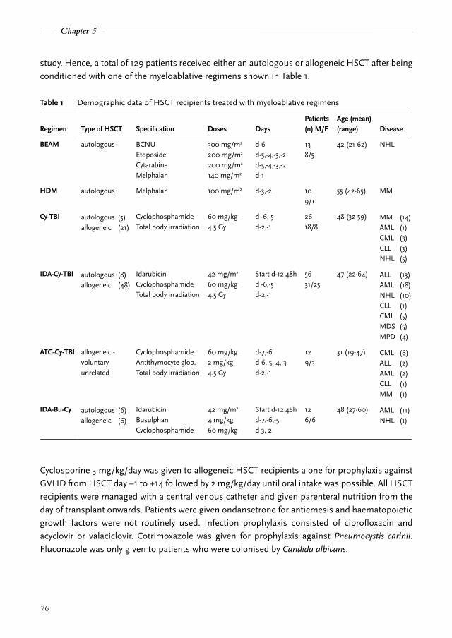
76
_____ Chapter 5 _______________________________________________________________________________________________________________ ___________________________________________________________ Prospective evaluation of gut mucosal barrier injury _____
77
study. Hence, a total of 129 patients received either an autologous or allogeneic HSCT after being conditioned with one of the myeloablative regimens shown in Table 1.
Table 1 Demographic data of HSCT recipients treated with myeloablative regimens
Regimen Type of HSCT Specification Doses Days Patients (n) M/F
Age (mean) (range) Disease
BEAM autologous BCNU Etoposide CytarabineMelphalan
300 mg/m2 200 mg/m2 200 mg/m2 140 mg/m2
d-6 d-5,-4,-3,-2 d-5,-4,-3,-2 d-1
138/5
42 (21-62) NHL
HDM autologous Melphalan 100 mg/m2 d-3,-2 109/1
55 (42-65) MM
Cy-TBI autologous (5)allogeneic (21)
CyclophosphamideTotal body irradiation
60 mg/kg 4.5 Gy
d -6,-5 d-2,-1
2618/8
48 (32-59) MM (14)AML (1)CML (3)CLL (3)NHL (5)
IDA-Cy-TBI autologous (8)allogeneic (48)
IdarubicinCyclophosphamideTotal body irradiation
42 mg/m2
60 mg/kg 4.5 Gy
Start d-12 48h d -6,-5 d-2,-1
56 31/25
47 (22-64) ALL (13)AML (18)NHL (10)CLL (1)CML (5)MDS (5)MPD (4)
ATG-Cy-TBI allogeneic -voluntary unrelated
CyclophosphamideAntithymocyte glob.Total body irradiation
60 mg/kg 2 mg/kg 4.5 Gy
d-7,-6 d-6,-5,-4,-3d-2,-1
129/3
31 (19-47) CML (6)ALL (2)AML (2)CLL (1)MM (1)
IDA-Bu-Cy autologous (6)allogeneic (6)
IdarubicinBusulphanCyclophosphamide
42 mg/m2
4 mg/kg60 mg/kg
Start d-12 48hd-7,-6,-5d-3,-2
126/6
48 (27-60) AML (11)NHL (1)
Cyclosporine 3 mg/kg/day was given to allogeneic HSCT recipients alone for prophylaxis against GVHD from HSCT day –1 to +14 followed by 2 mg/kg/day until oral intake was possible. All HSCT recipients were managed with a central venous catheter and given parenteral nutrition from the day of transplant onwards. Patients were given ondansetrone for antiemesis and haematopoietic growth factors were not routinely used. Infection prophylaxis consisted of ciprofloxacin and acyclovir or valaciclovir. Cotrimoxazole was given for prophylaxis against Pneumocystis carinii. Fluconazole was only given to patients who were colonised by Candida albicans.
76
_____ Chapter 5 _______________________________________________________________________________________________________________ ___________________________________________________________ Prospective evaluation of gut mucosal barrier injury _____
77
Mucositis assessmentPatients were monitored daily for the presence of oral mucositis for lesions, erythema, oedema, pain, bleeding, dryness and viscous mucous with each item being allocated a score of 0 (or normal) to 3 (or severe). These values were summed to yield a daily oral mucositis score (DMS) with 3 grades: DMS grade I (1-7 points) for mild mucositis, grade II (8-14 points) for moderate mucositis and grade III (>14 points) for severe mucositis. DMS grade II is comparable with WHO grade 3 [7]. Gut toxicity was determined from the frequency of vomiting and diarrhoea, and the occurrence of nausea, abdominal complaints, faecal incontinency and the volume of diarrhoea. Each item was scored from 0 to 3 and the values were summed to yield the daily gut score (DGS) with grade I (1-6 points) representing mild toxicity, grade II (7-12 points) moderate toxicity and grade III (>12 points) severe toxicity. All gut toxicities developing following chemotherapy were attributed to mucositis unless there was a more plausible alternative cause such as an adverse drug reaction or a proven infection.
Sugar permeability test An isotonic solution was prepared by dissolving 5 g lactulose (LAC), 1 g L-rhamnose (RHA), 0.2 g 3-O-methylglucose (OMG), 0.5 g D-xylose (XYL) in 100 ml demineralised water. The test solution was produced according to good manufacturing practice by our own hospital pharmacy. After an overnight fast, subjects emptied their bladders and then drank the solution. A light breakfast with a drink was allowed after 2 hours. Urine was collected for 5 hours after which the total volume passed was recorded, and aliquots of approximately 5 mL were stored in -80°C until analysis was performed. The first sugar permeability test was performed immediately before starting myeloablative therapy and formed the baseline. The test was repeated every Wednesday until discharge to allow changes in MBI to be followed adequately, while avoiding any potential interference from clinically apparent acute GVHD.
Determination of the sugarsLactulose is a disaccharide and is absorbed by paracellular transport that is, via open tight junctions and villous tip extrusion zone, so any increase in urinary excretion represents loss of integrity. The other sugars are monosaccharides with L-rhamnose being used to monitor passive transcellular diffusion, D-xylose to detect carrier-mediated transcellular transport and 3-O-methylglucose to measure ATP-dependent transcellular transport. A decrease in urinary excretion of any of these sugars represents a smaller surface area implying a loss of epithelial cell function or number. The concentration of each sugar was determined by gas chromatography employing flame ionisation for detection and D-glucoheptose as an internal standard [8]. Data were collected on-line using Star Chromatography software (Varian, Tempe, Arizona, USA) and peak areas were calculated and adjusted to the internal standard. The concentration of each sugar was then estimated in µmol/L using standard calibration curves and the urinary recovery of each sugar was calculated as a percentage of oral intake. The coefficients of variance for lactulose, D-xylose, L-rhamnose and 3-O-methylglucose were 2.3%, 3.7%, 3.1% and 2.3% respectively. Urinary excretion

78
_____ Chapter 5 _______________________________________________________________________________________________________________ ___________________________________________________________ Prospective evaluation of gut mucosal barrier injury _____
79
of two sugars, for example lactulose and rhamnose, was expressed as a ratio to minimise the influence of extraneous factors such as gastric emptying and bowel transit time. The lactulose/rhamnose ratio (LAC/RHA) was used as an index of intestinal integrity with a LAC/RHA ratio of 0.02 or less being considered normal while a higher LAC/RHA ratio represents perturbed gut integrity. Similarly, the L-rhamnose/3-O-methylglucose (RHA/OMG) and D-xylose/3-O-methylglucose (XYL/OMG) ratios were used, respectively, as indices of absorptive capacity and mediated cellular transport with ratios of less than 0.28 [9] and 0.65 [10] respectively, representing diminished absorptive capacity. Normal volunteers tested in the same manner as our patients resulted in the following mean ratios: LAC/RHA 0.03±0.01[s.d.], RHA/OMG 0.26±0.004 [s.d.], XYL/OMG 0.71±0.12 [s.d.] (Numico Research BV, data on file).
Data analysisThe sugar ratios and corresponding mean DMS and DGS scores were calculated, using all available data for the analysis, which was undertaken with SPSS 10. Missing data were replaced with the values for the day before or after the sugar test was carried out. An analysis of variance was used to test differences between and within groups. The Mann-Whitney U test was used for significance calculations of differences in mean DMS and DGS between baseline and consecutive samples within a treatment group and between treatment groups. The sugar concentrations were transformed to logarithms in order to obtain a normal distribution. The urinary recovery was used to calculate the ratios and expressed as the mean with 95% confidence interval and a 2-sided unpaired student’s t-test was used to calculate significant differences with respect to baseline. A p-value < 0.05 was considered statistically significant.
Results
Baseline dataThe LAC/RHA ratios were not significantly different between the patients in the various treatment groups (LAC/RHA ratio; 0.04 - 0.06) but were higher than normal. The ratios of XYL/OMG (0.57 - 0.69) and RHA/OMG (0,20 – 0,25) were also not significantly different between the patient groups but were lower than normal, except for the RHA/OMG ratios of patients treated with IDA-Bu-Cy 0,39 [0.09-0.70]. The sugar ratios were not significantly different between patients who had been previously received chemotherapy and those who had not, including patients with CML (data not shown).
Differences in MBI Gut integrity measured by LAC/RHA ratioThe mean LAC/RHA ratios of patients given Cy-TBI, ATG-Cy-TBI, BEAM and HDM were significantly higher (p < 0.003) by week 2 than at baseline but 1 week later for those treated with idarubicin (p < 0.0001). (Figure 1) Furthermore, at week 2 the mean LAC/RHA ratios of patients
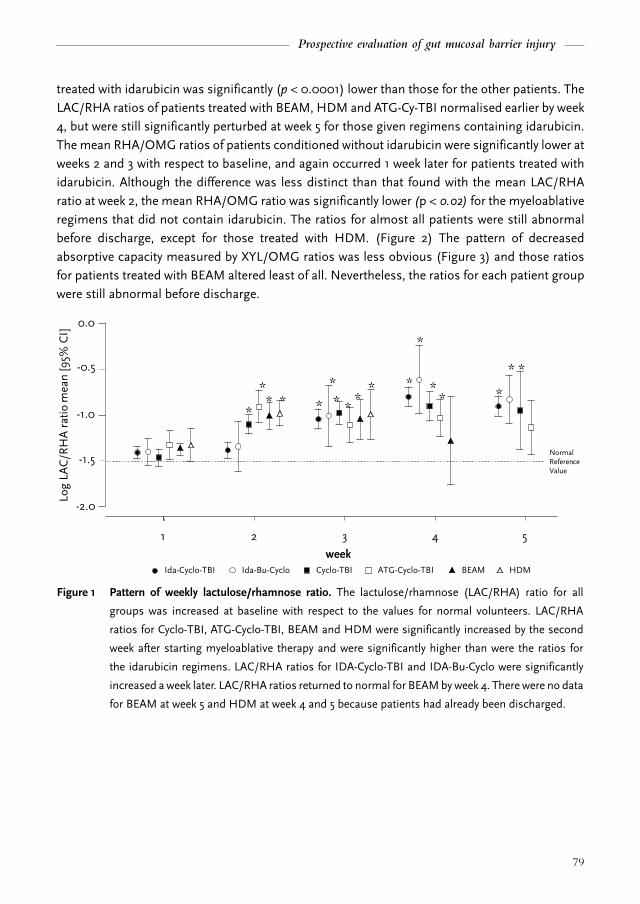
78
_____ Chapter 5 _______________________________________________________________________________________________________________ ___________________________________________________________ Prospective evaluation of gut mucosal barrier injury _____
79
treated with idarubicin was significantly (p < 0.0001) lower than those for the other patients. The LAC/RHA ratios of patients treated with BEAM, HDM and ATG-Cy-TBI normalised earlier by week 4, but were still significantly perturbed at week 5 for those given regimens containing idarubicin.The mean RHA/OMG ratios of patients conditioned without idarubicin were significantly lower at weeks 2 and 3 with respect to baseline, and again occurred 1 week later for patients treated with idarubicin. Although the difference was less distinct than that found with the mean LAC/RHA ratio at week 2, the mean RHA/OMG ratio was significantly lower (p < 0.02) for the myeloablative regimens that did not contain idarubicin. The ratios for almost all patients were still abnormal before discharge, except for those treated with HDM. (Figure 2) The pattern of decreased absorptive capacity measured by XYL/OMG ratios was less obvious (Figure 3) and those ratios for patients treated with BEAM altered least of all. Nevertheless, the ratios for each patient group were still abnormal before discharge.
Figure 1 Pattern of weekly lactulose/rhamnose ratio. The lactulose/rhamnose (LAC/RHA) ratio for all
groups was increased at baseline with respect to the values for normal volunteers. LAC/RHA
ratios for Cyclo-TBI, ATG-Cyclo-TBI, BEAM and HDM were significantly increased by the second
week after starting myeloablative therapy and were significantly higher than were the ratios for
the idarubicin regimens. LAC/RHA ratios for IDA-Cyclo-TBI and IDA-Bu-Cyclo were significantly
increased a week later. LAC/RHA ratios returned to normal for BEAM by week 4. There were no data
for BEAM at week 5 and HDM at week 4 and 5 because patients had already been discharged.
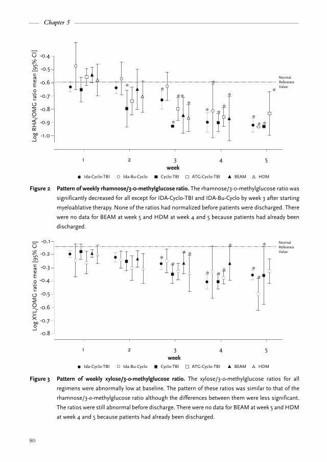
80
_____ Chapter 5 _______________________________________________________________________________________________________________ ___________________________________________________________ Prospective evaluation of gut mucosal barrier injury _____
81
Figure 2 Pattern of weekly rhamnose/3-0-methylglucose ratio. The rhamnose/3-0-methylglucose ratio was
significantly decreased for all except for IDA-Cyclo-TBI and IDA-Bu-Cyclo by week 3 after starting
myeloablative therapy. None of the ratios had normalized before patients were discharged. There
were no data for BEAM at week 5 and HDM at week 4 and 5 because patients had already been
discharged.
week
Figure 3 Pattern of weekly xylose/3-0-methylglucose ratio. The xylose/3-0-methylglucose ratios for all
regimens were abnormally low at baseline. The pattern of these ratios was similar to that of the
rhamnose/3-o-methylglucose ratio although the differences between them were less significant.
The ratios were still abnormal before discharge. There were no data for BEAM at week 5 and HDM
at week 4 and 5 because patients had already been discharged.
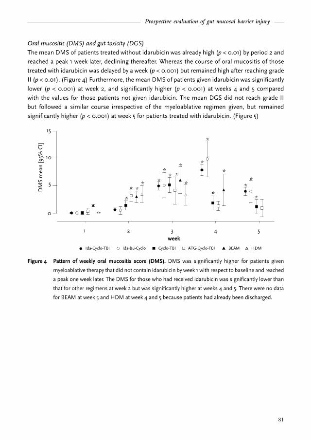
80
_____ Chapter 5 _______________________________________________________________________________________________________________ ___________________________________________________________ Prospective evaluation of gut mucosal barrier injury _____
81
Oral mucositis (DMS) and gut toxicity (DGS)The mean DMS of patients treated without idarubicin was already high (p < 0.01) by period 2 and reached a peak 1 week later, declining thereafter. Whereas the course of oral mucositis of those treated with idarubicin was delayed by a week (p < 0.001) but remained high after reaching grade II (p < 0.01). (Figure 4) Furthermore, the mean DMS of patients given idarubicin was significantly lower (p < 0.001) at week 2, and significantly higher (p < 0.001) at weeks 4 and 5 compared with the values for those patients not given idarubicin. The mean DGS did not reach grade II but followed a similar course irrespective of the myeloablative regimen given, but remained significantly higher (p < 0.001) at week 5 for patients treated with idarubicin. (Figure 5)
Figure 4 Pattern of weekly oral mucositis score (DMS). DMS was significantly higher for patients given
myeloablative therapy that did not contain idarubicin by week 1 with respect to baseline and reached
a peak one week later. The DMS for those who had received idarubicin was significantly lower than
that for other regimens at week 2 but was significantly higher at weeks 4 and 5. There were no data
for BEAM at week 5 and HDM at week 4 and 5 because patients had already been discharged.
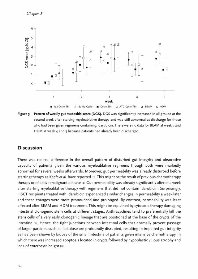
82
_____ Chapter 5 _______________________________________________________________________________________________________________ ___________________________________________________________ Prospective evaluation of gut mucosal barrier injury _____
83
Figure 5 Pattern of weekly gut mucositis score (DGS). DGS was significantly increased in all groups at the
second week after starting myeloablative therapy and was still abnormal at discharge for those
who had been given regimens containing idarubicin. There were no data for BEAM at week 5 and
HDM at week 4 and 5 because patients had already been discharged.
Discussion
There was no real difference in the overall pattern of disturbed gut integrity and absorptive capacity of patients given the various myeloablative regimens though both were markedly abnormal for several weeks afterwards. Moreover, gut permeability was already disturbed before starting therapy as Keefe et al. have reported [11]. This might be the result of previous chemotherapy therapy or of active malignant disease [4]. Gut permeability was already significantly altered a week after starting myeloablative therapy with regimens that did not contain idarubicin. Surprisingly, HSCT recipients treated with idarubicin experienced similar changes in permeability a week later and these changes were more pronounced and prolonged. By contrast, permeability was least affected after BEAM and HDM treatment. This might be explained by cytotoxic therapy damaging intestinal clonogenic stem cells at different stages. Anthracyclines tend to preferentially kill the stem cells of a very early clonogenic lineage that are positioned at the base of the crypts of the intestine [12]. Hence, the tight junctions between intestinal cells that normally prevent passage of larger particles such as lactulose are profoundly disrupted, resulting in impaired gut integrity as has been shown by biopsy of the small intestine of patients given intensive chemotherapy, in which there was increased apoptosis located in crypts followed by hypoplastic villous atrophy and loss of enterocyte height [13].
82
_____ Chapter 5 _______________________________________________________________________________________________________________ ___________________________________________________________ Prospective evaluation of gut mucosal barrier injury _____
83
Alterations in gut integrity preceded the changes in absorptive capacity and tended to normalise earlier. This indicates that tight junctions are loosened before functional enterocytes are reduced in number and that gut integrity tends to be restored before absorptive capacity returns to normal. It is known that tight junctions between intestinal cells are already restored before they become fully matured enterocytes capable of normal absorption [14]. The different times to normalisation after the various myeloablative regimens suggest that not only is inhibition of intestinal clonogenic stem cell growth important but also that different myeloablative regimens affect the capacity for repairing the mucosa differently, which is mediated by the trefoil factor and other intestinal growth factors produced by goblet cells [15, 16].
Wardley et al. [2] showed that the myeloablative regimen and not the disease or stem cell source determined the severity of oral mucositis with the highest peak of mucositis occurring after HDM. However their analysis did not include patients treated with myeloablative regimens containing idarubicin [2]. In our study, gut toxicity preceded oral mucositis in all treatment groups probably because DGS measures direct cytotoxic effects like nausea and vomiting that occur soon after starting myeloablative therapy even with proper antiemetic therapy with 5-HT
3 receptor
antagonists. This suggests that DMS, DGS and sugar permeability tests do actually measure different features of MBI. As we noted earlier [4], diarrhoea or abnormal permeability might also be early manifestations of acute GVHD of the gut [17], although our follow up did not allow us to determine this. Only gut biopsy could confirm this, but these patients are considered too fragile to perform this invasive procedure. Our main interest was to look for differences in integrity and absorptive capacity shortly after starting myeloablative therapy to coincide with neutropenia, which usually resolves before clinically overt acute GVHD occurs.
In conclusion, our study clearly showed that while sugar permeability tests could not distinguish between the MBI induced by different myeloablative regimens, they can determine the onset of disruption and dysfunction of the mucosal barrier of the small intestines and might therefore prove useful for testing interventions designed to ameliorate or prevent this complication.
Acknowledgements
We like to thank M. Balvers and B. van’t Land for technical assistance.
84
_____ Chapter 5 _______________________________________________________________________________________________________________
References
1. Parulekar W, Mackenzie R, Bjarnason G, Jordan RC. Scoring oral mucositis. Oral Oncol 1998; 34(1):63-71.2. Wardley AM, Jayson GC, Swindell R, Morgenstern GR, Chang J, Bloor R et al. Prospective evaluation of oral
mucositis in patients receiving myeloablative conditioning regimens and haematopoietic progenitor rescue. Br J Haematol 2000; 110:292-299
3. Rapoport AP, Miller Watelet LF, Linder T, Rberly S, Raubertas RF, Lipp J et al. Analysis of factors that correlate with mucositis in recipients of autologous and allogeneic stem-cell transplants. J Clin Oncol 1999; 17:2446-2453.
4. Blijlevens N.M.A., van’t Land B., Donnelly JP, M’Rabet L., DePauw BE. Measuring mucosal damage induced by cytotoxic therapy. Support Care Cancer 2004; 12:227-233.
5. Schaap N, Schattenberg A, Bar B, Preijers F, Geurts van Kessel A, van der Maazen R et al. Outcome of transplantation for standard-risk leukaemia with grafts depleted of lymphocytes after conditioning with an intensified regimen. Br J Haematol 1997; 98:750-759.
6. Muus P, Donnelly P, Schattenberg A, Linssen P, Minderman H, Dompeling E et al. Idarubicin-related side effects in recipients of T-cell-depleted allogeneic bone marrow transplants are schedule dependent. Semin Oncol 1993; 206 (Suppl 8):47-52.
7. Donnelly JP, Muus P, Schattenberg A, de-Witte T, Horrevorts A, DePauw BE. A scheme for daily monitoring of oral mucositis in allogeneic BMT recipients. Bone Marrow Transplant 1992; 96:409-413.
8. Jansen G, Muskiet FAJ., Schierbeek H., Berger R., Van melle G. Capillary gas chromatographic profiling of urinary, plasma and erythrocyte sugars and polyols as their trimethylsilyl derivates, preceded by a simple and rapid prepurification method. Clinica Chimica Acta 1986; 157:277-294.
9. Johnston JD, Harvey CJ, Menzies IS, Treacher DF. Gastrointestinal permeability and absorptive capacity in sepsis. Crit Care Med 1996; 24:1144-1149.
10. Lim SG, Menzies IS, Lee CA, Johnson MA, Pounder RE. Intestinal permeability and function in patients infected with Human Immunodeficiency Virus: a comparison with Coeliac Disease. Scand J Gastroenterol 1993; 28:573-580.
11. Keefe DM, Cummins AG, Dale BM, Kotasek D, Robb TA, Sage RE. Effect of high-dose chemotherapy on intestinal permeability in humans. Clin Sci Colch 1997; 924:385-389.
12. Ijiri K, Potten CS. Response of intestinal cells of differing topographical and hierarchical status to ten cytotoxic drugs and five sources of radiation. Br J Cancer 1983; 47:175-185.
13. Keefe DMK, Brealey J, Goland GJ, Cummins AG. Chemotherapy for cancer causes apoptosis that precedes hypoplasia in crypts of the small intestine in humans. Gut 2000; 47:632-637.
14. Cummins AG, Thompson FM, Butler RN, Cassidy JC, Gillis D, Lorenzetti M et al. Improvement in intestinal permeability precedes morphometric recovery of the small intestine in coeliac disease. Clinical Science 2001; 100:379-386.
15. Verburg M., Renes I.B., van Nispen D.J.P.M., Ferdinandusse S., Jorritsma M., Buller. et al. Specific responses in rat small intestinal epithelial mRNA expression and protein levels during chemotherapeutic damage and regeneration. J Histochem Cytochem 2002; 5011:1525-1536.
16. Beck P.L., Wong J.F., Li Y., Swaminathan S., Xavier R.J., Devaney K.L. et al. Chemotherapy- and radiotherapy-induced intestinal damage is regulated by intestinal trefoil factor. Gastroenterology 2004; 126:796-808.
17. Fegan C, Poynton CH, Whittaker JA. The gut mucosal barrier in bone marrow transplantation. Bone Marrow Transplant 1990; 56:373-377.
Citrulline: a potentially simple quantitative marker of intestinal epithelial damage following
myeloablative therapy
Chapter 6
N.M.A. Blijlevens1, L.C.H.W. Lutgens2, A.V.M.B. Schattenberg1 and J.P. Donnelly1
1Department of Hematology, University Medical Center St. Radboud, Nijmegen2Department of Radiation Therapy and Oncology, MAASTRO Clinic, Maastricht
The Netherlands
Bone Marrow Transplant. 2004;34(3):193-196

86
_____ Chapter 6 _______________________________________________________________________________________________________________ ____________________________________________________________ Citrulline: a marker of intestinal epithelial damage _____
87
86
_____ Chapter 6 _______________________________________________________________________________________________________________ ____________________________________________________________ Citrulline: a marker of intestinal epithelial damage _____
87
Summary
We noted a significant decline in the serum concentrations of citrulline of 32 haematopoietic stem cell transplant recipients following intensive myeloablative therapy during the first 3 weeks after transplant when patients have oral mucositis, a markedly disturbed gut integrity (L/R ratio) and are most at risk of infection and other severe complications. Closer inspection of the citrulline concentrations of 12 patients confirmed that the decline did indeed correspond with the onset of oral mucositis and altered gut integrity. Since serum citrulline is a reliable biochemical marker of small bowel enterocyte mass in humans with villous-atrophy associated diseases, it may prove a useful marker for intestinal mucosal damage induced by chemotherapy, allowing the relationship between gut mucosal damage and post-transplant complications including infections to be explored more readily.
Introduction
Mucositis is a manifestation of mucosal barrier injury and is the most frequent cause of morbidity associated with the myeloablative conditioning treatment to prepare for an haematopoietic stem cell transplantation (HSCT) [1]. Oral mucositis is easy to recognise whereas detection of intestinal mucosal injury relies essentially on non-specific symptoms like nausea, vomiting, diarrhoea and abdominal cramps, which affect almost every HSCT recipient. An alternative means of recognising gut mucosal barrier injury is required if it is to be better understood and managed. Alterations in permeability and loss of epithelial surface are features of gut injury that can be detected by sugar permeability tests which are non-invasive and generally accepted as a surrogate marker of gut damage in patients treated for cancer [2]. However, sugar permeability tests are cumbersome and wholly dependent on patient compliance. A marker of intestinal injury that could be determined in blood would be ideal but none has been described so far. Citrulline might prove valuable in this regard since it is an end-product of glutamine metabolism of small intestinal enterocytes, cells which lack the necessary cytosolic enzymes to convert the amino acid to arginine [3]. Blood concentrations of citrulline directly reflect the cell mass of the small intestine since the small intestine is the principal source of the amino acid and it is not metabolised by the liver [4]. In mice, citrulline levels were used to quantify acute small bowel epithelial damage after single-dose whole body irradiation [5]. The high-dose chemotherapy used to prepare patients to receive an HSCT injures the small intestine by inducing crypt apoptosis, hypoplastic villous atrophy and loss of enterocytes [6]. We determined the amino-acid profiles in the serum of a cohort of 32 HSCT recipients who had participated in a prospective randomised placebo-controlled study approved by the local Ethics Committee, the aim of which was to investigate the possible benefits of parenteral nutrition (PN) supplemented with glutamine-dipeptide. As citrulline was included, we were able to relate the concentrations temporally with oral mucositis, gut toxicity and sugar permeability tests (SPT).
88
_____ Chapter 6 _______________________________________________________________________________________________________________ ____________________________________________________________ Citrulline: a marker of intestinal epithelial damage _____
89
Patients and methods
Patients32 patients (12 F; 20 M) with a mean age of 48 years (range 25-65) had given their informed consent to participate in the prospective study and all had received an HLA-matched T-cell depleted sibling HSCT to treat a haematological malignancy (AML (nine), ALL (seven), NHL (seven), MDS (three), CML (two), MPD (two), CLL (one), MF (one). The preparative regimen consisted of idarubicin to lower the risk of relapse [7, 8] given at a dose of 42 mg/m2 by continuous infusion over 48 hours starting 12 days before transplant (HSCT day –12), followed by 120 mg/kg cyclophosphamide (60 mg/kg per day on HSCT days – 6 & - 5) and 9 Gy of total body irradiation (4.5 Gy per day on HSCT days –2 & -1). Ciclosporin 3 mg/kg/day was given as prophylaxis against GVHD by continuous infusion from HSCT day –1 to +14 followed by 2 mg/kg/day until oral intake was possible. Haematopoietic growth factors were not used. Anti-infective prophylaxis consisted of ciprofloxacin and acyclovir or valaciclovir. Co-trimoxazole (sulphamethoxazole-trimethoprim) was given as Pneumocystis carinii prophylaxis. No prophylactic systemic antifungal agents were given. Cefipime or ceftazidime was administered empirically at the onset of fever. All recipients were given PN supplemented by either glutamine-dipeptide or placebo on HSCT day –6. Glomerular filtration rate was determined on admission by measuring renal creatinine clearance (mL/min) and was estimated thereafter from the serum creatinine using the formula of Cockroft. & Gault [9].
Mucositis assessmentPatients were monitored daily for the presence of oral mucositis evidenced by lesions, erythema, oedema, pain, bleeding, dryness and the production of viscous mucous with each item being allocated a score of 0 or normal to 3 or severe. The separate values were summed to yield a daily oral mucositis score (DMS) with 3 grades: DMS grade I (1-7 points) mild mucositis, grade II (8-14 points) moderate mucositis and grade III (>14 points) severe mucositis [10]. Gut toxicity was scored likewise from 0 to 3 based on the frequency of vomiting and diarrhoea, as well as the occurrence of nausea, abdominal complaints, faecal incontinence and volume. Summation of the separate score yielded the daily gut score (DGS) grade I (1-6 points) represents mild toxicity, grade II (7-12 points) moderate toxicity and grade III (>12 points) severe toxicity. All gut toxicities following chemotherapy were ascribed to mucositis unless there was a more plausible alternative cause such as an adverse drug reaction or proven toxigenic infection.
Sugar permeability test (SPT)An isotonic solution was prepared by dissolving 5 g lactulose (LAC), 1 g L-rhamnose (RHA), 0.2 g 3-O-methylglucose (OMG), 0.5 g D-xylose (XYL) in 100 mL demineralised water, according to good manufacturing practice, by our own hospital pharmacy. After an overnight fast, the subjects emptied their bladders and then drank the solution. A light breakfast with a drink was allowed after 2 hours. Urine was collected for 5 hours after which the total volume passed was recorded,
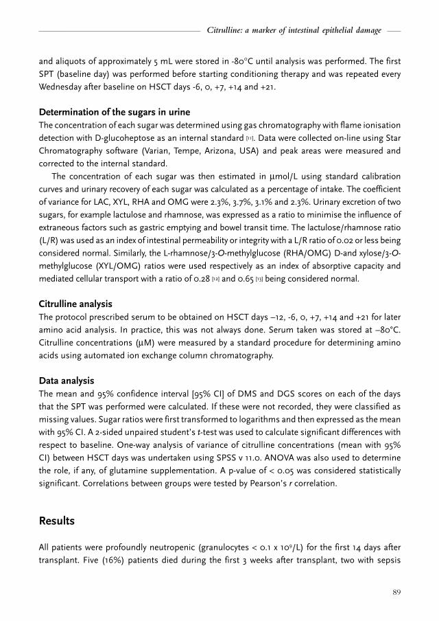
88
_____ Chapter 6 _______________________________________________________________________________________________________________ ____________________________________________________________ Citrulline: a marker of intestinal epithelial damage _____
89
and aliquots of approximately 5 mL were stored in -80°C until analysis was performed. The first SPT (baseline day) was performed before starting conditioning therapy and was repeated every Wednesday after baseline on HSCT days -6, 0, +7, +14 and +21.
Determination of the sugars in urineThe concentration of each sugar was determined using gas chromatography with flame ionisation detection with D-glucoheptose as an internal standard [11]. Data were collected on-line using Star Chromatography software (Varian, Tempe, Arizona, USA) and peak areas were measured and corrected to the internal standard.
The concentration of each sugar was then estimated in µmol/L using standard calibration curves and urinary recovery of each sugar was calculated as a percentage of intake. The coefficient of variance for LAC, XYL, RHA and OMG were 2.3%, 3.7%, 3.1% and 2.3%. Urinary excretion of two sugars, for example lactulose and rhamnose, was expressed as a ratio to minimise the influence of extraneous factors such as gastric emptying and bowel transit time. The lactulose/rhamnose ratio (L/R) was used as an index of intestinal permeability or integrity with a L/R ratio of 0.02 or less being considered normal. Similarly, the L-rhamnose/3-O-methylglucose (RHA/OMG) D-and xylose/3-O-methylglucose (XYL/OMG) ratios were used respectively as an index of absorptive capacity and mediated cellular transport with a ratio of 0.28 [12] and 0.65 [13] being considered normal.
Citrulline analysisThe protocol prescribed serum to be obtained on HSCT days –12, -6, 0, +7, +14 and +21 for later amino acid analysis. In practice, this was not always done. Serum taken was stored at –80°C. Citrulline concentrations (µM) were measured by a standard procedure for determining amino acids using automated ion exchange column chromatography.
Data analysisThe mean and 95% confidence interval [95% CI] of DMS and DGS scores on each of the days that the SPT was performed were calculated. If these were not recorded, they were classified as missing values. Sugar ratios were first transformed to logarithms and then expressed as the mean with 95% CI. A 2-sided unpaired student’s t-test was used to calculate significant differences with respect to baseline. One-way analysis of variance of citrulline concentrations (mean with 95% CI) between HSCT days was undertaken using SPSS v 11.0. ANOVA was also used to determine the role, if any, of glutamine supplementation. A p-value of < 0.05 was considered statistically significant. Correlations between groups were tested by Pearson’s r correlation.
Results
All patients were profoundly neutropenic (granulocytes < 0.1 x 10⁹/L) for the first 14 days after transplant. Five (16%) patients died during the first 3 weeks after transplant, two with sepsis
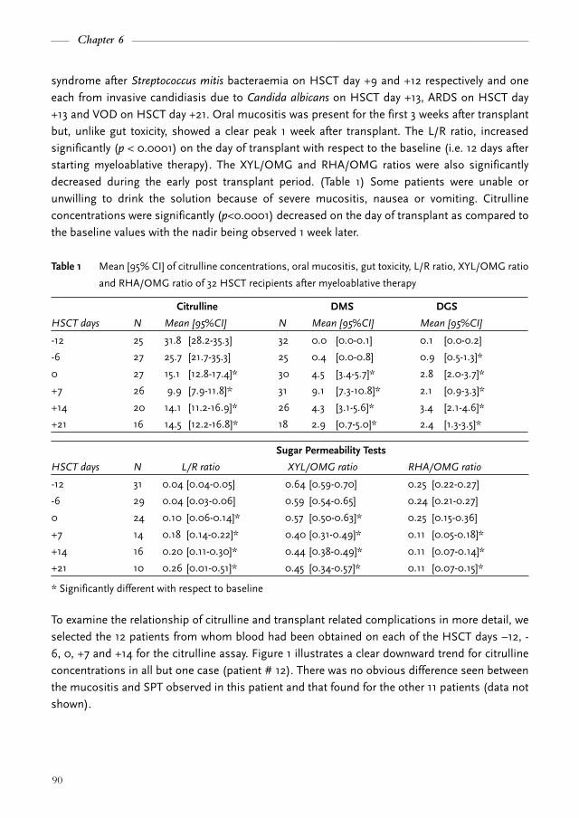
90
_____ Chapter 6 _______________________________________________________________________________________________________________ ____________________________________________________________ Citrulline: a marker of intestinal epithelial damage _____
91
syndrome after Streptococcus mitis bacteraemia on HSCT day +9 and +12 respectively and one each from invasive candidiasis due to Candida albicans on HSCT day +13, ARDS on HSCT day +13 and VOD on HSCT day +21. Oral mucositis was present for the first 3 weeks after transplant but, unlike gut toxicity, showed a clear peak 1 week after transplant. The L/R ratio, increased significantly (p < 0.0001) on the day of transplant with respect to the baseline (i.e. 12 days after starting myeloablative therapy). The XYL/OMG and RHA/OMG ratios were also significantly decreased during the early post transplant period. (Table 1) Some patients were unable or unwilling to drink the solution because of severe mucositis, nausea or vomiting. Citrulline concentrations were significantly (p<0.0001) decreased on the day of transplant as compared to the baseline values with the nadir being observed 1 week later.
Table 1 Mean [95% CI] of citrulline concentrations, oral mucositis, gut toxicity, L/R ratio, XYL/OMG ratio
and RHA/OMG ratio of 32 HSCT recipients after myeloablative therapy
Citrulline DMS DGS
HSCT days N Mean [95%CI] N Mean [95%CI] Mean [95%CI]
-12 25 31.8 [28.2-35.3] 32 0.0 [0.0-0.1] 0.1 [0.0-0.2]
-6 27 25.7 [21.7-35.3] 25 0.4 [0.0-0.8] 0.9 [0.5-1.3]*
0 27 15.1 [12.8-17.4]* 30 4.5 [3.4-5.7]* 2.8 [2.0-3.7]*
+7 26 9.9 [7.9-11.8]* 31 9.1 [7.3-10.8]* 2.1 [0.9-3.3]*
+14 20 14.1 [11.2-16.9]* 26 4.3 [3.1-5.6]* 3.4 [2.1-4.6]*
+21 16 14.5 [12.2-16.8]* 18 2.9 [0.7-5.0]* 2.4 [1.3-3.5]*
Sugar Permeability Tests
HSCT days N L/R ratio XYL/OMG ratio RHA/OMG ratio
-12 31 0.04 [0.04-0.05] 0.64 [0.59-0.70] 0.25 [0.22-0.27]
-6 29 0.04 [0.03-0.06] 0.59 [0.54-0.65] 0.24 [0.21-0.27]
0 24 0.10 [0.06-0.14]* 0.57 [0.50-0.63]* 0.25 [0.15-0.36]
+7 14 0.18 [0.14-0.22]* 0.40 [0.31-0.49]* 0.11 [0.05-0.18]*
+14 16 0.20 [0.11-0.30]* 0.44 [0.38-0.49]* 0.11 [0.07-0.14]*
+21 10 0.26 [0.01-0.51]* 0.45 [0.34-0.57]* 0.11 [0.07-0.15]*
* Significantly different with respect to baseline
To examine the relationship of citrulline and transplant related complications in more detail, we selected the 12 patients from whom blood had been obtained on each of the HSCT days –12, -6, 0, +7 and +14 for the citrulline assay. Figure 1 illustrates a clear downward trend for citrulline concentrations in all but one case (patient # 12). There was no obvious difference seen between the mucositis and SPT observed in this patient and that found for the other 11 patients (data not shown).
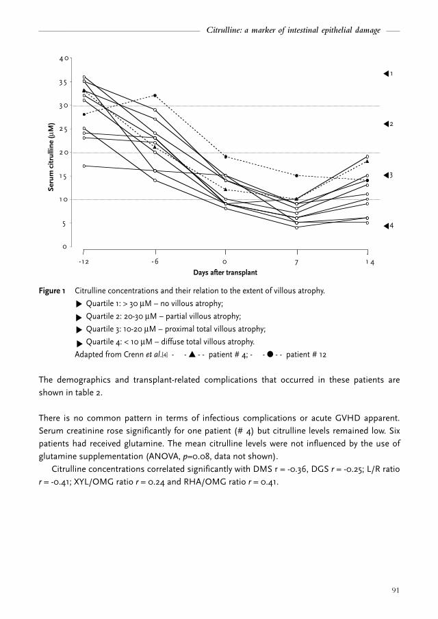
90
_____ Chapter 6 _______________________________________________________________________________________________________________ ____________________________________________________________ Citrulline: a marker of intestinal epithelial damage _____
91
Figure 1 Citrulline concentrations and their relation to the extent of villous atrophy.
Quartile 1: > 30 µM – no villous atrophy;
Quartile 2: 20-30 µM – partial villous atrophy;
Quartile 3: 10-20 µM – proximal total villous atrophy;
Quartile 4: < 10 µM – diffuse total villous atrophy.
Adapted from Crenn et al.[4] - - - - patient # 4; - - - - patient # 12
The demographics and transplant-related complications that occurred in these patients are shown in table 2.
There is no common pattern in terms of infectious complications or acute GVHD apparent. Serum creatinine rose significantly for one patient (# 4) but citrulline levels remained low. Six patients had received glutamine. The mean citrulline levels were not influenced by the use of glutamine supplementation (ANOVA, p=0.08, data not shown).
Citrulline concentrations correlated significantly with DMS r = -0.36, DGS r = -0.25; L/R ratio r = -0.41; XYL/OMG ratio r = 0.24 and RHA/OMG ratio r = 0.41.
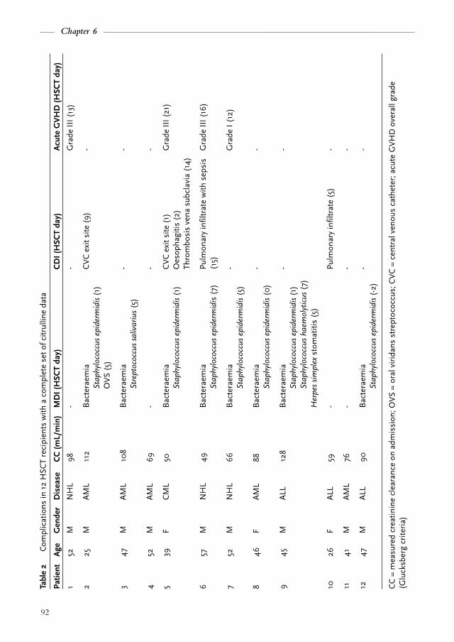
92
_____ Chapter 6 _______________________________________________________________________________________________________________ ____________________________________________________________ Citrulline: a marker of intestinal epithelial damage _____
93
Tabl
e 2
Com
plic
atio
ns in
12
HSC
T re
cipi
ents
with
a c
ompl
ete
set o
f citr
ullin
e da
ta
Patie
ntA
geG
ende
rD
isea
seC
C (m
L/m
in)
MD
I (H
SCT
day)
CD
I (H
SCT
day)
Acu
te G
VH
D (H
SCT
day)
152
MN
HL
98-
-G
rade
III (
13)
225
MA
ML
112
Bac
tera
emia
Stap
hylo
cocc
us e
pide
rmid
is (
1)
OV
S (5
)
CV
C e
xit s
ite (
9)-
347
MA
ML
108
Bac
tera
emia
Stre
ptoc
occu
s sa
livar
ius
(5)
--
452
MA
ML
69-
--
539
FC
ML
50B
acte
raem
ia
St
aphy
loco
ccus
epi
derm
idis
(1)
CV
C e
xit s
ite (
1)O
esop
hagi
tis (
2)Th
rom
bosi
s ve
na s
ubcl
avia
(14
)
Gra
de II
I (21
)
657
MN
HL
49B
acte
raem
ia
St
aphy
loco
ccus
epi
derm
idis
(7)
Pulm
onar
y in
filtr
ate
with
sep
sis
(15)
Gra
de II
I (16
)
752
MN
HL
66B
acte
raem
ia
St
aphy
loco
ccus
epi
derm
idis
(5)
-G
rade
I (1
2)
846
FA
ML
88B
acte
raem
ia
St
aphy
loco
ccus
epi
derm
idis
(0
)-
-
945
MA
LL12
8B
acte
raem
ia
St
aphy
loco
ccus
epi
derm
idis
(1)
St
aphy
loco
ccus
hae
mol
ytic
us (
7)H
erpe
s si
mpl
ex s
tom
atiti
s (5
)
--
1026
FA
LL59
-Pu
lmon
ary
infil
trat
e (5
)-
1141
MA
ML
76-
--
1247
MA
LL90
Bac
tera
emia
Stap
hylo
cocc
us e
pide
rmid
is (
-2)
--
CC
= m
easu
red
crea
tinin
e cl
eara
nce
on a
dmis
sion
; OV
S =
oral
vir
idan
s st
rept
ococ
cus;
CV
C =
cen
tral
ven
ous
cath
eter
; acu
te G
VH
D o
vera
ll gr
ade
(Glu
cksb
erg
crite
ria)
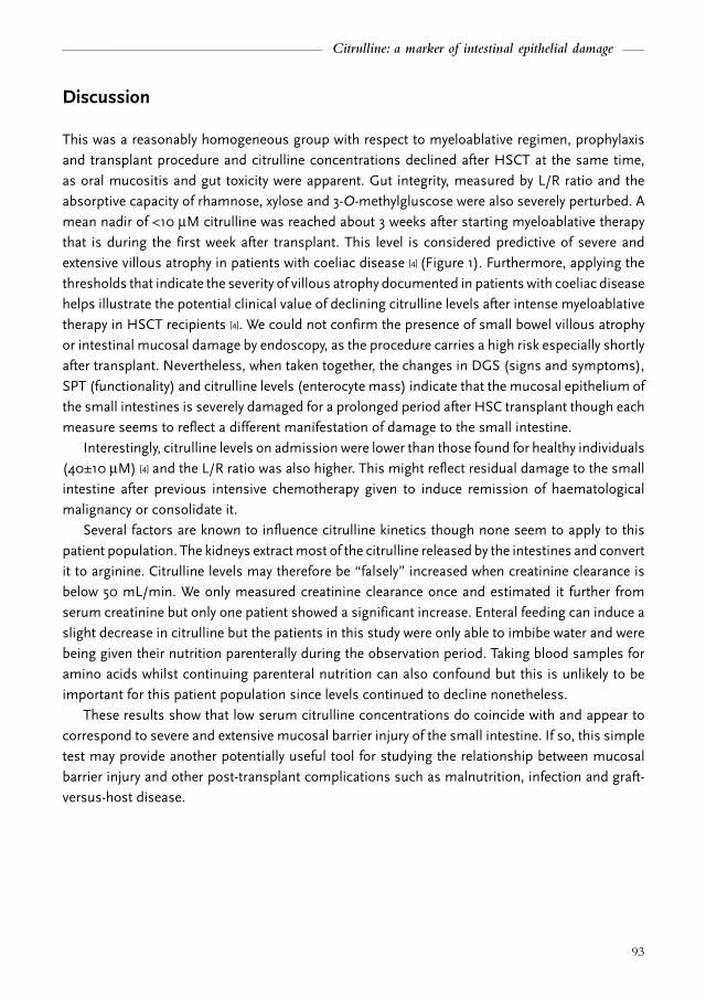
92
_____ Chapter 6 _______________________________________________________________________________________________________________ ____________________________________________________________ Citrulline: a marker of intestinal epithelial damage _____
93
Discussion
This was a reasonably homogeneous group with respect to myeloablative regimen, prophylaxis and transplant procedure and citrulline concentrations declined after HSCT at the same time, as oral mucositis and gut toxicity were apparent. Gut integrity, measured by L/R ratio and the absorptive capacity of rhamnose, xylose and 3-O-methylgluscose were also severely perturbed. A mean nadir of <10 µM citrulline was reached about 3 weeks after starting myeloablative therapy that is during the first week after transplant. This level is considered predictive of severe and extensive villous atrophy in patients with coeliac disease [4] (Figure 1). Furthermore, applying the thresholds that indicate the severity of villous atrophy documented in patients with coeliac disease helps illustrate the potential clinical value of declining citrulline levels after intense myeloablative therapy in HSCT recipients [4]. We could not confirm the presence of small bowel villous atrophy or intestinal mucosal damage by endoscopy, as the procedure carries a high risk especially shortly after transplant. Nevertheless, when taken together, the changes in DGS (signs and symptoms), SPT (functionality) and citrulline levels (enterocyte mass) indicate that the mucosal epithelium of the small intestines is severely damaged for a prolonged period after HSC transplant though each measure seems to reflect a different manifestation of damage to the small intestine.
Interestingly, citrulline levels on admission were lower than those found for healthy individuals (40±10 µM) [4] and the L/R ratio was also higher. This might reflect residual damage to the small intestine after previous intensive chemotherapy given to induce remission of haematological malignancy or consolidate it.
Several factors are known to influence citrulline kinetics though none seem to apply to this patient population. The kidneys extract most of the citrulline released by the intestines and convert it to arginine. Citrulline levels may therefore be “falsely” increased when creatinine clearance is below 50 mL/min. We only measured creatinine clearance once and estimated it further from serum creatinine but only one patient showed a significant increase. Enteral feeding can induce a slight decrease in citrulline but the patients in this study were only able to imbibe water and were being given their nutrition parenterally during the observation period. Taking blood samples for amino acids whilst continuing parenteral nutrition can also confound but this is unlikely to be important for this patient population since levels continued to decline nonetheless.
These results show that low serum citrulline concentrations do coincide with and appear to correspond to severe and extensive mucosal barrier injury of the small intestine. If so, this simple test may provide another potentially useful tool for studying the relationship between mucosal barrier injury and other post-transplant complications such as malnutrition, infection and graft-versus-host disease.
94
_____ Chapter 6 _______________________________________________________________________________________________________________
References
1. Elting LS, Cooksley C, Chambers M, Cantor SB, Manzullo E., Rubenstein EB. The burdens of cancer therapy: clinical and economic outcomes of chemotherapy-induced mucositis. Cancer 2003; 98:1531-1539.
2. Blijlevens NMA., Donnelly JP, De Pauw BE. Mucosal barrier injury: biology, pathology, clinical counterparts and consequences of intensive treatment for haematological malignancy: an overview. Bone Marrow Transplant 2000; 25:1269-1278.
3. Wu G. Synthesis of citrulline and arginine from proline in enterocytes of postnatal pigs. Am J Physiol 1997; 272:313-318.
4. Crenn P, Vahedi K, Lavergne-Slove A, Cynober L, Matuchansky C, Messing B. Plasma citrulline: a marker of enterocyte mass in villous atrophy-associated small bowel disease. Gastroenterology 2003; 124:1210-1219.
5. Lutgens L.C.H.W., Deutz N.E.P, Gueulette J., Cleutjens J.P.M., Berger M.P.F., Wouters B.G., von-Meyenfeldt MF, Lambin P. citrulline: a physiologic marker enabling quantitation and monitoring of epithelial radiation-induced small bowel damage. Int J Radiat Oncol Biol Phys 2003; 57:1067-1074.
6. Keefe DMK, Brealey J, Goland GJ, Cummins AG. Chemotherapy for cancer causes apoptosis that precedes hypoplasia in crypts of the small intestine in humans. Gut 2000; 47:632-637.
7. Schaap N, Schattenberg A, Bar B, Preijers F, Geurts van Kessel A, van der Maazen R, De Boo T, De Witte T. Outcome of transplantation for standard-risk leukaemia with grafts depleted of lymphocytes after conditioning with an intensified regimen. British Journal of Haematology 1997; 98:750-759.
8. Muus P, Donnelly P, Schattenberg A, Linssen P, Minderman H, Dompeling E, de-Witte T. Idarubicin-related side effects in recipients of T-cell-depleted allogeneic bone marrow transplants are schedule dependent. Semin Oncol 1993; 20:47-52.
9. Cockcroft D.W., Gault M.H. Prediction of creatinine clearance from serum creatinine. Nephron 1976; 16:31-41.10. Donnelly JP, Muus P, Schattenberg A, de-Witte T, Horrevorts A, DePauw BE. A scheme for daily monitoring of oral
mucositis in allogeneic BMT recipients. Bone Marrow Transplant 1992; 9:409-413.11. Jansen G, Muskiet FAJ., Schierbeek H., Berger R., Van melle G. Capillary gas chromatographic profiling of urinary,
plasma and erythrocyte sugars and polyols as their trimethylsilyl derivates, preceded by a simple and rapid prepurification method. Clinica Chimica Acta 1986; 157:277-294.
12. Johnston JD, Harvey CJ, Menzies IS, Treacher DF. Gastrointestinal permeability and absorptive capacity in sepsis. Crit Care Med 1996; 24:1144-1149.
13. Lim SG, Menzies IS, Lee CA, Johnson MA, Pounder RE. Intestinal permeability and function in patients infected with Human Immunodeficiency Virus: a comparison with Coeliac Disease. Scand J Gastroenterol 1993; 28:573-580.

Monitoring myelo-ablative therapy induced small bowel toxicity by serum citrulline
concentration: a comparison with sugar permeability tests
Chapter 7
L.C.H.W. Lutgens1, N.M.A. Blijlevens2, N.E.P. Deutz3, J.P. Donnelly2, P. Lambin1, B.E. de Pauw2
1Department of Radiation Therapy and Oncology, MAASTRO Clinic, Maastricht2Department of Hematology, University Medical Center St. Radboud, Nijmegen
3Department of Surgery, Maastricht University, Maastricht
The Netherlands
Cancer 2005: 103:191-199

96
_____ Chapter 7 _______________________________________________________________________________________________________________ _____________________________________________ Monitoring small bowel toxicity by serum citrulline concentration _____
97
96
_____ Chapter 7 _______________________________________________________________________________________________________________ _____________________________________________ Monitoring small bowel toxicity by serum citrulline concentration _____
97
Summary
Intestinal mucositis is an important cause of cancer treatment related morbidity and mortality, carrying a serious economic burden. Currently, objective parameters are lacking that would enable the monitoring of gut damage in routine clinical practice, thus hindering the development of clinical studies designed to investigate potential new strategies aimed at reducing or preventing this side effect. The authors investigated the characteristics of serum citrulline concentration compared with sugar permeability tests with respect to its use as a marker for cancer treatment-induced small bowel injury.
In this prospective study, 10 patients with haematological malignancies who were receiving myeloablative therapy had gut toxicity assessed with sugar permeability tests. Serum citrulline concentration also were determined using archival serum samples. The association between both parameters and their respective characteristics were analyzed and compared with data from the literature.
Sensitivity and specificity were better for the citrulline assay compared with sugar permeability tests. Maximum gut damage assessed with the citrulline assay was observed 1-2 weeks earlier compared with the sugar permeability test. Similarly, citrulline indicated recovery of gut damage at 3 weeks after transplantation, whereas most sugar permeability tests remained abnormal.
The simplicity of the method, the low costs and the lack of drawbacks to the method make the citrulline assay the first choice for measuring and monitoring treatment-related gut damage and provides an objective parameter for cancer treatment-related gut toxicity.
Introduction
An increased use of multiple treatment modalities is characteristic for current developments in curative cancer treatment. This strategy has yielded superior treatment results in a variety of solid tumors, although treatment related acute toxicity is increased as well [1-5]. In patients with haematological malignancies, epithelial gut damage has been suggested as a causal factor in the life-threatening graft-versus-host disease that occurs after myeloablative therapy [6]. In general, severe mucositis has a detrimental effect on treatment outcome due to necessary reductions in treatment intensity and/or treatment interruption. In addition to these detrimental acute effects, epithelial gut damage has also been suggested as one of several mechanisms that contribute to late treatment-related sequellae [7]. Taking into account mucositis-associated complications, such as a longer hospital stay, an increased number of infectious events and nutritional support, Elting et al. [8] recently estimated an incremental cost of $ 2725 and $ 5565 per chemotherapy cycle associated grade 1-2 mucositis and grade 3-4 mucositis, respectively. Hence, it is expected that preventing and/or reducing epithelial gut damage will have significant clinical and socioeconomic impact. However, clinical studies addressing treatment-induced gut damage have been hampered by the fact that objective parameters are lacking to enable monitoring of damage in routine clinical practice.
98
_____ Chapter 7 _______________________________________________________________________________________________________________ _____________________________________________ Monitoring small bowel toxicity by serum citrulline concentration _____
99
Previously, clinical symptoms were used most frequently as surrogate endpoint for gut damage. In addition to the fact that toxicity grading systems are not used uniformly by investigators [9] they are undergoing adjustment on a regular basis. More important, however, is that clinical symptoms correlate poorly with objective parameters of gut damage such as sugar permeability tests (SPT) [10-12], altered histomorphology [13] or treatment-related parameters [9], illustrating the complexicity of the pathophysiology of clinical symptoms related to cytotoxic treatment-induced small bowel damage [14-16].
Currently, morphologic parameters and SPT’s are the objective endpoints used in pre-clinical and clinical studies. However, both methods are unsuited for monitoring purposes in routine clinical practice. In a preclinical study, we recently demonstrated that plasma citrulline concentration is a radiation dose-dependent parameter that correlates with radiation-induced small bowel morphologic endpoints, such as mucosal surface or crypt regeneration [17]. In a subsequent clinical study, we demonstrated that plasma citrulline concentration is correlated with small bowel radiation dose and volume parameters [9]. Similarly, we observed decreased citrulline levels in response to myelo-ablative therapy [18]. In addition to surgery [19], celiac and non-celiac disease [20] and acute cellular rejection following small bowel transplantation [21, 22], cytotoxic treatment was identified as another event associated with small bowel epithelial cell loss that can be monitored by plasma citrulline.
The objective of the current study was to compare the citrulline assay with a currently used method for measuring small bowel damage, that is, the SPT. For this purpose, we analyzed a group of 10 patients who were uniformly treated in a prospective pharmacokinetic study for a haematological malignancy. We hypothesized that changes in citrullinaemia are correlated with changes in absorption of sugar probes associated with the loss of mucosal surface and with changes in gut permeation as assessed with multiple sugar probes.
Materials and methods
SubjectsPatient characteristics are summarized in Table 1. Between June 1999 and December 2000, 10 patients with a mean age of 43 years (age range 22 – 59 years) underwent haematopoietic stem cell transplantation (HSCT) for a haematological malignancy at the department of Haematology, University Medical Centre St. Radboud, Nijmegen. All patients provided informed consent to participate in a prospective study investigating the pharmacokinetics and safety of 14 days of intravenous itraconazole nanocrystals in patients who were undergoing a HLA-matched partial T-cell depleted sibling bone marrow transplantation. The conditioning regimen consisted of idarubicin given at a dose of 42 mg/m2 by continuous infusion over 48 hours starting 12 days before transplantation (HSCT day –12), followed by 120 mg/kg cyclophosphamide (60 mg/kg per day on HSCT day –6 and –5) and 9 Gy of total body irradiation (TBI) in 2 fractions (4.5 Gy per fraction on HSCT day –2 and –1). The total duration of the study period was 34 days, from
98
_____ Chapter 7 _______________________________________________________________________________________________________________ _____________________________________________ Monitoring small bowel toxicity by serum citrulline concentration _____
99
HSCT day –12 until HSCT day +21. For prophylaxis against graft-versus-host disease, patients were administered cyclosporine at 3 mg/kg per day by continuous infusion from HSCT day –1 to HSCT day +14 followed by 2 mg/kg per day until oral intake was possible. Parenteral nutritional support was started on HSCT day –7 and was sustained until the daily oral intake was sufficient. No agents were applied that are believed to ameliorate alimentary tract mucositis (that is, amifostine, glutamine, keratinocyte growth factor). Anti-infective prophylaxis consisted of ciprofloxacin, acyclovir and sulphamethoxazole-trimethoprim. Antifungal prophylaxis started on HSCT day –6 consisting of intravenous itraconazole 200 mg twice daily for 2 days followed by 200 mg once daily for 10 days. All patients were treated with meropenem pre-emptively from the day after transplant until neutrophil recovery (> 0.5x10⁹/L). The glomerular filtration rate was determined on admission by measuring renal creatinine clearance (in mL per minute) and was estimated thereafter from the serum creatinine level using the formula from Cockroft & Gault [23].
Clinical toxicity assessmentGut toxicity was scored daily using six items, that is, frequency of emesis and diarrhea, occurrence of nausea, abdominal complaints, fecal incontinence and fecal volume [12]. Each of these items was allocated a score between 0 (normal) and 3 (severe). Summation of the scores yielded a daily gut score (DGS) with 3 grades: mild toxicity (DGS grade I; 1-6 points), moderate toxicity (DGS II; 7 – 12 points) and severe toxicity (DGS grade III; 13 – 18 points). All gut toxicities after cytotoxic treatment were ascribed to mucositis, unless there was a more plausible alternative cause, such as an adverse drug reaction or proven toxigenic infection.
Sugar permeability test (SPT)An isotonic solution was prepared by dissolving 5 g lactulose (LAC), 1 g L-rhamnose (RHA), 0.5 gram D-xylose (XYL) and 0.2 gram 3-O-methyl-glucose (OMG) in 100 mL demineralised water, according to good manufacturing practice by the pharmacy of the University Medical Centre St. Radboud. After an overnight fast, the subjects emptied their bladders and then drank the test solution. A light breakfast with a drink was allowed after 2 hours. Urine was collected for 5 hours. The total volume was recorded and aliquots of 5 mL were stored in -80°C until analysis. The first (baseline) SPT was performed on HSCT day –12 before starting conditioning therapy and was repeated on HSCT day –7, 0, +7, +14 and +21, yielding 6 time points in 5 weeks per patient.
Urinary excretion ratios of differential sugar permeability testsUrinary excretion was determined using gas chromatography with flame ionization detection with D-glucoheptose as an internal standard [24]. Data were collected online using Star Chromatography software (Varian, Tempe, Arizona, USA) and peak areas were measured and corrected for the internal standard. Urinary recovery of each sugar was expressed as a percentage of the intake and the lactulose/rhamnose ratio (L/R), the D-xylose/3-O-methyl-glucose ratio (XYL/OMG) and the rhamnose/3-O-methyl-glucose ratio (RHA/OMG) were calculated. The L/R ratio is considered a measure of intestinal permeability, whereas the XYL/OMG and RHA/OMG ratios are used as
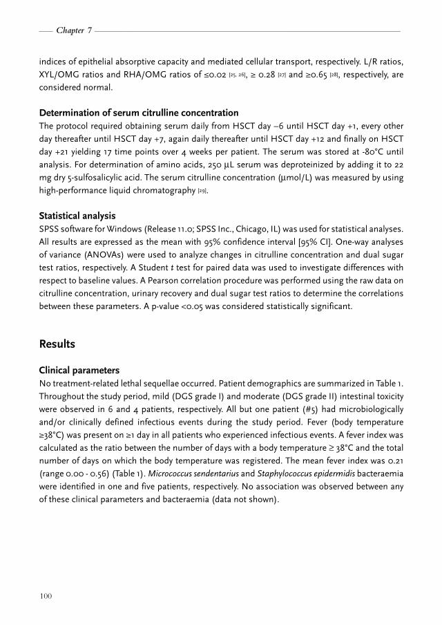
100
_____ Chapter 7 _______________________________________________________________________________________________________________ _____________________________________________ Monitoring small bowel toxicity by serum citrulline concentration _____
101
indices of epithelial absorptive capacity and mediated cellular transport, respectively. L/R ratios, XYL/OMG ratios and RHA/OMG ratios of ≤0.02 [25, 26], ≥ 0.28 [27] and ≥0.65 [28], respectively, are considered normal.
Determination of serum citrulline concentrationThe protocol required obtaining serum daily from HSCT day –6 until HSCT day +1, every other day thereafter until HSCT day +7, again daily thereafter until HSCT day +12 and finally on HSCT day +21 yielding 17 time points over 4 weeks per patient. The serum was stored at -80°C until analysis. For determination of amino acids, 250 µL serum was deproteinized by adding it to 22 mg dry 5-sulfosalicylic acid. The serum citrulline concentration (µmol/L) was measured by using high-performance liquid chromatography [29].
Statistical analysisSPSS software for Windows (Release 11.0; SPSS Inc., Chicago, IL) was used for statistical analyses. All results are expressed as the mean with 95% confidence interval [95% CI]. One-way analyses of variance (ANOVAs) were used to analyze changes in citrulline concentration and dual sugar test ratios, respectively. A Student t test for paired data was used to investigate differences with respect to baseline values. A Pearson correlation procedure was performed using the raw data on citrulline concentration, urinary recovery and dual sugar test ratios to determine the correlations between these parameters. A p-value <0.05 was considered statistically significant.
Results
Clinical parametersNo treatment-related lethal sequellae occurred. Patient demographics are summarized in Table 1. Throughout the study period, mild (DGS grade I) and moderate (DGS grade II) intestinal toxicity were observed in 6 and 4 patients, respectively. All but one patient (#5) had microbiologically and/or clinically defined infectious events during the study period. Fever (body temperature ≥38°C) was present on ≥1 day in all patients who experienced infectious events. A fever index was calculated as the ratio between the number of days with a body temperature ≥ 38°C and the total number of days on which the body temperature was registered. The mean fever index was 0.21 (range 0.00 - 0.56) (Table 1). Micrococcus sendentarius and Staphylococcus epidermidis bacteraemia were identified in one and five patients, respectively. No association was observed between any of these clinical parameters and bacteraemia (data not shown).
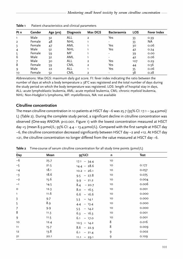
100
_____ Chapter 7 _______________________________________________________________________________________________________________ _____________________________________________ Monitoring small bowel toxicity by serum citrulline concentration _____
101
Table 1 Patient characteristics and clinical parameters
Pt n Gender Age (yrs) Diagnosis Max DGS Bacteraemia LOS Fever Index
1 Male 32 ALL 2 Yes 33 0.392 Female 48 NHL 1 - 33 NA3 Female 47 AML 1 Yes 30 0.064 Male 52 NHL 1 Yes 42 0.245 Female 59 MF 1 - 39 0.006 Male 32 AML 1 - 42 0.067 Male 30 ALL 2 Yes 107 0.298 Female 59 CML 2 Yes 44 0.569 Male 22 ALL 1 Yes 35 0.0610 Female 52 CML 2 - 38 0.28
Abbreviations: Max DGS: maximum daily gut score. FI: fever index indicating the ratio between the number of days at which a body temperature ≥ 38°C was registered and the total number of days during the study period on which the body temperature was registered. LOS: length of hospital stay in days, ALL: acute lymphoblastic leukemia, AML: acute myeloid leukemia, CML: chronic myeloid leukemia, NHL: Non-Hodgkin’s lymphoma, MF: myelofibrosis, NA: not available
Citrulline concentrationThe mean citrulline concentration in 10 patients at HSCT day –6 was 25.7 (95% CI: 17.1 – 34.4 µmol/L) (Table 2). During the complete study period, a significant decline in citrulline concentration was observed (One-way ANOVA: p<0.001. Figure 1) with the lowest concentration measured at HSCT day +5 (mean 8.9 µmol/L; 95% CI: 4.4 – 13.4 µmol/L). Compared with the first sample at HSCT day –6, the citrulline concentration decreased significantly between HSCT day –2 and +12. At HSCT day +21, the citrulline concentration no longer differed from the value measured at HSCT day –6.
Table 2 Time-course of serum citrulline concentration for all study time points (µmol/L)
Day Mean 95%CI n Test
–6 25.7 17.1 – 34.4 10–5 21.5 14.4 – 28.6 10 0.177 –4 18.1 10.2 – 26.1 10 0.057 –3 18.6 9.5 – 27.8 10 0.075 –2 15.6 9.9 – 21.2 10 0.004 –1 14.5 8.4 – 20.7 10 0.006 0 12.3 8.0 – 16.5 10 0.001 1 11.6 6.6 – 16.6 10 0.000 3 9.7 5.3 – 14.1 10 0.000 5 8.9 4.4 – 13.4 10 0.000 7 9.9 5.5 – 14.2 10 0.000 8 11.3 6.3 – 16.3 10 0.001 9 11.5 6.1 – 17.0 10 0.001 10 12.4 10.5 – 14.2 8 0.016 11 15.7 8.6 – 22.9 8 0.009 12 13.8 6.1 – 21.4 9 0.002 21 20.1 11.1 – 29.1 9 0.109
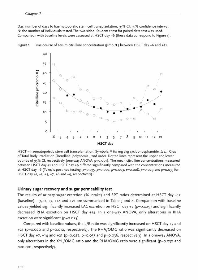
102
_____ Chapter 7 _______________________________________________________________________________________________________________ _____________________________________________ Monitoring small bowel toxicity by serum citrulline concentration _____
103
Day: number of days to haematopoietic stem cell transplantation, 95% CI: 95% confidence interval, N: the number of individuals tested.The two-sided, Student t test for paired data test was used. Comparison with baseline levels were assessed at HSCT day –6 (these data correspond to Figure 1).
Figure 1 Time-course of serum citrulline concentration (µmol/L) between HSCT day –6 and +21.
0
5
10
15
20
25
30
35
40
-6 -5 -4 -3 -2 -1 0 1 3 5 7 8 9 10 11 12 21
HSCT day
Citr
ullin
e (m
icro
mol
/L)
HSCT = haematopoietic stem cell transplantation. Symbols: ◊ 60 mg /kg cyclophosphamide. ∆ 4.5 Gray of Total Body Irradiation. Trendline: polynomial, 2nd order. Dotted lines represent the upper and lower bounds of 95% CI, respectively (one-way ANOVA; p<0.001). The mean citrulline concentrations measured between HSCT day +1 and HSCT day +9 differed significantly compared with the concentrations measured at HSCT day –6 (Tukey’s post-hoc testing: p=0.035, p=0.007, p=0.003, p=0.008, p=0.029 and p=0.035 for HSCT day +1, +3, +5, +7, +8 and +9, respectively).
Urinary sugar recovery and sugar permeability testThe results of urinary sugar excretion (% intake) and SPT ratios determined at HSCT day –12 (baseline), –7, 0, +7, +14 and +21 are summarized in Table 3 and 4. Comparison with baseline values yielded significantly increased LAC excretion on HSCT day +7 (p=0.029) and significantly decreased RHA excretion on HSCT day +14. In a one-way ANOVA, only alterations in RHA excretion were significant (p=0.025).
Compared with baseline values, the L/R ratio was significantly increased on HSCT day +7 and +21 (p=0.020 and p=0.012, respectively). The RHA/OMG ratio was significantly decreased on HSCT day +7, +14 and +21 (p=0.027, p=0.033 and p=0.036, respectively). In a one-way ANOVA, only alterations in the XYL/OMG ratio and the RHA/OMG ratio were significant (p=0.032 and p<0.001, respectively).
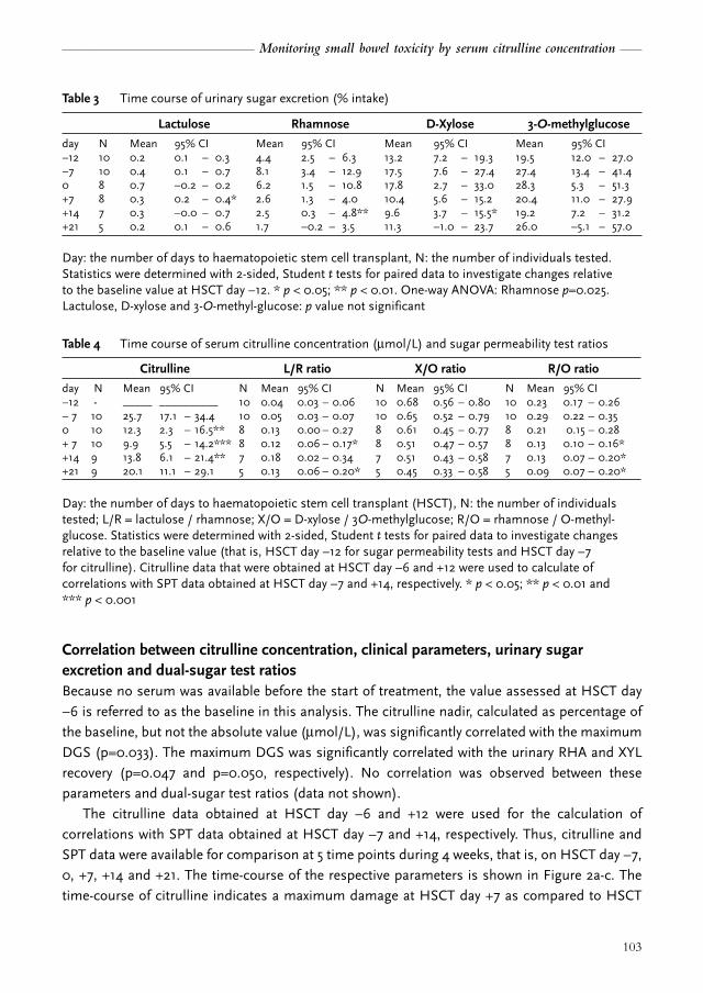
102
_____ Chapter 7 _______________________________________________________________________________________________________________ _____________________________________________ Monitoring small bowel toxicity by serum citrulline concentration _____
103
Table 3 Time course of urinary sugar excretion (% intake)
Lactulose Rhamnose D-Xylose 3-O-methylglucose
day N Mean 95% CI Mean 95% CI Mean 95% CI Mean 95% CI–12 10 0.2 0.1 – 0.3 4.4 2.5 – 6.3 13.2 7.2 – 19.3 19.5 12.0 – 27.0–7 10 0.4 0.1 – 0.7 8.1 3.4 – 12.9 17.5 7.6 – 27.4 27.4 13.4 – 41.40 8 0.7 –0.2 – 0.2 6.2 1.5 – 10.8 17.8 2.7 – 33.0 28.3 5.3 – 51.3+7 8 0.3 0.2 – 0.4* 2.6 1.3 – 4.0 10.4 5.6 – 15.2 20.4 11.0 – 27.9+14 7 0.3 –0.0 – 0.7 2.5 0.3 – 4.8** 9.6 3.7 – 15.5* 19.2 7.2 – 31.2+21 5 0.2 0.1 – 0.6 1.7 –0.2 – 3.5 11.3 –1.0 – 23.7 26.0 –5.1 – 57.0
Day: the number of days to haematopoietic stem cell transplant, N: the number of individuals tested. Statistics were determined with 2-sided, Student t tests for paired data to investigate changes relative to the baseline value at HSCT day –12. * p < 0.05; ** p < 0.01. One-way ANOVA: Rhamnose p=0.025. Lactulose, D-xylose and 3-O-methyl-glucose: p value not significant
Table 4 Time course of serum citrulline concentration (µmol/L) and sugar permeability test ratios
Citrulline L/R ratio X/O ratio R/O ratio
day N Mean 95% CI N Mean 95% CI N Mean 95% CI N Mean 95% CI–12 - _____ __________ 10 0.04 0.03 – 0.06 10 0.68 0.56 – 0.80 10 0.23 0.17 – 0.26– 7 10 25.7 17.1 – 34.4 10 0.05 0.03 – 0.07 10 0.65 0.52 – 0.79 10 0.29 0.22 – 0.350 10 12.3 2.3 – 16.5** 8 0.13 0.00 – 0.27 8 0.61 0.45 – 0.77 8 0.21 0.15 – 0.28 + 7 10 9.9 5.5 – 14.2*** 8 0.12 0.06 – 0.17* 8 0.51 0.47 – 0.57 8 0.13 0.10 – 0.16*+14 9 13.8 6.1 – 21.4** 7 0.18 0.02 – 0.34 7 0.51 0.43 – 0.58 7 0.13 0.07 – 0.20*+21 9 20.1 11.1 – 29.1 5 0.13 0.06 – 0.20* 5 0.45 0.33 – 0.58 5 0.09 0.07 – 0.20*
Day: the number of days to haematopoietic stem cell transplant (HSCT), N: the number of individuals tested; L/R = lactulose / rhamnose; X/O = D-xylose / 3O-methylglucose; R/O = rhamnose / O-methyl-glucose. Statistics were determined with 2-sided, Student t tests for paired data to investigate changes relative to the baseline value (that is, HSCT day –12 for sugar permeability tests and HSCT day –7 for citrulline). Citrulline data that were obtained at HSCT day –6 and +12 were used to calculate of correlations with SPT data obtained at HSCT day –7 and +14, respectively. * p < 0.05; ** p < 0.01 and *** p < 0.001
Correlation between citrulline concentration, clinical parameters, urinary sugar excretion and dual-sugar test ratiosBecause no serum was available before the start of treatment, the value assessed at HSCT day –6 is referred to as the baseline in this analysis. The citrulline nadir, calculated as percentage of the baseline, but not the absolute value (µmol/L), was significantly correlated with the maximum DGS (p=0.033). The maximum DGS was significantly correlated with the urinary RHA and XYL recovery (p=0.047 and p=0.050, respectively). No correlation was observed between these parameters and dual-sugar test ratios (data not shown).
The citrulline data obtained at HSCT day –6 and +12 were used for the calculation of correlations with SPT data obtained at HSCT day –7 and +14, respectively. Thus, citrulline and SPT data were available for comparison at 5 time points during 4 weeks, that is, on HSCT day –7, 0, +7, +14 and +21. The time-course of the respective parameters is shown in Figure 2a-c. The time-course of citrulline indicates a maximum damage at HSCT day +7 as compared to HSCT

104
_____ Chapter 7 _______________________________________________________________________________________________________________ _____________________________________________ Monitoring small bowel toxicity by serum citrulline concentration _____
105
day +14 for the urinary recovery of the individual sugar probes and, to some extent, for the L/R ratio and compared with HSCT day +21 for the XYL/OMG and RHA/OMG ratios. A recovery of intestinal damage during the study period is indicated by the citrulline, RHA, XYL and OMG urinary recovery data and the L/R ratio in contrast to LAC recovery and the XYL/OMG ratio and the RHA/OMG ratio. It is interesting to note that urinary recovery of all sugar probes was increased 1 week after initiation of the conditioning regimen. (Figure 2a-c)
The L/R ratio and the urinary recovery of all four sugars were significantly correlated with each other, although they were not correlated with the citrulline concentration. The citrulline concentration (µmol/L) was correlated with the RHA/OMG ratio (p=0.025). No correlation was observed with the L/R and XYL/OMG ratios.
Figure 2 Tim
e course of serum citrulline concentration (A
), urinary sugar excretion (B) and dual-sugar test ratios (C
) detemined on H
SCT day –12, -7,
0, +7, +14 and +21.
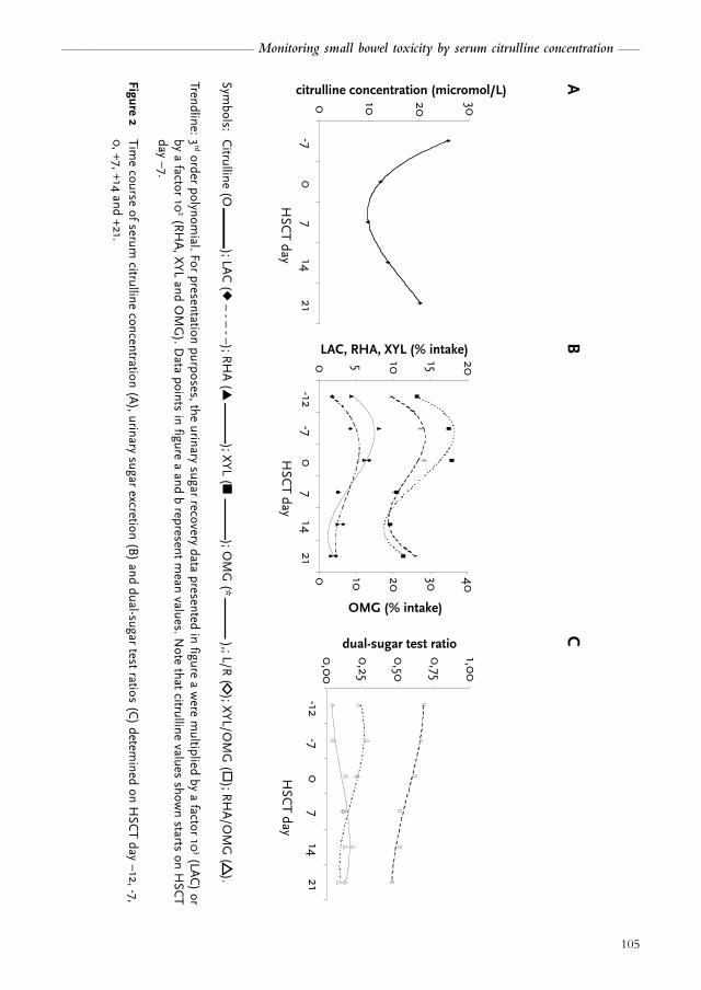
104
_____ Chapter 7 _______________________________________________________________________________________________________________ _____________________________________________ Monitoring small bowel toxicity by serum citrulline concentration _____
105
Symbols:
Citrulline (O
); LAC
( _ - _ - _); R
HA
( ); X
YL ( ); O
MG
(* ),; L/R (
); XYL/O
MG
(o); R
HA
/OM
G (
).
Trendline: 3rd order polynom
ial. For presentation purposes, the urinary sugar recovery data presented in figure a were m
ultiplied by a factor 103 (LA
C) or
by a factor 102 (R
HA
, XYL and O
MG
). Data points in figure a and b represent m
ean values. Note that citrulline values show
n starts on HSC
T day –7.
Figure 2 Tim
e course of serum citrulline concentration (A
), urinary sugar excretion (B) and dual-sugar test ratios (C
) detemined on H
SCT day –12, -7,
0, +7, +14 and +21.
AB
C
106
_____ Chapter 7 _______________________________________________________________________________________________________________ _____________________________________________ Monitoring small bowel toxicity by serum citrulline concentration _____
107
Discussion
In the current study, further support was provided for the use of serum citrulline concentration as a marker for cytotoxic treatment-induced gut damage [9, 17, 18]. We observed a discrepancy between the time course of two objective endpoints for epithelial gut damage (that is, the serum citrulline concentration and the sugar permeability test). Compared with the sugar probe results, the time-course for citrulline is in agreement with known kinetics of epithelial cell loss after radiotherapy [30] and chemotherapy [13].
Gut permeability testing is a noninvasive method for the assessment of altered gut barrier function as an endpoint for intestinal damage [25, 26]. Currently, the SPT is the objective endpoint used in clinical studies to quantify gut damage [12, 31] and to assess treatment effects aimed at mucoprotection [32]. By simultaneously using multiple sugar probes, which are chosen on behalf of different permeation pathways, information about intestinal absorptive capacity is obtained in addition to information about intestinal permeability. It is generally accepted that alterations in the L/R ratio provide an index for the intestinal permeability, whereas the recovery of RHA, XYL and OMG and their ratios provide an index for the intestinal absorptive capacity [26, 33]. Following myeloablative therapy, increased gut permeability (L/R ratio) and decreased absorptive capacity (RHA/OMG ratio) was observed (Table 4) in agreement with previous results [12, 18] and with reports from the literature [10, 13]. Remarkably, the decrease of the XYL/OMG ratio was not statistically significant compared with previous findings from a group of 56 similarly treated patients in our department [12]. Similarly, urinary recovery data did not display major changes (Table 3). The small number of patients in the present series and the relatively large standard variations may explain this finding.
We performed a series of preclinical and clinical experiments to develop an objective parameter that would enable us to monitor gut damage in routine clinical practice [9, 17]. Cytotoxic treatment induces intestinal epithelial cell loss [13, 30]. Plasma citrulline concentration is a surrogate endpoint for the small intestinal epithelial cell mass, irrespective of the underlying cause of cell loss, such as surgical resection [19, 34, 35], celiac and non-celiac disease [20] or cellular rejection after small bowel transplantation [21, 22]. We identified plasma citrulline concentration as a simple and sensitive marker enabling quantitation of cytotoxic treatment-induced small bowel epithelial cell loss [9, 17]. This was confirmed in patients subjected to myeloablative therapy [18]. In the current study, the citrulline concentration was determined in archival serum samples obtained during an observation period of 5 weeks in a series of 10 patients who were uniformly treated with intensive myeloablative therapy. The results were in complete agreement with previous data obtained in similarly treated patients [18] and clearly demonstrated the reproducibility and simplicity of the assay and its ability to enable close monitoring of this type of treatment-induced gut damage, as illustrated in Figure 1.
In contrast to Crenn et al. [20] we were not able to correlate the citrulline level with the degree of mucosal atrophy. Instead, clinical symptoms [12] associated with mucosal atrophy were used as a surrogate endpoint, in addition to sugar permeability tests. The absolute citrulline concentration
106
_____ Chapter 7 _______________________________________________________________________________________________________________ _____________________________________________ Monitoring small bowel toxicity by serum citrulline concentration _____
107
(µmol/L) did not correlate with these endpoints whereas the normalized citrulline level (% baseline) correlated with the maximal gut toxicity observed during the observation period. A similar correlation was observed between the urinary recovery of RHA and XYL (% intake) and maximal gut toxicity. However, there were several limitations to the use of these surrogate endpoints in the current study. First, compared with the serum citrulline concentration, which is considered to be highly dependent upon the small intestinal cell mass [36, 37], the gut toxicity score we used does not specifically address small intestinal toxicity but merely gastrointestinal toxicity in general. Second, mucosal denudation is merely one of several events underlying clinical symptoms related to cytotoxic treatment [13-16].
Comparison of the citrulline assay results with the SPT results yielded some striking differences between both endpoints. The citrulline level (µmol/L or % baseline) did not correlate with gut permeability (L/R ratio). Except for a correlation with the RHA/OMG ratio, no other correlation was observed with parameters indicative for the absorptive capacity used in this study (that is, XYL/OMG ratio or the urinary recovery of RHA, XYL and OMG, respectively). Furthermore, the time course of both endpoints was essentially different (Figure 2). The maximum damage and recovery were recorded between 1-2 weeks earlier using the citrulline assay compared with the sugar probe test results. In contrast to the time course of urinary sugar probe recovery, the time course observed for citrulline was in complete agreement with the time course observed for other function tests related to a reduced number of epithelial cells [17] and with known kinetics of injured gut epithelium after radiotherapy [30] or chemotherapy [13]. Remarkably, the RHA recovery, which was considered the most appropriate estimate of the mucosal surface available for absorption [33] and citrulline concentration during the initial study period, yielded contradictory results concerning the relations with mucosal atrophy and known kinetics of this mucosal event [13, 30]. The initial increase in the sugar probe recovery (and in RHA recovery in particular) may be explained by alterations in the permeation pathway or by altered pre-absorptive factors, such as gastric emptying or small bowel transit time, and both factors are frequently affected by this type of treatment.
Unfortunately, no serum was available for citrulline analysis at HSCT day –12. However, the citrulline concentrations measured at HSCT day –6, (that is, 1 week after initiation of the conditioning regimen) were comparable to baseline citrulline concentrations in a previous study (mean: 30.9 µmol/L; 95% CI: 26.6 – 35.2 µmol/L) [9]. Hence, it is reasonable to assume that a change, if any, occurring in the citrulline level during the first week of the conditioning regimen would be a minor decline. Finally, the average 95% CI was significantly greater for LAC, RHA, XYL and OMG compared with citrulline (p=0.001) (Table 2-3). Although the sugar tests showed remarkable changes during the study period (Figure 2), statistical significance was moderate (Table 2-3) compared with citrulline.
Gastrointestinal mucositis is a common side effect of cancer treatment that, not infrequently, is persistent [38], which may cause a major economic burden [8]. In addition to tumor control, data on normal tissue damage are required to assess the therapeutic value of treatment regimens. In patients with small bowel damage, clinical signs and symptoms expressed in toxicity scores are
108
_____ Chapter 7 _______________________________________________________________________________________________________________ _____________________________________________ Monitoring small bowel toxicity by serum citrulline concentration _____
109
the current standard. For the reasons described above, there is an urgent need for an objective parameter. Ideally, such a marker should be easily accessed and should be independent of medication and metabolic events, such as diet and nutritional status. Previous attempts have been made to develop simple blood tests for measuring gut damage [39, 40]. However, to date, none are used in clinical practice. We suggest that serum citrulline fulfils most, if not all, of these requirements. In addition to yielding highly reproducible results and functioning as a sensitive and specific marker for epithelial cell loss, the citrulline assay can be applied as a simple blood test and is relatively inexpensive.
The results of the current study, in which serum citrulline concentration was used as a marker for cytotoxic treatment-induced intestinal damage are highly reproducible. Overall, the citrulline concentration appears to be a quantitative parameter that is independent of the underlying cause for epithelial cell loss [9, 17-21]. Compared to sugar permeability tests, the citrulline assay is more sensitive and more specific for measuring small bowel epithelial cell loss. In addition, this assay enables more accurate assessment of induction and repair kinetics of damage. The simplicity of the method, the low costs and the lack of methodological drawbacks, as discussed earlier, make this assay the first choice for measuring and monitoring treatment-related gut damage. In addition, the citrulline assay provides an objective parameter to the clinician that enables standardized assessment of treatment-related morbidity.
References
1. Han SC, Kim DH, Higgins SA, Carcangiu ML, Kacinski BM. Chemoradiation as primary or adjuvant treatment for locally advanced carcinoma of the vulva. Int J Radiat Oncol Biol Phys 2000; 47 (5): 1235-44.
2. Green JA, Kirwan JM, Tierney JF, Symonds P, Fresco L, Collingwood M, et al. Survival and recurrence after concomitant chemotherapy and radiotherapy for cancer of the uterine cervix: a systematic review and meta-analysis. Lancet 2001; 358 (9284): 781-6.
3. Pahlman L, Hohenberger W, Gunther K, Fietkau R, Metzger U. Is radiochemotherapy necessary in the treatment of rectal cancer? Eur J Cancer 1998; 34 (4): 438-48.
4. Curran WJ, Jr. Evolving chemoradiation treatment strategies for locally advanced non-small-cell lung cancer. Oncology 2003; 17 (12 Suppl 13): 7-14.
5. Stehman FB, Rose PG, Greer BE, Roy M, Plante M, Penalver M, et al. Innovations in the treatment of invasive cervical cancer. Cancer 2003; 98 (9 Suppl): 2052-63.
6. Hill GR, Crawford JM, Cooke KR, Brinson YS, Pan L, Ferrara JL. Total body irradiation and acute graft-versus-host disease: the role of gastrointestinal damage and inflammatory cytokines. Blood 1997; 90 (8): 3204-13.
7. Richter KK, Langberg CW, Sung CC, Hauer Jensen M. Increased transforming growth factor beta (TGF-beta) immunoreactivity is independently associated with chronic injury in both consequential and primary radiation enteropathy. Int J Radiat Oncol Biol Phys 1997; 39 (1): 187-95.
8. Elting LS, Cooksley C, Chambers M, Cantor SB, Manzullo E, Rubenstein EB. The burdens of cancer therapy. Clinical and economic outcomes of chemotherapy-induced mucositis. Cancer 2003; 98 (7): 1531-9.
9. Lutgens L, Deutz N, Granzier-Peeters M, Beets-Tan R, De Ruysscher D, Gueulette J, et al. Plasma citrulline concentration: a surrogate endpoint for radiation-induced mucosal atrophy of the small bowel. A feasibility study in 23 patients. Int J Radiat Oncol Biol Phys 2004; 60: 275-285.
10. Johansson JE, Ekman T. Gastro-intestinal toxicity related to bone marrow transplantation: disruption of the intestinal barrier precedes clinical findings. Bone Marrow Transplant 1997; 19 (9): 921-5.
108
_____ Chapter 7 _______________________________________________________________________________________________________________ _____________________________________________ Monitoring small bowel toxicity by serum citrulline concentration _____
109
11. Johansson JE, Ekman T. Gut mucosa barrier preservation by orally administered IgA-IgG to patients undergoing bone marrow transplantation: a randomised pilot study. Bone Marrow Transplant 1999; 24 (1): 35-9.
12. Blijlevens NM, van’t Land B, Donnelly JP, M’Rabet L, de Pauw BE. Measuring mucosal damage induced by cytotoxic therapy. Support Care Cancer 2004; 12 (4): 227-33.
13. Keefe DM, Brealey J, Goland GJ, Cummins AG. Chemotherapy for cancer causes apoptosis that precedes hypoplasia in crypts of the small intestine in humans. Gut 2000; 47 (5): 632-7.
14. Thomson AB, Keelan M, Thiesen A, Clandinin MT, Ropeleski M, Wild GE. Small bowel review: diseases of the small intestine. Dig Dis Sci 2001; 46 (12): 2555-66.
15. Griffiths NM. The example of gastrointestinal damage induced by ionising radiation: are there accessible markers? Cell Mol Biol 2001; 47 (3): 427-35.
16. MacNaughton WK. Review article: new insights into the pathogenesis of radiation-induced intestinal dysfunction. Aliment Pharmacol Ther 2000; 14 (5): 523-8.
17. Lutgens LC, Deutz NE, Gueulette J, Cleutjens JP, Berger MP, Wouters BG, et al. Citrulline: A physiologic marker enabling quantitation and monitoring of epithelial radiation-induced small bowel damage. Int J Radiat Oncol Biol Phys 2003; 57 (4): 1067-74.
18. Blijlevens NMA, Lutgens LCHW, Schattenberg A, Donnelly JP. Citrulline: a potentially simple quantitative marker of intestinal epithelial damage following myeloablative therapy. Bone Marrow Transplant 2004; 34 (3):193-196
19. Crenn P, Coudray Lucas C, Thuillier F, Cynober L, Messing B. Postabsorptive plasma citrulline concentration is a marker of absorptive enterocyte mass and intestinal failure in humans. Gastroenterology 2000; 119 (6): 1496-505.
20. Crenn P, Vahedi K, Lavergne Slove A, Cynober L, Matuchansky C, Messing B. Plasma citrulline: A marker of enterocyte mass in villous atrophy-associated small bowel disease. Gastroenterology 2003; 124 (5): 1210-9.
21. Gondolesi G, Fishbein T, Chehade M, Tschernia A, Magid M, Kaufman S, et al. Serum citrulline is a potential marker for rejection of intestinal allografts. Transplant Proc 2002; 34 (3): 918-20.
22. Pappas PA, Saudubray JM, Tzakis AG, Rabier D, Carreno MR, Gomez Marin O, et al. Serum citrulline as a marker of acute cellular rejection for intestinal transplantation. Transplant Proc 2002; 34 (3): 915-7.
23. Cockcroft DW, Gault MH. Prediction of creatinine clearance from serum creatinine. Nephron 1976; 16 (1): 31-41.24. Jansen G, Muskiet FA, Schierbeek H, Berger R, van der Slik W. Capillary gas chromatographic profiling of urinary,
plasma and erythrocyte sugars and polyols as their trimethylsilyl derivatives, preceded by a simple and rapid prepurification method. Clin Chim Acta 1986; 157 (3): 277-93.
25. Bjarnason I, MacPherson A, Hollander D. Intestinal permeability: an overview. Gastroenterology 1995; 108 (5): 1566-81.
26. Travis S, Menzies I. Intestinal permeability: functional assessment and significance. Clin Sci 1992; 82 (5): 471-88.27. Johnston JD, Harvey CJ, Menzies IS, Treacher DF. Gastrointestinal permeability and absorptive capacity in sepsis.
Crit Care Med 1996; 24 (7): 1144-9. 28. Lim SG, Menzies IS, Lee CA, Johnson MA, Pounder RE. Intestinal permeability and function in patients infected
with human immunodeficiency virus. A comparison with coeliac disease. Scand J Gastroenterol 1993; 28 (7): 573-80.
29. van Eijk HM, Rooyakkers DR, Deutz NE. Rapid routine determination of amino acids in plasma by high-performance liquid chromatography with a 2-3 microns Spherisorb ODS II column. J Chromatogr 1993; 620 (1): 143-8.
30. Becciolini A, Balzi M, Potten CS. Radiation effects on proliferation and differentiation in the rat small intestine. In: Hendry JH, ed. Radiation and gut., ed. 1 Amsterdam: Elsevier Science B.V., 1995: 85-143.
31. Blijlevens NM, Donnelly JP, de Pauw BE. Impaired gut function as risk factor for invasive candidiasis in neutropenic patients. Br J Haematol 2002; 117 (2): 259-64.
32. Yoshida S, Matsui M, Shirouzu Y, Fujita H, Yamana H, Shirouzu K. Effects of glutamine supplements and radiochemotherapy on systemic immune and gut barrier function in patients with advanced esophageal cancer. Ann Surg 1998; 227 (4): 485-91.
33. Menzies IS, Zuckerman MJ, Nukajam WS, Somasundaram SG, Murphy B, Jenkins AP, et al. Geography of intestinal permeability and absorption. Gut 1999; 44 (4): 483-9.
34. Wakabayashi Y, Yamada E, Yoshida T, Takahashi N. Effect of intestinal resection and arginine-free diet on rat physiology. Am J Physiol 1995; 269 (2 Pt 1): G313-8.
35. Chen K, Nezu R, Sando K, Haque SM, Iiboshi Y, Masunari A, et al. Influence of glutamine-supplemented parenteral nutrition on intestinal amino acid metabolism in rats after small bowel resection. Surg Today 1996; 26 (8): 618-23.
110
_____ Chapter 7 _______________________________________________________________________________________________________________
36. Hoogenraad N, Totino N, Elmer H, Wraight C, Alewood P, Johns RB. Inhibition of intestinal citrulline synthesis causes severe growth retardation in rats. Am J Physiol 1985; 249 (6 Pt 1): G792-9.
37. Wakabayashi Y, Yamada E, Hasegawa T, Yamada R. Enzymological evidence for the indispensability of small intestine in the synthesis of arginine from glutamate. I. Pyrroline-5-carboxylate synthase. Arch Biochem Biophys 1991; 291 (1):1-8.
38. Yeoh E, Horowitz M, Russo A, Muecke T, Robb T, Chatterton B. The effects of abdominal irradiation for seminoma of the testis on gastrointestinal function. J Gastroenterol Hepatol 1995; 10 (2): 125-30.
39. Overgaard J, Matsui M. Effect of radiation on glucose absorption in the mouse jejunum in vivo. Radiother Oncol 1990; 18 (1): 71-7.
40. Sorensen SH, Proud FJ, Rutgers HC, Markwell P, Adam A, Batt RM. A blood test for intestinal permeability and function: a new tool for the diagnosis of chronic intestinal disease in dogs. Clin Chim Acta 1997; 264 (1): 103-15.
A randomised, double-blinded, placebo-controlled pilot study of parenteral glutamine for allogeneic
stem cell transplant patients
Chapter 8
N.M.A. Blijlevens1, J.P. Donnelly1, A.H.J. Naber2, A.V.M.B. Schattenberg1 and B.E. De Pauw1
Department of Hematology1 and Department of Gastroenterology and Hepatology2,
University Medical Center St. Radboud, Nijmegen
The Netherlands
Support Care Cancer 2005 Mar 15; [Epub ahead of print]

112
_____ Chapter 8 _______________________________________________________________________________________________________________ _____________________________________________________________________________ A pilot study of parenteral glutamine _____
113
112
_____ Chapter 8 _______________________________________________________________________________________________________________ _____________________________________________________________________________ A pilot study of parenteral glutamine _____
113
Summary
We conducted a prospective, randomised, double-blinded, placebo-controlled pilot study of parenteral nutrition (PN) supplemented with 0.57 g/kg glutamine-dipeptide in a homogeneous group of 32 allogeneic stem cell transplant (HSCT) recipients to determine its effect on mucosal barrier injury (MBI). All patients had been prepared with idarubicin, cyclophosphamide and total body irradiation. PN (by continuous infusion) started on HSCT day –6 for a median of 19 days. MBI measured by sugar permeability tests, daily mucositis score, daily gut score, and citrulline concentrations was not reduced by glutamine-dipeptide. However, the daily gut score was significantly lower for the glutamine group on HSCT +7 (p = 0.001) whilst citrulline was lower (p = 0.03) for the placebo group on HSCT day +21. Albumin was significantly lower in the placebo group on HSCT day +21 (32±4 versus 37±3, p = 0.001) whilst CRP was higher (74±48 versus 34±38, p = 0.003). Other transplant-related complications (infections, acute graft-versus-host disease) were less common although this did not reach statistical significance nor translate into a reduced length of hospital stay or lower mortality. These results indicate that it would be worthwhile conducting a larger trial to see whether or not giving glutamine-dipeptide reduces the 100-day allogeneic transplant-related complications.
Introduction
Glutamine can behave as an essential amino acid in clinical settings where there is marked metabolic stress such as occurs after haematopoeitic stem cell transplant (HSCT). The amino acid is a key substrate involved in many metabolic processes including inter-organ nitrogen transfer, protein synthesis, gluconeogenesis, acid-base homeostasis and nucleic acid biosynthesis. Glutamine is also a substrate for glutathione synthesis thereby playing a role as an antioxidant and scavenger and is the major fuel of intestinal mucosal cells and immune cells, including those located in the gut-associated lymphoid tissue [1]. Glutamine supplementation in animals reduced bacterial translocation and mucosal damage after chemotherapy or radiotherapy, making supplementation an attractive option for reducing post-transplant complications [2]. However, the results of clinical studies amongst recipients of autologous or allogeneic HSCT given glutamine-dipeptide orally or parenterally are contradictory. Some indicate a trend towards reduced mortality, shorter length of hospital stay (LOS), lower rejection and infection rate, and less use of parenteral nutrition (PN) [3]. Autologous HSCT recipients reported that oral administration of glutamine leads to less severe oral mucositis and a shorter duration as they had less oral pain and required less opioid medication [4]. However, interpretation of the results is hampered by the heterogeneity of the patient populations. There has also been a lack of an objective tool for measuring mucosal barrier injury (MBI) not only of the mouth but also of the digestive tract as a whole [5], despite the fact that MBI is the most frequent cause of morbidity associated with the myeloablative conditioning treatment to prepare for an HSCT [6;7] and increases the costs of
114
_____ Chapter 8 _______________________________________________________________________________________________________________ _____________________________________________________________________________ A pilot study of parenteral glutamine _____
115
health care resources [8]. Oral mucositis is relatively easy to recognise [9] whereas the detection of intestinal mucosal injury has relied essentially on non-specific symptoms such as nausea, vomiting, diarrhoea and abdominal cramps, which affect almost every HSCT recipient and do not necessary reflect MBI. Sugar permeability tests provide an objective assessment of gut integrity and absorptive capacity, reflecting alterations in permeability and loss of epithelial surface that are features of gut MBI [10]. We recently reported that serum citrulline concentrations of HSCT recipients who participated in a pilot study of glutamine-dipeptide supplementation could also be used to monitor intestinal mucosal damage after myeloablative therapy [11]. We now report in full the results of the prospective, randomised, double-blinded, placebo-controlled pilot study to determine whether glutamine-dipeptide supplementation reduces mucosal barrier injury as measured by a daily mucositis score (DMS), daily gut score (DGS), sugar permeability tests (SPT) and serum citrulline.
Patients and methods
PatientsFrom July 1999 to July 2002, patients admitted to receive an HLA-matched T-cell depleted sibling graft were asked to participate in the study provided they were between 18-65 years of age and were to receive myeloablative therapy with idarubicin to lower the risk of relapse given at a dose of 42 mg/m2 by continuous infusion over 48 hours starting 12 days before transplant (HSCT day –12), 120 mg/kg cyclophosphamide (60 mg/kg per day on HSCT days –6 & - 5) and 9 Gray of total body irradiation (4.5 Gy per day on HSCT days –2 & -1) [12]. In addition, patients had to be free of an inborn-error of amino acid metabolism, insulin-dependent diabetes and inflammatory bowel disease. All patients had to give their informed consent in writing before randomisation. The local ethics committee approved the protocol.
TreatmentPatients were randomly allocated to receive either standard Aminomix (Fresenius-Kabi, Nederland BV, ‘s-Hertogenbosch, The Netherlands) parenterally or Aminomix in which a portion of the amino acids were replaced by 200 ml glutamine-dipeptide, L-alanyl-L-glutamine (Dipeptiven, Fresenius-Kabi) to obtain an isonitrogenous and isocaloric solution equivalent to 0.57 g/kg/d glutamine-dipeptide. Randomisation was performed by the hospital pharmacy and, except for the hospital pharmacist, all other personnel involved in the trial were blinded, as were the patients. Parenteral nutrition was administered from HSCT day –6 onwards until bone marrow recovery (granulocyte count above 0.5 x 10⁹/L), removal of central venous catheter for any reason or because of intolerance.
114
_____ Chapter 8 _______________________________________________________________________________________________________________ _____________________________________________________________________________ A pilot study of parenteral glutamine _____
115
Supportive careCyclosporin 3 mg/kg/day was given as prophylaxis against graft-versus-host disease (GVHD) by continuous infusion from HSCT day –1 to +14 followed by 2 mg/kg/day until oral intake was possible. Patients received continuously intravenous morphine on demand to control mucositis-related pain. Erythrocyte transfusions were given when the haemoglobin concentration was less than 6 mmol/L and platelets were transfused for bleeding or when platelet counts fell below 15 x 109/L. Haematopoietic growth factors were not used. Anti-infective prophylaxis consisted of ciprofloxacin and acyclovir or valaciclovir. Co-trimoxazole (sulphamethoxazole-trimethoprim) was given for prophylaxis against Pneumocystis carinii. Fluconazole was only given to patients colonised with Candida albicans and no other systemic antifungal agents were given for prophylaxis. Cefipime or ceftazidime was administered empirically at the onset of fever. Full blood counts together with renal and liver function tests and C-reactive protein (CRP) were performed daily. The glomerular filtration rate was determined on admission by measuring renal creatinine clearance (millilitres per minute) and was estimated thereafter from the serum creatinine using the formula of Cockroft & Gault [13]. Baseline body mass index (BMI) was calculated and albumin was measured weekly.
Citrulline assayThe protocol-prescribed serum was obtained on HSCT days –12, -6, 0, +7, +14 and +21 for later amino acid analysis. In practice, this was not always done. Serum taken was stored at –80°C. Citrulline concentrations (micromolar) were measured by a standard procedure for determining amino acids using automated ion-exchange column chromatography after completion of the study. The citrulline concentrations of the population have been recently published [11].
Mucositis assessmentPatients were monitored daily for the presence of oral mucositis evidenced by lesions, erythema, oedema, pain, bleeding, dryness and the production of viscous mucous with each item being allocated a score of 0 (normal) to 3 (severe). The separate values were summed to yield a daily oral mucositis score (DMS) with 3 grades: 1-7 for DMS grade I or mild mucositis, 8-14 for grade II or moderate mucositis and ≥ 15 for grade III or severe mucositis. DMS grade II is comparable with WHO grade III [14]. Gut toxicity was scored likewise from 0 to 3 based on the frequency of vomiting and diarrhoea, as well as the occurrence of nausea, abdominal complaints, faecal incontinence and volume. Summation of the separate score yielded the daily gut score (DGS) with 1-6 for grade I or mild toxicity, 7-12 for grade II or moderate toxicity and ≥ 13 for grade III or severe toxicity. All gut toxicities following chemotherapy were ascribed to mucositis unless there was a more plausible alternative explanation, such as an adverse drug reaction or proven Clostridium difficile infection.
116
_____ Chapter 8 _______________________________________________________________________________________________________________ _____________________________________________________________________________ A pilot study of parenteral glutamine _____
117
Sugar permeability test (SPT)An isotonic solution was prepared by dissolving 5 g lactulose (LAC), 1 g L-rhamnose (RHA), 0.2 g 3-O-methylglucose (OMG), 0.5 g D-xylose (XYL) in 100 mL demineralised water according to good manufacturing practice by our own hospital pharmacy. After overnight fasting, subjects emptied their bladders and then drank the solution. A light breakfast with a drink was allowed after 2 hours. Urine was collected for 5 hours after which the total volume passed was recorded, and aliquots of approximately 5 mL were stored in -80°C until assayed. The first SPT (baseline day) was performed before starting conditioning therapy and others were determined every Wednesday after baseline on HSCT days –12, -6, 0, +7, +14 and +21.
Determination of the sugars in urineThe concentration of each sugar was determined using gas chromatography with flame ionisation detection with D-glucoheptose as an internal standard [15]. Data were collected on-line using Star Chromatography software (Varian, Tempe, Arizona, USA) and peak areas were measured and corrected to the internal standard. The concentration of each sugar was then estimated in micromoles per litre using standard calibration curves and urinary recovery of each sugar was calculated as a percentage of intake. The coefficient of variance for LAC, XYL, RHA and OMG were 2.3%, 3.7%, 3.1% and 2.3%, respectively. Urinary excretion of two sugars, for example lactulose and rhamnose, was expressed as a ratio to minimise the influence of extraneous factors such as gastric emptying and bowel transit time. The lactulose/rhamnose ratio (L/R) was used as an index of intestinal permeability or integrity with a L/R ratio of 0.02 or less being considered normal. Similarly, the L-rhamnose/3-O-methylglucose (RHA/OMG) and D-xylose/3-O-methylglucose (XYL/OMG) ratios were used, respectively, as indices of absorptive capacity and mediated cellular transport, with a ratio of 0.28 [16] and 0.65 [17] being considered normal.
Transplant related complicationsEvery event that occurred during the study until 3 months post-transplant was recorded and classified as either an infectious event (IE) or non-infectious event (NIE), irrespective of its relation to study treatment. The infectious events were sub-divided into microbiologically defined infections (MDI) with or without bacteraemia, clinically defined infections (CDI) or unexplained fever. Non-infectious events were rated as mild, moderate or severe. Acute GVHD was scored using the Glucksberg criteria. Any event resulting in admission to the ICU or having a significant mortality risk despite optimal therapy or resulting in death was considered a serious adverse event.
Data analysisThe primary end point of the study was to investigate the effect of glutamine-dipeptide supplementation on gut MBI measured by SPT. Secondary endpoints were differences in DMS, DGS, citrulline and morphine use (mean daily dose), time to granulocyte recovery (> 0.5 x 10⁹/L (days), length of hospital stay (LOS in days), and the frequency of transplant-related

116
_____ Chapter 8 _______________________________________________________________________________________________________________ _____________________________________________________________________________ A pilot study of parenteral glutamine _____
117
complications estimated by the number of events per patient divided by LOS. The mean and standard deviation (SD) of DMS, DGS scores and citrulline concentrations on each of the days that the SPT was performed were calculated. If these items were not recorded on the specified day, the scores obtained on the day before or after were used. The sugar ratios were first transformed to logarithms and then expressed as the mean with SD. ANOVA was used to determine the impact, if any, of glutamine supplementation. Comparison of variables between the two groups was done using the Student’s t-test or Mann-Whitney U test. All analyses were done using SPSS v 11.0 with a p-value of < 0.05 being considered statistically significant.
Results
Patient characteristicsThirty-five patients were recruited but three failed to meet the inclusion criteria and were not entered into the study: one patient was not transplanted because of relapsed disease, another developed a cerebral lesion whilst the third was given another myeloablative regimen. Twenty of the 32 patients were men, and the mean age of the population was 48 (range 25-65) years. All patients had been treated for a haematological malignancy – acute myeloid leukaemia (9), acute lymphoblastic leukaemia (7), non-Hodgkin’s lymphoma (7), myelodysplastic syndrome (3), chronic myelogenous leukaemia (2), myeloproliferative disorder (2), chronic lymphocytic leukaemia (1), chronic myelofibrosis (1). (Table 1)
Table 1 Characteristics of patients at baseline
Glutamine-dipeptide Placebo P value
Number of patients 16 16 ns
Age (mean + range in years) 49 (25-64) 48 (28-57) ns
Women (n) 5 7 ns
BMI (mean) 26 27 ns
BMI > 25 (n) 10 11 ns
Acute leukaemia (n) 7 9 ns
Albumin (mean ± SD) 45 (± 2) 44 (± 3) ns
C-reactive protein (mean ± SD) 11 (± 14) 18 (± 18) ns
Creatinine clearance (ml/min) (mean ± SD) 99 (± 54) 79 (± 30) p = 0.2
BMI Body Mass Index, ns not significant
There was no difference between the treatment and placebo groups in terms of baseline BMI, the proportion considered overweight (BMI > 25), creatinine clearance (ml/min), CRP or albumin concentrations. Also DMS, DGS, citrulline and the ratios of L/R, XYL/OMG and RHA/OMG were similar (Table 2).
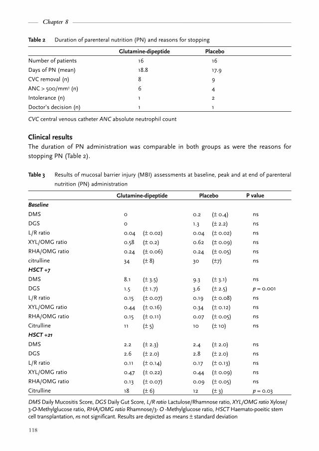
118
_____ Chapter 8 _______________________________________________________________________________________________________________ _____________________________________________________________________________ A pilot study of parenteral glutamine _____
119
Table 2 Duration of parenteral nutrition (PN) and reasons for stopping
Glutamine-dipeptide Placebo
Number of patients 16 16
Days of PN (mean) 18.8 17.9
CVC removal (n) 8 9
ANC > 500/mm3 (n) 6 4
Intolerance (n) 1 2
Doctor’s decision (n) 1 1
CVC central venous catheter ANC absolute neutrophil count
Clinical resultsThe duration of PN administration was comparable in both groups as were the reasons for stopping PN (Table 2).
Table 3 Results of mucosal barrier injury (MBI) assessments at baseline, peak and at end of parenteral
nutrition (PN) administration
Glutamine-dipeptide Placebo P value
Baseline
DMS 0 0.2 (± 0.4) ns
DGS 0 1.3 (± 2.2) ns
L/R ratio 0.04 (± 0.02) 0.04 (± 0.02) ns
XYL/OMG ratio 0.58 (± 0.2) 0.62 (± 0.09) ns
RHA/OMG ratio 0.24 (± 0.06) 0.24 (± 0.05) ns
citrulline 34 (± 8) 30 (±7) ns
HSCT +7
DMS 8.1 (± 3.5) 9.3 (± 3.1) ns
DGS 1.5 (± 1.7) 3.6 (± 2.5) p = 0.001
L/R ratio 0.15 (± 0.07) 0.19 (± 0.08) ns
XYL/OMG ratio 0.44 (± 0.16) 0.34 (± 0.12) ns
RHA/OMG ratio 0.15 (± 0.11) 0.07 (± 0.05) ns
Citrulline 11 (± 5) 10 (± 10) ns
HSCT +21
DMS 2.2 (± 2.3) 2.4 (± 2.0) ns
DGS 2.6 (± 2.0) 2.8 (± 2.0) ns
L/R ratio 0.11 (± 0.14) 0.17 (± 0.13) ns
XYL/OMG ratio 0.47 (± 0.22) 0.44 (± 0.09) ns
RHA/OMG ratio 0.13 (± 0.07) 0.09 (± 0.05) ns
Citrulline 18 (± 6) 12 (± 3) p = 0.03
DMS Daily Mucositis Score, DGS Daily Gut Score, L/R ratio Lactulose/Rhamnose ratio, XYL/OMG ratio Xylose/3-O-Methylglucose ratio, RHA/OMG ratio Rhamnose/3- O -Methylglucose ratio, HSCT Haemato-poeitic stem cell transplantation, ns not significant. Results are depicted as means ± standard deviation
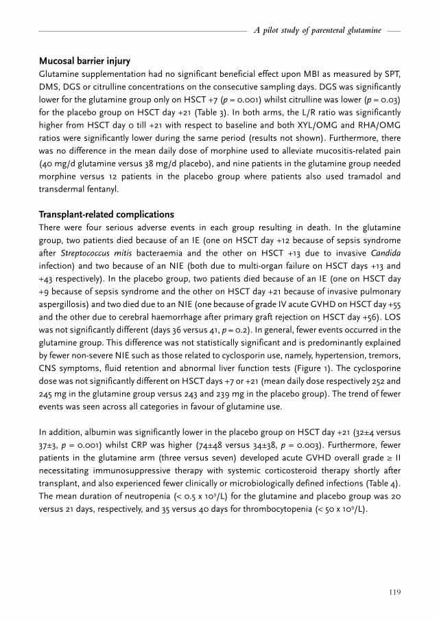
118
_____ Chapter 8 _______________________________________________________________________________________________________________ _____________________________________________________________________________ A pilot study of parenteral glutamine _____
119
Mucosal barrier injuryGlutamine supplementation had no significant beneficial effect upon MBI as measured by SPT, DMS, DGS or citrulline concentrations on the consecutive sampling days. DGS was significantly lower for the glutamine group only on HSCT +7 (p = 0.001) whilst citrulline was lower (p = 0.03) for the placebo group on HSCT day +21 (Table 3). In both arms, the L/R ratio was significantly higher from HSCT day 0 till +21 with respect to baseline and both XYL/OMG and RHA/OMG ratios were significantly lower during the same period (results not shown). Furthermore, there was no difference in the mean daily dose of morphine used to alleviate mucositis-related pain (40 mg/d glutamine versus 38 mg/d placebo), and nine patients in the glutamine group needed morphine versus 12 patients in the placebo group where patients also used tramadol and transdermal fentanyl.
Transplant-related complications There were four serious adverse events in each group resulting in death. In the glutamine group, two patients died because of an IE (one on HSCT day +12 because of sepsis syndrome after Streptococcus mitis bacteraemia and the other on HSCT +13 due to invasive Candida infection) and two because of an NIE (both due to multi-organ failure on HSCT days +13 and +43 respectively). In the placebo group, two patients died because of an IE (one on HSCT day +9 because of sepsis syndrome and the other on HSCT day +21 because of invasive pulmonary aspergillosis) and two died due to an NIE (one because of grade IV acute GVHD on HSCT day +55 and the other due to cerebral haemorrhage after primary graft rejection on HSCT day +56). LOS was not significantly different (days 36 versus 41, p = 0.2). In general, fewer events occurred in the glutamine group. This difference was not statistically significant and is predominantly explained by fewer non-severe NIE such as those related to cyclosporin use, namely, hypertension, tremors, CNS symptoms, fluid retention and abnormal liver function tests (Figure 1). The cyclosporine dose was not significantly different on HSCT days +7 or +21 (mean daily dose respectively 252 and 245 mg in the glutamine group versus 243 and 239 mg in the placebo group). The trend of fewer events was seen across all categories in favour of glutamine use.
In addition, albumin was significantly lower in the placebo group on HSCT day +21 (32±4 versus 37±3, p = 0.001) whilst CRP was higher (74±48 versus 34±38, p = 0.003). Furthermore, fewer patients in the glutamine arm (three versus seven) developed acute GVHD overall grade ≥ II necessitating immunosuppressive therapy with systemic corticosteroid therapy shortly after transplant, and also experienced fewer clinically or microbiologically defined infections (Table 4). The mean duration of neutropenia (< 0.5 x 10⁹/L) for the glutamine and placebo group was 20 versus 21 days, respectively, and 35 versus 40 days for thrombocytopenia (< 50 x 109/L).
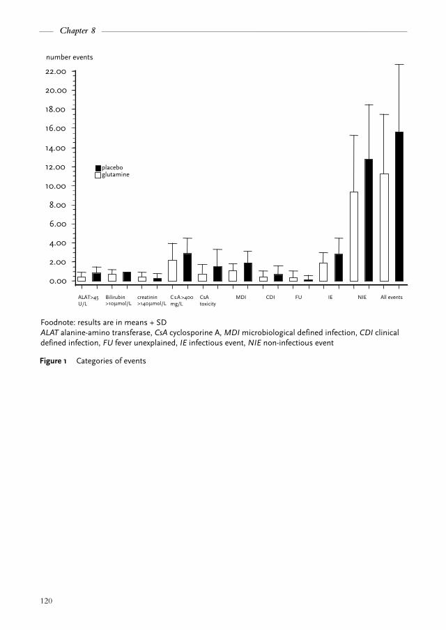
120
_____ Chapter 8 _______________________________________________________________________________________________________________ _____________________________________________________________________________ A pilot study of parenteral glutamine _____
121
Figure 1 Categories of events
ALAT>45 U/L
0.00
2.00
4.00
6.00
8.00
10.00
12.00
14.00
16.00
18.00
20.00
22.00
Bilirubin>10µmol/L
creatinin>140µmol/L
CsA >400mg/L
CsA toxicity
MDI CDI FU IE NIE All events
placebo glutamine
number events
Foodnote: results are in means + SDALAT alanine-amino transferase, CsA cyclosporine A, MDI microbiological defined infection, CDI clinical defined infection, FU fever unexplained, IE infectious event, NIE non-infectious event
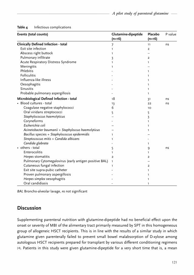
120
_____ Chapter 8 _______________________________________________________________________________________________________________ _____________________________________________________________________________ A pilot study of parenteral glutamine _____
121
Table 4 Infectious complications
Events (total counts) Glutamine-dipeptide (n=16)
Placebo (n=16)
P value
Clinically Defined Infection - total Exit site infection Abscess right buttock Pulmonary infiltrate Acute Respiratory Distress Syndrome Meningitis Phlebitis Folliculitis Influenza-like illness Oesophagitis Sinusitis Probable pulmonary aspergillosis
711311------
112–21–111111
ns
Microbiological Defined Infection - total• Blood cultures - total Coagulase negative staphylococci Oral viridans streptococci Staphylococcus haemolyticus Coryneforms Escherichia coli Acinetobacter baumanii + Staphyloccus haemolyticus Bacillus species + Staphylococcus epidermidis Streptococcus mitis + Candida albicans Candida glabrata• others - total Enterocolitis Herpes stomatitis Pulmonary Cytomegalovirus (early antigen positive BAL) Cutaneous fungal infection Exit site supra-pubic catheter Proven pulmonary aspergillosis Herpes simplex oesophagitis Oral candidiasis
181365–-––11-51211----
31221053111–-1912–21111
nsns
ns
BAL Broncho-alveolar lavage, ns not significant
Discussion
Supplementing parenteral nutrition with glutamine-dipeptide had no beneficial effect upon the onset or severity of MBI of the alimentary tract primarily measured by SPT in this homogeneous group of allogeneic HSCT recipients. This is in line with the results of a similar study in which glutamine given parenterally failed to prevent small bowel malabsorption of D-xylose among autologous HSCT recipients prepared for transplant by various different conditioning regimens [18]. Patients in this study were given glutamine-dipeptide for a very short time that is, a mean
122
_____ Chapter 8 _______________________________________________________________________________________________________________ _____________________________________________________________________________ A pilot study of parenteral glutamine _____
123
of 3 days compared with those in our study. Only DGS was significantly lower for the patients given glutamine on HSCT day +7 but DGS measured other symptoms than those strictly related to MBI and therefore does not give a true reflection of MBI [10]. In the current study, there were significant differences in citrulline, albumin and CRP apparent on HSCT day +21 favouring glutamine-dipeptide supplementation. This suggests that glutamine might have a beneficial effect upon repair of damaged tissues, such as those of the small intestines since citrulline concentrations start to return to normal levels around this time after HSCT, indicating recovery of small intestinal cells [19]. In the allogeneic setting, acute GVHD also tends to occur then, though citrulline concentrations were not different between recipients with or without acute GVHD (data not shown). Strikingly, fewer patients given glutamine-dipeptide developed severe acute GVHD during the first 3 months post-transplant. Lymphocyte recovery tends to be significantly quicker after glutamine supplementation because glutamine is the major fuel of immunoreactive cells, but neither in our study nor in the autologous HSCT recipients was the duration of neutropenia influenced by glutamine-dipeptide [20;21].
In general, the patients receiving glutamine faired better (Figure 1). The number of non-infectious and infectious events was higher in the placebo group but not significantly, probably because of the small size of the group of patients. Two older randomised placebo-controlled studies done predominantly in allogeneic HSCT recipients prepared with various regimens also indicated a reduction in infections and LOS after glutamine-dipeptide (0.57 g/kg/day) [22;23]. All these data taken together would justify a larger multicentre study to assess whether glutamine reduces post-transplant-related complications.
The lack of impact on MBI measured objectively and the apparent reduction in post-transplant complications at first appears inconsistent. However, glutamine is an essential component of glutathione (GSH) production that, in turn, is hampered by the conditioning regimen resulting in depletion of GSH [3]. Glutamine is the rate-limiting factor in hepatic and intestinal GSH production, and GSH is one of the main antioxidant protecting tissues in general and hepatocytes and endothelial cells in particular from the free radicals generated by conditioning therapy [24]. Glutamine given parenterally was shown to preserve the protein C and albumin levels of HSCT recipients [25]. We found fewer liver function abnormalities, higher albumin levels and less toxicities related to cyclosporin among patients receiving glutamine-dipeptide just as Brown et al. had reported suggesting that glutamine preserves hepatic function and has a role in reducing tissue damage [25]. This effect can also be mediated by the inflammatory response which, is able to down-regulate the kinetics of drugs that are metabolised by the cytochrome P450 system [26]. So, differences in inflammation could account for alterations in cyclosporine pharmacokinetics [27].
In conclusion, glutamine exerts a possible beneficial effect by preventing oxidative tissue damage and improving the restoration of damaged tissue. It therefore seems warranted to conduct a large multicentre trial to determine whether glutamine is capable of reducing transplant-related complications 3 months after allogeneic HSCT.
122
_____ Chapter 8 _______________________________________________________________________________________________________________ _____________________________________________________________________________ A pilot study of parenteral glutamine _____
123
Acknowledgements
The funding of this study was supported by an educational grant of Fresenius-Kabi Nederland BV, ‘s Hertogenbosch, The Netherlands. All parenteral solutions, except glutamine-dipeptide (Dipeptiven), were purchased from the distributor.
References
1. Lacey JM, Wilmore DW. Is glutamine a conditionally essential amino acid? Nutr Rev 1990; 48(8):297-309.2. Klimberg VS, McClellan JL. Claude H. Organ, Jr. Honorary Lectureship. Glutamine, cancer, and its therapy. Am J
Surg 1996; 172(5):418-424.3. Ziegler TR. Glutamine supplementation in cancer patients receiving bone marrow transplantation and high dose
chemotherapy. J Nutr 2001; 131:2578S-2584S.4. Anderson PM, Ramsay NKC, Shu XO, Rydholm N, Rogosheske J, Nicklow R et al. Effect of low-dose oral glutamine
on painful stomatitis during bone marrow transplantation. Bone Marrow Transplant 1998; 22(4):339-344.5. Blijlevens NMA., Donnelly JP, De Pauw BE. Mucosal barrier injury: biology, pathology, clinical counterparts and
consequences of intensive treatment for haematological malignancy: an overview. Bone Marrow Transplant 2000; 25:1269-1278.
6. Elting LS, Cooksley C, Chambers M, Cantor SB, Manzullo E., Rubenstein EB. The burdens of cancer therapy: clinical and economic outcomes of chemotherapy-induced mucositis. Cancer 2003; 98:1531-1539.
7. Rapoport AP, Miller Watelet LF, Linder T, Rberly S, Raubertas RF, Lipp J et al. Analysis of factors that correlate with mucositis in recipients of autologous and allogeneic stem-cell transplants. J Clin Oncol 1999; 17:2446-2453.
8. Sonis ST, Oster G, Fuchs H, Bellm L, Bradford WZ, Edelsberg J et al. Oral mucositis and the clinical and economic outcomes of hematopoietic stem-cell transplantation. J Clin Oncol 2001; 19(8):2201-2205.
9. Donnelly JP, Muus P, Schattenberg A, de-Witte T, Horrevorts A, DePauw BE. A scheme for daily monitoring of oral mucositis in allogeneic BMT recipients. Bone Marrow Transplant 1992; 9(6):409-413.
10. Blijlevens N.M.A., van’t Land B., Donnelly JP, M’Rabet L., DePauw BE. Measuring mucosal damage induced by cytotoxic therapy. Support Care Cancer 2004; 12:227-233.
11. Blijlevens N.M.A., Lutgens L.C.H.W., Schattenberg A.V.M.B.., De Pauw B.E. Citrulline: a potentially simple quantitative marker of intestinal epithelial damage following myeloablative therapy. Bone Marrow Transplant 2004; 34(3):193-196.
12. Schaap N, Schattenberg A, Bar B, Preijers F, Geurts van Kessel A, van der Maazen R et al. Outcome of transplantation for standard-risk leukaemia with grafts depleted of lymphocytes after conditioning with an intensified regimen. British Journal of Haematology 1997; 98:750-759.
13. Cockcroft D.W., Gault M.H. Prediction of creatinine clearance from serum creatinine. Nephron 1976; 16(1):31-41.14. Donnelly JP, Muus P, Schattenberg A, de-Witte T, Horrevorts A, DePauw BE. A scheme for daily monitoring of oral
mucositis in allogeneic BMT recipients. Bone Marrow Transplant 1992; 9(6):409-413.15. Jansen G, Muskiet FAJ., Schierbeek H., Berger R., Van melle G. Capillary gas chromatographic profiling of urinary,
plasma and erythrocyte sugars and polyols as their trimethylsilyl derivates, preceded by a simple and rapid prepurification method. Clinica Chimica Acta 1986; 157:277-294.
16. Johnston JD, Harvey CJ, Menzies IS, Treacher DF. Gastrointestinal permeability and absorptive capacity in sepsis. Crit Care Med 1996; 24:1144-1149.
17. Lim SG, Menzies IS, Lee CA, Johnson MA, Pounder RE. Intestinal permeability and function in patients infected with Human Immunodeficiency Virus: a comparison with Coeliac Disease. Scand J Gastroenterol 1993; 28:573-580.
18. Pytlik R, Benes P, Patorkova M, Chocenska E, Gregora E, Prochazka B et al. Standardized parenteral alanyl-glutamine dipeptide supplementation is not beneficial in autologous transplant patients: a randomized, double-blind, placebo controlled study. Bone Marrow Transplant 2004; 30:953-961.
124
_____ Chapter 8 _______________________________________________________________________________________________________________
19. Lutgens LC, Blijlevens NM, Deutz NE, Donnelly JP, Lambin P, de Pauw BE. Monitoring myeloablative therapy-induced small bowel toxicity by serum citrulline concentration: a comparison with sugar permeability tests. Cancer 2005; 103(1):191-199.
20. Canovas G., Leon-Sanz M., Gomez P., Angeles Valero M., Gomis P., La Huerta J.J. Oral glutamin supplements in autologous hematopoietic transplants: impact on gastro-intestinal toxicity and plasma protein levels. Haematologica 2000; 85(11):1229-1230.
21. Piccirillo N., De Matteis S., Laurenti L., Chiusolo P., Sora F., Pittiruti M. et al. Glutamine-enriched parenteral nutrition after autologous peripheral blood stem cell transplantation: effects on immune reconstitution and mucositis. Haematologica 2003; 88(02):192-200.
22. Ziegler TR, Young LS, Benfell K, Scheltinga M, Hortos K, Bye R et al. Clinical and metabolic efficacy of glutamine-supplemented parenteral nutrition after bone marrow transplantation. A randomized, double-blind, controlled study. Ann Intern Med 1992; 116(10):821-828.
23. Schloerb PR, Amare M. Total parenteral nutrition with glutamine in bone marrow transplantation and other clinical applications (a randomized, double-blind study) [see comments]. JPEN J Parenter Enteral Nutr 1993; 17(5):407-413.
24. Jonas CR, Puckett A.B., Jones D.P., Griffith DP, Szesycki E.E., Bergman G.F. et al. Plasma antioxidant status after high-dose chemotherapy: a randomized trial of parenteral nutrition in bone marrow transplantation patients. Am J Clin Nutr 2000; 72:181-189.
25. Brown SA, Goringe A, Fegan C, Davies SV, Giddings J, Whittaker JA et al. Parenteral glutamine protects hepatic function during bone marrow transplantation. Bone Marrow Transplant 1998; 22(3):281-284.
26. Slaviero KA, Clarke SJ, Rivory LP. Inflammatory response: an unrecognised source of variability in the pharmacokinetics and pharmacodynamics of cancer chemotherapy. Lancet Oncol 2003; 4(4):224-232.
27. Chen YL, Le V, V, Leneveu A, Dreyfus F, Stheneur A, Florentin I et al. Acute-phase response, interleukin-6, and alteration of cyclosporine pharmacokinetics. Clin Pharmacol Ther 1994; 55(6):649-660.
Inflammatory response to mucosal barrier injury after myeloablative therapy in allogeneic
stem cell transplant recipients
Chapter 9
N.M.A. Blijlevens1, J.P. Donnelly1 and B.E. De Pauw2
Department of Hematology1 and Department of Blood Transfusion and Immunology2,
University Medical Center St. Radboud, Nijmegen
The Netherlands
Submitted to Bone Marrow Transplant.

126
_____ Chapter 9 _______________________________________________________________________________________________________________ ____________________________ Inflammatory response to mucosal barrier injury after myeloablative therapy _____
127
126
_____ Chapter 9 _______________________________________________________________________________________________________________ ____________________________ Inflammatory response to mucosal barrier injury after myeloablative therapy _____
127
Summary
We noted a significant increase of IL-8, LBP and CRP mirroring the pattern of mucosal barrier injury as measured by gut integrity (Lactulose/Rhamnose ratio), daily mucositis score (DMS) and serum citrulline concentrations of 32 haematopoietic stem cell transplant recipients following intensive myeloablative therapy. Concentrations of IL-8, LBP and CRP were already significantly elevated before the onset of fever or bacteraemia due to oral viridans streptococci in the first week after transplant during profound neutropenia. These markers reached their peak when citrulline concentrations reached their nadir, the highest scores of DMS were attained and when there was significantly decreased gut integrity. This suggests that the degree of mucosal barrier injury rather than bacteraemia due to oral viridans streptococci determines the intensity of the inflammatory response.
Introduction
Mucosal Barrier Injury (MBI) of the digestive tract induced by intensive cytotoxic therapy is a complex pathobiological process [1]. It begins immediately after starting anticancer therapy with the activation of the nuclear transcription factor NF-κB resulting in the production and release of pro-inflammatory cytokines by various cells including lymphocytes, macrophages, endothelial cells and gut epithelial cells [2;3]. Gut epithelial cells produce not only pro-inflammatory cytokines like TNF-α, IL-1α, IL-6 but also the chemokines Monocyte Chemoattractant Protein-1 (MCP-1) and IL-8, antimicrobial peptides (defensins, cathelicidins, lactoferrin) and the acute phase protein, LPS-Binding Protein (LBP) [3;4, 5]. The release of pro-inflammatory cytokines and the blocking of clonogenic epithelial stem cells renewal results in apoptosis and necrosis, which manifests itself as epithelial atrophy and ulcerative lesions [6]. The latter are a potential portal of entry for oral viridans (alpha-haemolytic) streptococci (OVS), which are universal commensals of the oral cavity placing haematopoietic stem cell transplant (HSCT) recipients at risk of developing bacteraemia due to these bacteria [7]. Our standard myeloablative regimen of idarubicin, cyclophosphamide and TBI to prepare patients for a T-cell depleted HSCT [8] is known to induce prolonged and severe MBI of the oral cavity and digestive tract [9]. This is invariably accompanied by fever and bacteraemia due to viridans streptococci occurs frequently [10] as well as the continued release of C-reactive protein (CRP) (unpublished observation). We had prospectively routinely determined CRP concentrations throughout admission of 32 HSCT recipients participating in a prospective randomised placebo-controlled study of glutamine-dipeptide enriched parenteral nutrition and had collected serum and plasma samples for measuring IL-8 and LBP later. We were particularly interested in investigating the production of IL-8 and LBP together with CRP to determine the pro-inflammatory response of the gut during the process of MBI using oral mucositis, sugar permeability tests (SPT) and serum citrulline concentrations [11].
128
_____ Chapter 9 _______________________________________________________________________________________________________________ ____________________________ Inflammatory response to mucosal barrier injury after myeloablative therapy _____
129
Patients and methods
Patients32 patients (12 F; 20 M) with a mean age of 48 years (range 25-65) had given their informed consent to participate in the prospective study of glutamine-enriched total parenteral nutrition. All had received an HLA-matched T-cell depleted sibling HSCT to treat a haematological malignancy – acute myeloid leukaemia (9), acute lymphoblastic leukaemia (7), non-Hodgkin’s lymphoma (7), myelodysplastic syndrome (3), chronic myelogenous leukaemia (2), myeloproliferative disorder (2), chronic lymphocytic leukaemia (1), chronic myelofibrosis (1). On admission, a triple lumen intravascular catheter (IVC) was inserted into the subclavian vein. The myeloablative regimen started on HSCT day –12. Idarubicin was given at a dose of 42 mg/m2 by continuous infusion over 48 hours, followed by 120 mg/kg cyclophosphamide (60 mg/kg/d) on HSCT days – 6 & - 5 and TBI (4.5 Gy/d) on HSCT days –2 & -1. All recipients were given parenteral nutrition on HSCT day –6 supplemented with either glutamine-dipeptide or placebo. Cyclosporin 3 mg/kg/day was given by continuous infusion as prophylaxis against GVHD for the first 2 weeks after transplant followed by 2 mg/kg/d until oral intake was possible. Haematopoietic growth factors were not used. Anti-infective prophylaxis was administered orally and consisted of ciprofloxacin, acyclovir or valaciclovir and co-trimoxazole (sulphamethoxazole-trimethoprim). Fluconazole was only given orally to patients colonised with Candida albicans. No other systemic antifungal agents were given for prophylaxis. Full blood counts with absolute neutrophil count (ANC) together with renal and liver function tests were performed daily. Oral gargle samples and faeces was obtained twice weekly for culture to detect the presence of Gram-negative bacilli and yeasts. Cefepime or ceftazidime was administered empirically at the onset of fever (≥38.5 °C once or >38 °C sustained for > 6 hours during neutropenia (ANC < 0.5x10⁹/L).
Blood culturesTen millilitres of blood was obtained from each lumen of the IVC for aerobic culture at approximately 9:00 every Monday and Thursday on HSCT days -12, -6, -2, +1, +5, +8, +12, +15, +19, + 22. At the onset of fever, two additional blood cultures were obtained from each lumen of the IVC together with two 20 mL samples drawn through two separate peripheral veins within 30 minutes of each other. Bacteraemia due to viridans streptococci was considered present when the organism was recovered from a single blood culture from any source. Isolates were identified as oral viridans streptococci on the basis of cultural appearance, Gram stain and morphology, resistance to optochin and the identification of Streptococcus mitis, Streptococcus oralis or Streptococcus sanguis using the API 20S STREP kit (bioMérieux sa, Marcy l’Etoile, France)
Inflammatory responseC-reactive proteinCRP was measured on HSCT –12 and daily from HSCT –7 to HSCT +22 using a nephelometric assay (Turbidtimer®, DADE-BEHRING). Values below 10 mg/L were considered normal.
128
_____ Chapter 9 _______________________________________________________________________________________________________________ ____________________________ Inflammatory response to mucosal barrier injury after myeloablative therapy _____
129
Interleukin-8 and lipopolysaccharide binding proteinPlasma was obtained on HSCT days –12, -6, 0, +7, +14 and +21 and stored at –80°C for later analysis. IL-8 and LBP concentrations were determined by a chemiluminescence immunoassay according to the manufacturer’s instructions (Immotile, Diagnostic Products Corporation, Los Angeles, USA). The cut-off value for IL-8 and LBP were 40 ng/L and 2 mg/L, respectively.
Mucosal barrier injuryCitrulline The protocol prescribed serum to be obtained on HSCT days –12, -6, 0, +7, +14 and +21 for later amino acid analysis. In practice, this was not always done. Serum taken was stored at –80°C. Citrulline concentrations (µM) were measured by a standard procedure for determining amino acids using automated ion exchange column chromatography after closing of the study [12].
Oral mucositis assessment Patients were monitored daily by trained nurses for the presence of oral mucositis evidenced by lesions, erythema, oedema, pain, bleeding, dryness and the production of viscous mucous with each item being allocated a score of 0 or normal to 3 or severe. The separate values were summed to yield a daily oral mucositis score (DMS) with 3 grades: DMS grade I (1-7 points) mild mucositis, grade II (8-14 points) moderate mucositis and grade III (>14 points) severe mucositis. The DMS grade is comparable with the WHO scale [13].
Sugar permeability test (SPT) The SPT was performed as previously described [9]. Briefly, after an overnight fast, the subjects emptied their bladders and then drank an isotonic solution containing 5 g lactulose, 1 g L-rhamnose, 0.2 g 3-O-methylglucose (OMG), 0.5 g D-xylose in 100 mL demineralised water. Urine was then collected for 5 hours after which the total volume passed was recorded, and aliquots of approximately 5 mL were stored in -80°C until analysis was performed. The first SPT (baseline day) was performed before starting conditioning therapy and was repeated every Wednesday after baseline on HSCT days –12, -6, 0, +7, +14 and +21. Urinary excretion was determined using gas chromatography and urinary recovery of each sugar was expressed as a percentage of the intake. The lactulose/rhamnose ratio (L/R) was used as an index of intestinal permeability or integrity with a L/R ratio of 0.02 or less being considered normal.
Statistical analysisSPSS for windows software (Release 12.1) was used for statistical analysis. All results are expressed as the mean with 95% CI. One-way analysis of variance (ANOVA) was used to analyse changes in the concentrations of CRP, IL-8, LBP and citrulline, sugar test recoveries, L/R ratio, ANC and maximum daily temperature, respectively. CRP was measured daily but only those samples taken on or within a day of SPT being performed were used. Student’s t test was done to investigate differences with respect to baseline values. The correlation between the citrulline concentration,
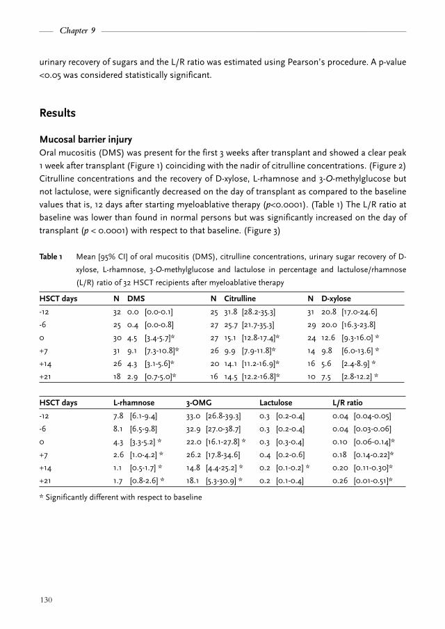
130
_____ Chapter 9 _______________________________________________________________________________________________________________ ____________________________ Inflammatory response to mucosal barrier injury after myeloablative therapy _____
131
urinary recovery of sugars and the L/R ratio was estimated using Pearson’s procedure. A p-value <0.05 was considered statistically significant.
Results
Mucosal barrier injuryOral mucositis (DMS) was present for the first 3 weeks after transplant and showed a clear peak 1 week after transplant (Figure 1) coinciding with the nadir of citrulline concentrations. (Figure 2) Citrulline concentrations and the recovery of D-xylose, L-rhamnose and 3-O-methylglucose but not lactulose, were significantly decreased on the day of transplant as compared to the baseline values that is, 12 days after starting myeloablative therapy (p<0.0001). (Table 1) The L/R ratio at baseline was lower than found in normal persons but was significantly increased on the day of transplant (p < 0.0001) with respect to that baseline. (Figure 3)
Table 1 Mean [95% CI] of oral mucositis (DMS), citrulline concentrations, urinary sugar recovery of D-
xylose, L-rhamnose, 3-O-methylglucose and lactulose in percentage and lactulose/rhamnose
(L/R) ratio of 32 HSCT recipients after myeloablative therapy
HSCT days N DMS N Citrulline N D-xylose
-12 32 0.0 [0.0-0.1] 25 31.8 [28.2-35.3] 31 20.8 [17.0-24.6]
-6 25 0.4 [0.0-0.8] 27 25.7 [21.7-35.3] 29 20.0 [16.3-23.8]
0 30 4.5 [3.4-5.7]* 27 15.1 [12.8-17.4]* 24 12.6 [9.3-16.0] *
+7 31 9.1 [7.3-10.8]* 26 9.9 [7.9-11.8]* 14 9.8 [6.0-13.6] *
+14 26 4.3 [3.1-5.6]* 20 14.1 [11.2-16.9]* 16 5.6 [2.4-8.9] *
+21 18 2.9 [0.7-5.0]* 16 14.5 [12.2-16.8]* 10 7.5 [2.8-12.2] *
HSCT days L-rhamnose 3-OMG Lactulose L/R ratio
-12 7.8 [6.1-9.4] 33.0 [26.8-39.3] 0.3 [0.2-0.4] 0.04 [0.04-0.05]
-6 8.1 [6.5-9.8] 32.9 [27.0-38.7] 0.3 [0.2-0.4] 0.04 [0.03-0.06]
0 4.3 [3.3-5.2] * 22.0 [16.1-27.8] * 0.3 [0.3-0.4] 0.10 [0.06-0.14]*
+7 2.6 [1.0-4.2] * 26.2 [17.8-34.6] 0.4 [0.2-0.6] 0.18 [0.14-0.22]*
+14 1.1 [0.5-1.7] * 14.8 [4.4-25.2] * 0.2 [0.1-0.2] * 0.20 [0.11-0.30]*
+21 1.7 [0.8-2.6] * 18.1 [5.3-30.9] * 0.2 [0.1-0.4] 0.26 [0.01-0.51]*
* Significantly different with respect to baseline
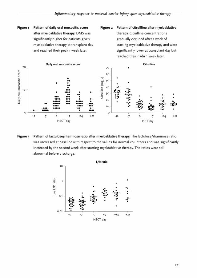
130
_____ Chapter 9 _______________________________________________________________________________________________________________ ____________________________ Inflammatory response to mucosal barrier injury after myeloablative therapy _____
131
Figure 1 Pattern of daily oral mucositis score
after myeloablative therapy. DMS was
significantly higher for patients given
myeloablative therapy at transplant day
and reached their peak 1 week later.
Figure 2 Pattern of citrullline after myeloablative
therapy. Citrulline concentrations
gradually declined after 1 week of
starting myeloablative therapy and were
significantly lower at transplant day but
reached their nadir 1 week later.
Figure 3 Pattern of lactulose/rhamnose ratio after myeloablative therapy. The lactulose/rhamnose ratio
was increased at baseline with respect to the values for normal volunteers and was significantly
increased by the second week after starting myeloablative therapy. The ratios were still
abnormal before discharge.
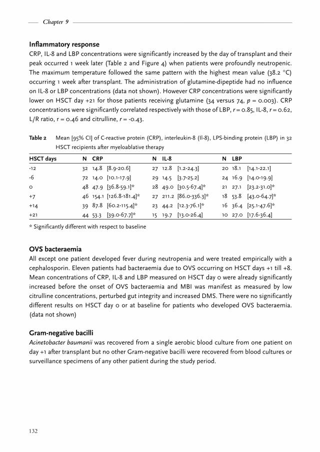
132
_____ Chapter 9 _______________________________________________________________________________________________________________ ____________________________ Inflammatory response to mucosal barrier injury after myeloablative therapy _____
133
Inflammatory responseCRP, IL-8 and LBP concentrations were significantly increased by the day of transplant and their peak occurred 1 week later (Table 2 and Figure 4) when patients were profoundly neutropenic. The maximum temperature followed the same pattern with the highest mean value (38.2 °C) occurring 1 week after transplant. The administration of glutamine-dipeptide had no influence on IL-8 or LBP concentrations (data not shown). However CRP concentrations were significantly lower on HSCT day +21 for those patients receiving glutamine (34 versus 74, p = 0.003). CRP concentrations were significantly correlated respectively with those of LBP, r = 0.85, IL-8, r = 0.62, L/R ratio, r = 0.46 and citrulline, r = -0.43.
Table 2 Mean [95% CI] of C-reactive protein (CRP), interleukin-8 (Il-8), LPS-binding protein (LBP) in 32
HSCT recipients after myeloablative therapy
HSCT days N CRP N IL-8 N LBP
-12 32 14.8 [8.9-20.6] 27 12.8 [1.2-24.3] 20 18.1 [14.1-22.1]
-6 72 14.0 [10.1-17.9] 29 14.5 [3.7-25.2] 24 16.9 [14.0-19.9]
0 48 47.9 [36.8-59.1]* 28 49.0 [30.5-67.4]* 21 27.1 [23.2-31.0]*
+7 46 154.1 [126.8-181.4]* 27 211.2 [86.0-336.3]* 18 53.8 [43.0-64.7]*
+14 39 87.8 [60.2-115.4]* 23 44.2 [12.3-76.1]* 16 36.4 [25.1-47.6]*
+21 44 53.3 [39.0-67.7]* 15 19.7 [13.0-26.4] 10 27.0 [17.6-36.4]
* Significantly different with respect to baseline
OVS bacteraemiaAll except one patient developed fever during neutropenia and were treated empirically with a cephalosporin. Eleven patients had bacteraemia due to OVS occurring on HSCT days +1 till +8. Mean concentrations of CRP, IL-8 and LBP measured on HSCT day 0 were already significantly increased before the onset of OVS bacteraemia and MBI was manifest as measured by low citrulline concentrations, perturbed gut integrity and increased DMS. There were no significantly different results on HSCT day 0 or at baseline for patients who developed OVS bacteraemia. (data not shown)
Gram-negative bacilliAcinetobacter baumanii was recovered from a single aerobic blood culture from one patient on day +1 after transplant but no other Gram-negative bacilli were recovered from blood cultures or surveillance specimens of any other patient during the study period.
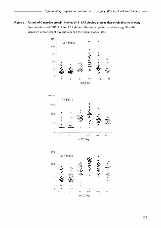
132
_____ Chapter 9 _______________________________________________________________________________________________________________ ____________________________ Inflammatory response to mucosal barrier injury after myeloablative therapy _____
133
Figure 4 Pattern of C-reactive protein, interleukin-8, LPS-binding protein after myeloablative therapy
Concentrations of CRP, IL-8 and LBP showed the similar pattern and were significantly
increased at transplant day and reached their peak 1 week later.
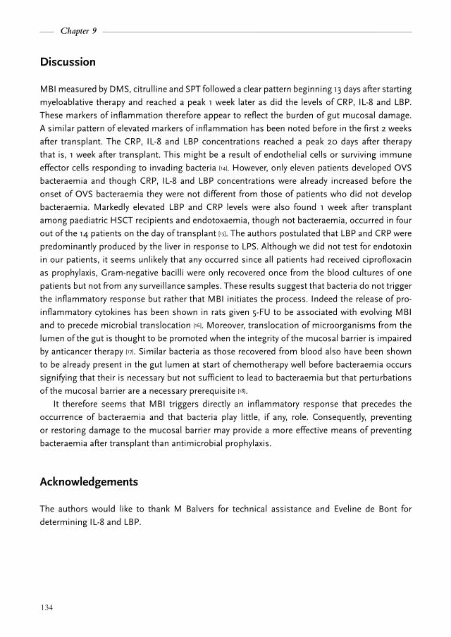
134
_____ Chapter 9 _______________________________________________________________________________________________________________ ____________________________ Inflammatory response to mucosal barrier injury after myeloablative therapy _____
135
Discussion
MBI measured by DMS, citrulline and SPT followed a clear pattern beginning 13 days after starting myeloablative therapy and reached a peak 1 week later as did the levels of CRP, IL-8 and LBP. These markers of inflammation therefore appear to reflect the burden of gut mucosal damage. A similar pattern of elevated markers of inflammation has been noted before in the first 2 weeks after transplant. The CRP, IL-8 and LBP concentrations reached a peak 20 days after therapy that is, 1 week after transplant. This might be a result of endothelial cells or surviving immune effector cells responding to invading bacteria [14]. However, only eleven patients developed OVS bacteraemia and though CRP, IL-8 and LBP concentrations were already increased before the onset of OVS bacteraemia they were not different from those of patients who did not develop bacteraemia. Markedly elevated LBP and CRP levels were also found 1 week after transplant among paediatric HSCT recipients and endotoxaemia, though not bacteraemia, occurred in four out of the 14 patients on the day of transplant [15]. The authors postulated that LBP and CRP were predominantly produced by the liver in response to LPS. Although we did not test for endotoxin in our patients, it seems unlikely that any occurred since all patients had received ciprofloxacin as prophylaxis, Gram-negative bacilli were only recovered once from the blood cultures of one patients but not from any surveillance samples. These results suggest that bacteria do not trigger the inflammatory response but rather that MBI initiates the process. Indeed the release of pro-inflammatory cytokines has been shown in rats given 5-FU to be associated with evolving MBI and to precede microbial translocation [16]. Moreover, translocation of microorganisms from the lumen of the gut is thought to be promoted when the integrity of the mucosal barrier is impaired by anticancer therapy [17]. Similar bacteria as those recovered from blood also have been shown to be already present in the gut lumen at start of chemotherapy well before bacteraemia occurs signifying that their is necessary but not sufficient to lead to bacteraemia but that perturbations of the mucosal barrier are a necessary prerequisite [18].
It therefore seems that MBI triggers directly an inflammatory response that precedes the occurrence of bacteraemia and that bacteria play little, if any, role. Consequently, preventing or restoring damage to the mucosal barrier may provide a more effective means of preventing bacteraemia after transplant than antimicrobial prophylaxis.
Acknowledgements
The authors would like to thank M Balvers for technical assistance and Eveline de Bont for determining IL-8 and LBP.
134
_____ Chapter 9 _______________________________________________________________________________________________________________ ____________________________ Inflammatory response to mucosal barrier injury after myeloablative therapy _____
135
References
1. Blijlevens NM, Donnelly JP, de Pauw BE. Mucosal barrier injury: biology, pathology, clinical counterparts and consequences of intensive treatment for haematological malignancy: an overview. Bone Marrow Transplant 2000; 25(12):1269-1278.
2. Hill GR, Ferrara JL. The primacy of the gastrointestinal tract as a target organ of acute graft-versus-host disease: rationale for the use of cytokine shields in allogeneic bone marrow transplantation. Blood 2000; 95(9):2754-2759.
3. van’t Land B, Blijlevens NM, Marteijn J, Timal S, Donnelly JP, de Witte TJ et al. Role of curcumin and the inhibition of NF-kappaB in the onset of chemotherapy-induced mucosal barrier injury. Leukemia 2004; 18(2):276-284.
4. Xun CQ, Thompson JS, Jennings CD, Brown SA, Widmer MB. Effect of total body irradiation, busulfan-cyclophosphamide, or cyclophosphamide conditioning on inflammatory cytokine release and development of acute and chronic graft-versus-host disease in H-2-incompatible transplanted SCID mice. Blood 1994; 83(8):2360-2367.
5. Otte JM, Podolsky DK. Functional modulation of enterocytes by gram-positive and gram-negative microorganisms. Am J Physiol Gastrointest Liver Physiol 2004; 286(4):G613-G626.
6. Keefe DM, Brealey J, Goland GJ, Cummins AG. Chemotherapy for cancer causes apoptosis that precedes hypoplasia in crypts of the small intestine in humans. Gut 2000; 47(5):632-637.
7. Ruescher TJ, Sodeifi A, Scrivani SJ, Kaban LB, Sonis ST. The impact of mucositis on alpha-hemolytic streptococcal infection in patients undergoing autologous bone marrow transplantation for hematologic malignancies. Cancer 1998; 82(11):2275-2281.
8. Schaap N, Schattenberg A, Bar B, Preijers F, Geurts vK, van der MR et al. Outcome of transplantation for standard-risk leukaemia with grafts depleted of lymphocytes after conditioning with an intensified regimen. Br J Haematol 1997; 98(3):750-759.
9. Blijlevens NM, van’t Land B, Donnelly JP, M’Rabet L, de Pauw BE. Measuring mucosal damage induced by cytotoxic therapy. Support Care Cancer 2004; 12(4):227-233.
10. Donnelly JP, Dompeling EC, Meis JF, de Pauw BE. Bacteremia due to oral viridans streptococci in neutropenic patients with cancer: cytostatics are a more important risk factor than antibacterial prophylaxis. Clin Infect Dis 1995; 20(2):469-470.
11. Lutgens LC, Blijlevens NM, Deutz NE, Donnelly JP, Lambin P, de Pauw BE. Monitoring myeloablative therapy-induced small bowel toxicity by serum citrulline concentration: a comparison with sugar permeability tests. Cancer 2005; 103(1):191-199.
12. Blijlevens NM, Lutgens LC, Schattenberg AV, Donnelly JP. Citrulline: a potentially simple quantitative marker of intestinal epithelial damage following myeloablative therapy. Bone Marrow Transplant 2004; 34(3):193-196.
13. Donnelly JP, Muus P, Schattenberg A, De Witte T, Horrevorts A, DePauw BE. A scheme for daily monitoring of oral mucositis in allogeneic BMT recipients. Bone Marrow Transplant 1992; 9(6):409-413.
14. Oude Nijhuis CS, Vellenga E, Daenen SM, Kamps WA, De Bont ES. Endothelial cells are main producers of interleukin 8 through Toll-like receptor 2 and 4 signaling during bacterial infection in leukopenic cancer patients. Clin Diagn Lab Immunol 2003; 10(4):558-563.
15. Levy O, Teixeira-Pinto A, White ML, Carroll SF, Lehmann L, Wypij D et al. Endotoxemia and elevation of lipopolysaccharide-binding protein after hematopoietic stem cell transplantation. Pediatr Infect Dis J 2003; 22(11):978-981.
16. Tsuji E, Hiki N, Nomura S, Fukushima R, Kojima J, Ogawa T et al. Simultaneous onset of acute inflammatory response, sepsis-like symptoms and intestinal mucosal injury after cancer chemotherapy. Int J Cancer 2003; 107(2):303-308.
17. Ellis M. Preventing microbial translocation in haematological malignancy. Br J Haematol 2004; 125(3):282-293.18. Frere P, Hermanne JP, Debouge MH, de Mol P, Fillet G, Beguin Y. Bacteremia after hematopoietic stem cell
transplantation: incidence and predictive value of surveillance cultures. Bone Marrow Transplant 2004; 33(7):745-749.


Mucosal Barrier Injury: A paradigm for dysregulation of the mucosal barrier and microbial flora
Chapter 10
N.M.A. Blijlevens and J.P. Donnelly
Department of Haematology, University Medical Center St. Radboud, Nijmegen
The Netherlands
Submitted

138
_____ Chapter 10 ____________________________________________________________________________________________________________ _____________________________________________________________ Mucosal Barrier Injury paradigm of dysregulation _____
139

138
_____ Chapter 10 ____________________________________________________________________________________________________________ _____________________________________________________________ Mucosal Barrier Injury paradigm of dysregulation _____
139
Summary
This review explores the role, if any, of the microbial commensal flora in the pathophysiology of the “mucosal barrier injury” induced by chemotherapy. Mucosal barrier injury (MBI) extends beyond oral mucositis to involve the entire digestive tract and is more than just a risk factor for severe infectious complications; it is a manifestation of the collapse of the relationship between the host and the endogenous microbial flora. Epithelial cells are an integral component of the anatomical and immunological mucosal barrier functioning as the front line of host defence against microorganisms. Clarification of how these cells recognise and interact with the microbial flora is critical for understanding the process of mucosal barrier injury and necessary for managing those patients at the greatest risk of developing MBI and the related infectious complications.
Introduction
Historically, the stomatotoxicity or oral mucositis induced by anticancer therapy was simply attributed to direct impairment of the proliferation of epithelial cells resulting in ulceration. Colonisation of the affected area by bacteria then exacerbated the mucositis. The subsequent disruption of the mucosal surfaces together with evolving neutropenia then opened the door to opportunistic infection which was often life-threatening [1]. A new model was proposed by Sonis describing four successive phases of oral mucositis (Figure 1) – an inflammatory (connective tissue) phase followed by an epithelial phase, an ulcerative/infective phase and finally a healing phase [2].
It was also postulated that bacterial colonisation of ulcerated surfaces up-regulated the pro-inflammatory cytokine response and lead directly to systemic infection. Since infection was thought to play an important role in the pathophysiology of mucositis it seemed reasonable to employ antimicrobial agents for prevention and treatment of oral mucositis but this proved futile [3]. The model of Sonis restricts itself to the oral cavity and microorganisms are not involved in the first phase of pro-inflammatory cytokine release. To better understand the role, if any, of the microflora in causing mucosal damage, it is necessary to postulate that “mucosal barrier injury” (MBI) affects the entire digestive tract. Particularly at the level of epithelial cells, as they are an integral component of the anatomical and immunological mucosal barrier. This barrier functions as the front line of host defence against microorganisms [4]. Intestinal epithelial cells are the key players in the innate immune response (Figure 2) and therefore, it is important to clarify how epithelial cells recognise and interact with the microflora as this is critical for understanding the process of MBI.
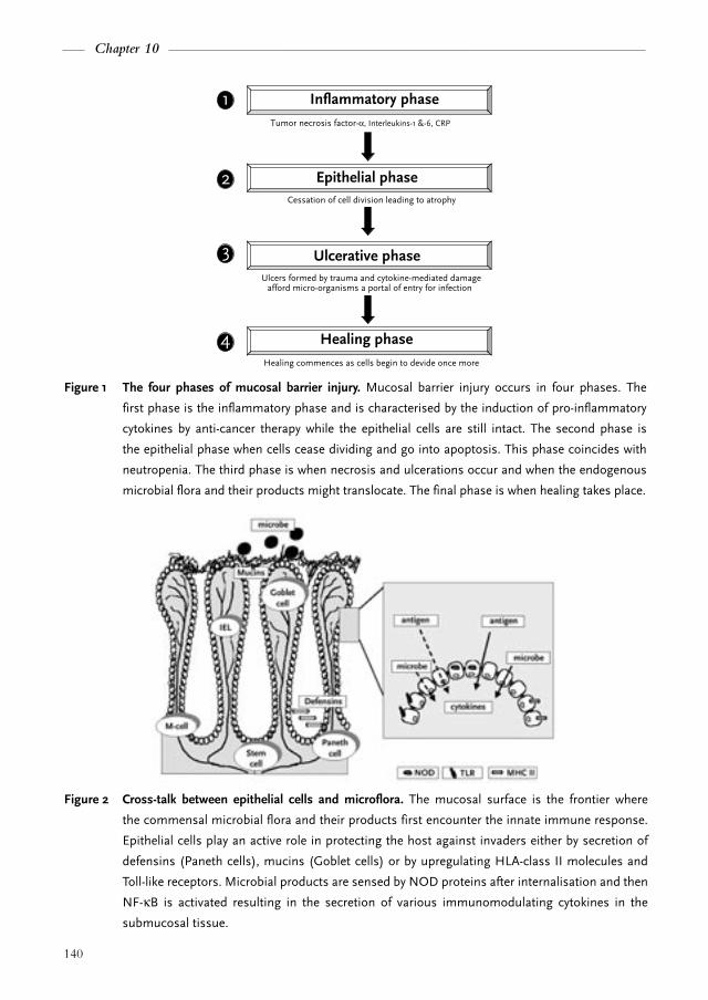
140
_____ Chapter 10 ____________________________________________________________________________________________________________ _____________________________________________________________ Mucosal Barrier Injury paradigm of dysregulation _____
141
Figure 1 The four phases of mucosal barrier injury. Mucosal barrier injury occurs in four phases. The
first phase is the inflammatory phase and is characterised by the induction of pro-inflammatory
cytokines by anti-cancer therapy while the epithelial cells are still intact. The second phase is
the epithelial phase when cells cease dividing and go into apoptosis. This phase coincides with
neutropenia. The third phase is when necrosis and ulcerations occur and when the endogenous
microbial flora and their products might translocate. The final phase is when healing takes place.
Figure 2 Cross-talk between epithelial cells and microflora. The mucosal surface is the frontier where
the commensal microbial flora and their products first encounter the innate immune response.
Epithelial cells play an active role in protecting the host against invaders either by secretion of
defensins (Paneth cells), mucins (Goblet cells) or by upregulating HLA-class II molecules and
Toll-like receptors. Microbial products are sensed by NOD proteins after internalisation and then
NF-κB is activated resulting in the secretion of various immunomodulating cytokines in the
submucosal tissue.
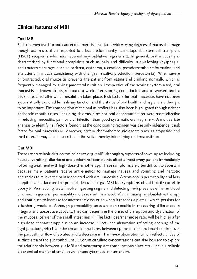
140
_____ Chapter 10 ____________________________________________________________________________________________________________ _____________________________________________________________ Mucosal Barrier Injury paradigm of dysregulation _____
141
Clinical features of MBI
Oral MBIEach regimen used for anti-cancer treatment is associated with varying degrees of mucosal damage though oral mucositis is reported to affect predominantly haematopoietic stem cell transplant (HSCT) recipients who have received myeloablative regimens [5]. In general, oral mucositis is characterised by functional complaints such as pain and difficulty in swallowing (dysphagia) and anatomic changes such as oedema, erythema, ulceration, pseudomembrane formation, and alterations in mucus consistency with changes in saliva production (xerostomia). When severe or protracted, oral mucositis prevents the patient from eating and drinking normally, which is frequently managed by giving parenteral nutrition. Irrespective of the scoring system used, oral mucositis is known to begin around a week after starting conditioning and to worsen until a peak is reached after which resolution takes place. Risk factors for oral mucositis have not been systematically explored but salivary function and the status of oral health and hygiene are thought to be important. The composition of the oral microflora has also been highlighted though neither antiseptic mouth rinses, including chlorhexidine nor oral decontamination were more effective in reducing mucositis, pain or oral infection than good systematic oral hygiene [6]. A multivariate analysis to identify risk factors found that the conditioning regimen was the only independent risk factor for oral mucositis [7]. Moreover, certain chemotherapeutic agents such as etoposide and methotrexate may also be secreted in the saliva thereby intensifying oral mucositis [8].
Gut MBI There are no reliable data on the incidence of gut MBI although symptoms of bowel upset including nausea, vomiting, diarrhoea and abdominal complaints affect almost every patient immediately following treatment with high-dose chemotherapy. These symptoms are often difficult to ascertain because many patients receive anti-emetics to manage nausea and vomiting and narcotic analgesics to relieve the pain associated with oral mucositis. Alterations in permeability and loss of epithelial surface are the principle features of gut MBI but symptoms of gut toxicity correlate poorly [9]. Permeability tests involve ingesting sugars and detecting their presence either in blood or urine. In general, permeability increases within a week after initiating myeloablative therapy and continues to increase for another 10 days or so when it reaches a plateau which persists for a further 3 weeks [9]. Although permeability tests are non-specific in measuring differences in integrity and absorptive capacity, they can determine the onset of disruption and dysfunction of the mucosal barrier of the small intestines [10]. The lactulose/rhamnose ratio will be higher after high-dose chemotherapy due to an increase in lactulose absorption reflecting opening of the tight junctions, which are the dynamic structures between epithelial cells that exert control over the paracellular flow of solutes and a decrease in rhamnose absorption which reflects a loss of surface area of the gut epithelium [11]. Serum citrulline concentrations can also be used to explore the relationship between gut MBI and post-transplant complications since citrulline is a reliable biochemical marker of small bowel enterocyte mass in humans [12].
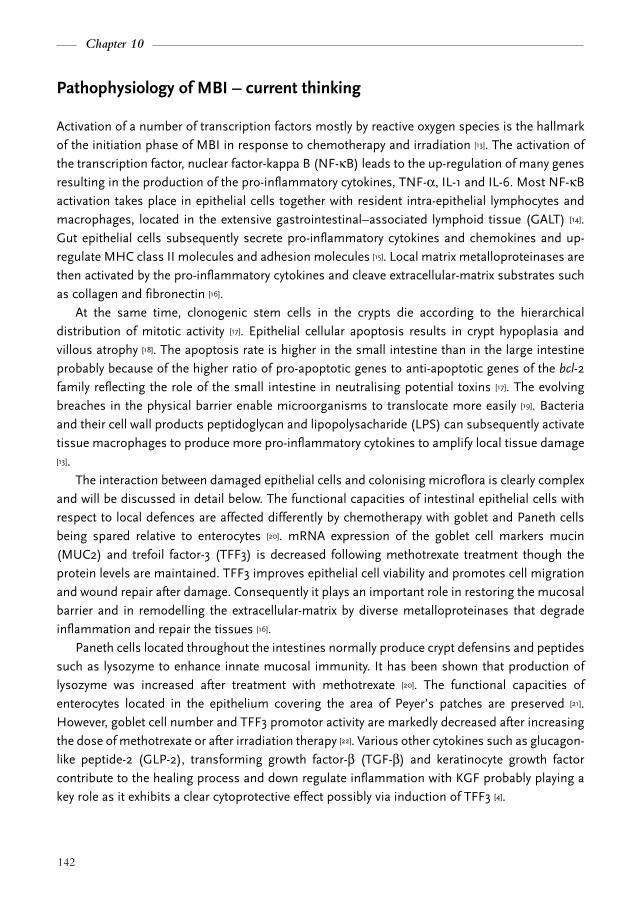
142
_____ Chapter 10 ____________________________________________________________________________________________________________ _____________________________________________________________ Mucosal Barrier Injury paradigm of dysregulation _____
143
Pathophysiology of MBI – current thinking
Activation of a number of transcription factors mostly by reactive oxygen species is the hallmark of the initiation phase of MBI in response to chemotherapy and irradiation [13]. The activation of the transcription factor, nuclear factor-kappa B (NF-κB) leads to the up-regulation of many genes resulting in the production of the pro-inflammatory cytokines, TNF-α, IL-1 and IL-6. Most NF-κB activation takes place in epithelial cells together with resident intra-epithelial lymphocytes and macrophages, located in the extensive gastrointestinal–associated lymphoid tissue (GALT) [14]. Gut epithelial cells subsequently secrete pro-inflammatory cytokines and chemokines and up-regulate MHC class II molecules and adhesion molecules [15]. Local matrix metalloproteinases are then activated by the pro-inflammatory cytokines and cleave extracellular-matrix substrates such as collagen and fibronectin [16].
At the same time, clonogenic stem cells in the crypts die according to the hierarchical distribution of mitotic activity [17]. Epithelial cellular apoptosis results in crypt hypoplasia and villous atrophy [18]. The apoptosis rate is higher in the small intestine than in the large intestine probably because of the higher ratio of pro-apoptotic genes to anti-apoptotic genes of the bcl-2 family reflecting the role of the small intestine in neutralising potential toxins [17]. The evolving breaches in the physical barrier enable microorganisms to translocate more easily [19]. Bacteria and their cell wall products peptidoglycan and lipopolysacharide (LPS) can subsequently activate tissue macrophages to produce more pro-inflammatory cytokines to amplify local tissue damage [13].
The interaction between damaged epithelial cells and colonising microflora is clearly complex and will be discussed in detail below. The functional capacities of intestinal epithelial cells with respect to local defences are affected differently by chemotherapy with goblet and Paneth cells being spared relative to enterocytes [20]. mRNA expression of the goblet cell markers mucin (MUC2) and trefoil factor-3 (TFF3) is decreased following methotrexate treatment though the protein levels are maintained. TFF3 improves epithelial cell viability and promotes cell migration and wound repair after damage. Consequently it plays an important role in restoring the mucosal barrier and in remodelling the extracellular-matrix by diverse metalloproteinases that degrade inflammation and repair the tissues [16].
Paneth cells located throughout the intestines normally produce crypt defensins and peptides such as lysozyme to enhance innate mucosal immunity. It has been shown that production of lysozyme was increased after treatment with methotrexate [20]. The functional capacities of enterocytes located in the epithelium covering the area of Peyer’s patches are preserved [21]. However, goblet cell number and TFF3 promotor activity are markedly decreased after increasing the dose of methotrexate or after irradiation therapy [22]. Various other cytokines such as glucagon-like peptide-2 (GLP-2), transforming growth factor-β (TGF-β) and keratinocyte growth factor contribute to the healing process and down regulate inflammation with KGF probably playing a key role as it exhibits a clear cytoprotective effect possibly via induction of TFF3 [4].
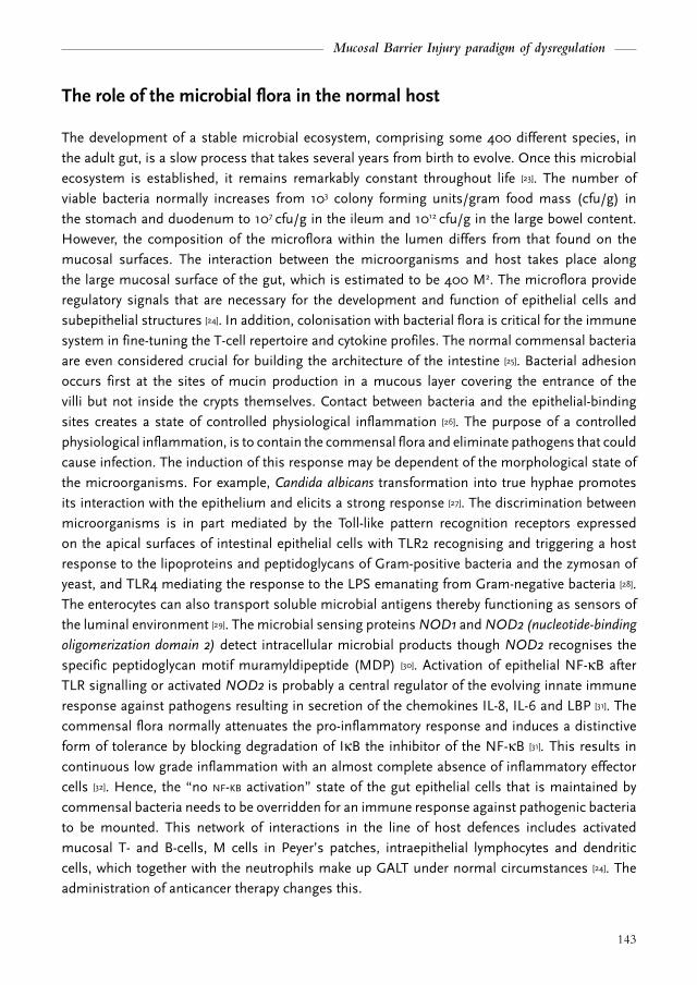
142
_____ Chapter 10 ____________________________________________________________________________________________________________ _____________________________________________________________ Mucosal Barrier Injury paradigm of dysregulation _____
143
The role of the microbial flora in the normal host
The development of a stable microbial ecosystem, comprising some 400 different species, in the adult gut, is a slow process that takes several years from birth to evolve. Once this microbial ecosystem is established, it remains remarkably constant throughout life [23]. The number of viable bacteria normally increases from 103 colony forming units/gram food mass (cfu/g) in the stomach and duodenum to 107 cfu/g in the ileum and 1012 cfu/g in the large bowel content. However, the composition of the microflora within the lumen differs from that found on the mucosal surfaces. The interaction between the microorganisms and host takes place along the large mucosal surface of the gut, which is estimated to be 400 M2. The microflora provide regulatory signals that are necessary for the development and function of epithelial cells and subepithelial structures [24]. In addition, colonisation with bacterial flora is critical for the immune system in fine-tuning the T-cell repertoire and cytokine profiles. The normal commensal bacteria are even considered crucial for building the architecture of the intestine [25]. Bacterial adhesion occurs first at the sites of mucin production in a mucous layer covering the entrance of the villi but not inside the crypts themselves. Contact between bacteria and the epithelial-binding sites creates a state of controlled physiological inflammation [26]. The purpose of a controlled physiological inflammation, is to contain the commensal flora and eliminate pathogens that could cause infection. The induction of this response may be dependent of the morphological state of the microorganisms. For example, Candida albicans transformation into true hyphae promotes its interaction with the epithelium and elicits a strong response [27]. The discrimination between microorganisms is in part mediated by the Toll-like pattern recognition receptors expressed on the apical surfaces of intestinal epithelial cells with TLR2 recognising and triggering a host response to the lipoproteins and peptidoglycans of Gram-positive bacteria and the zymosan of yeast, and TLR4 mediating the response to the LPS emanating from Gram-negative bacteria [28]. The enterocytes can also transport soluble microbial antigens thereby functioning as sensors of the luminal environment [29]. The microbial sensing proteins NOD1 and NOD2 (nucleotide-binding oligomerization domain 2) detect intracellular microbial products though NOD2 recognises the specific peptidoglycan motif muramyldipeptide (MDP) [30]. Activation of epithelial NF-κB after TLR signalling or activated NOD2 is probably a central regulator of the evolving innate immune response against pathogens resulting in secretion of the chemokines IL-8, IL-6 and LBP [31]. The commensal flora normally attenuates the pro-inflammatory response and induces a distinctive form of tolerance by blocking degradation of IκB the inhibitor of the NF-κB [31]. This results in continuous low grade inflammation with an almost complete absence of inflammatory effector cells [32]. Hence, the “no NF-KB activation” state of the gut epithelial cells that is maintained by commensal bacteria needs to be overridden for an immune response against pathogenic bacteria to be mounted. This network of interactions in the line of host defences includes activated mucosal T- and B-cells, M cells in Peyer’s patches, intraepithelial lymphocytes and dendritic cells, which together with the neutrophils make up GALT under normal circumstances [24]. The administration of anticancer therapy changes this.
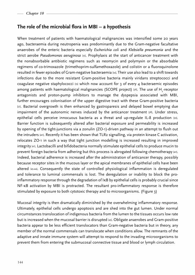
144
_____ Chapter 10 ____________________________________________________________________________________________________________ _____________________________________________________________ Mucosal Barrier Injury paradigm of dysregulation _____
145
The role of the microbial flora in MBI – a hypothesis
When treatment of patients with haematological malignancies was intensified some 20 years ago, bacteraemia during neutropenia was predominantly due to the Gram-negative facultative anaerobes of the enteric bacteria especially Escherichia coli and Klebsiella pneumonia and the strict aerobe Pseudomonas aeruginosa [33]. Prophylaxis at the start of anticancer treatment with the nonabsorbable antibiotic regimens such as neomycin and polymyxin or the absorbable regimens of co-trimoxazole (trimethoprim-sulfamethoxazole) and colistin or a fluoroquinolone resulted in fewer episodes of Gram-negative bacteraemia [34]. Their use also lead to a shift towards infections due to the more resistant Gram-positive bacteria mainly viridans streptococci and coagulase negative staphylococci [35] which now account for 3 of every 4 bacteraemic episodes among patients with haematological malignancies (SCOPE project) [36]. The use of H
2-receptor
antagonists and proton-pump inhibitors to manage the dyspepsia associated with MBI, further encourages colonisation of the upper digestive tract with these Gram-positive bacteria [37]. Bacterial overgrowth is then enhanced by gastroparesis and delayed bowel emptying due impairment of the autonomic nerves induced by the anticancer treatment [38]. Under stress, epithelial cells perceive innocuous bacteria as a threat and up-regulate IL-8 production [39]. Barrier function is subsequently altered after bacterial exposure and permeability is increased by opening of the tight-junctions via a zonulin (ZO-1)-driven pathway in an attempt to flush out the intruders [40]. Recently it has been shown that TLR2 signalling, via protein kinase C activation, relocates ZO-1 in such a way that tight junction modelling is increased resulting in improved integrity [41]. Lactobacilli and bifidobacteria normally stimulate epithelial cells to produce mucin to prevent foreign bacteria from adhering but this process is abrogated following chemotherapy [42]. Indeed, bacterial adherence is increased after the administration of anticancer therapy, possibly because receptor sites in the mucous layer or the apical membranes of epithelial cells have been altered [43,44]. Consequently the state of controlled physiological inflammation is deregulated and tolerance to luminal commensals is lost. The deregulation or inability to block the pro-inflammatory response through the degradation of IκB by epithelial cells is probably crucial since NF-κB activation by MBI is protracted. The resultant pro-inflammatory response is therefore stimulated by exposure to both cytotoxic therapy and to microorganisms. (Figure 3)
Mucosal integrity is then dramatically diminished by the overwhelming inflammatory response. Ultimately, epithelial cells undergo apoptosis and are shed into the gut lumen. Under normal circumstances translocation of indigenous bacteria from the lumen to the tissues occurs low rate but is increased when the mucosal barrier is disrupted [45]. Obligate anaerobes and Gram-positive bacteria appear to be less efficient translocators than Gram-negative bacteria but in theory, any member of the normal commensals can translocate when conditions allow. The remnants of the adaptive and innate immune system will attempt to respond to the invading microorganisms to prevent them from entering the submucosal connective tissue and blood or lymph circulation.
144
_____ Chapter 10 ____________________________________________________________________________________________________________ _____________________________________________________________ Mucosal Barrier Injury paradigm of dysregulation _____
145
Figure 3 Model for inducing mucosal barrier injury. Both cytoreductive therapy and the microbial flora
activate the transcription factor, NF-κB in epithelial cells resulting in an ongoing inflammatory
response aggravating MBI.
Infectious complications of MBI
Damaged mucosa increases the risk of infections and mortality especially among transplant recipients [46]. The duration of fever, parenteral narcotic use, total parenteral nutrition, antibiotic therapy and the total number of days in hospital correlate directly with the extent and severity of MBI. Gram-positive bacteraemia due to oral viridans streptococci (OVS) is related to MBI of the upper part of the digestive tract, whilst neutropenic enterocolitis is related to the lower part of the digestive tract. Gram-negative bacillary infections and those due to coagulase-negative staphylococci (CoNS) can be related to MBI at any site. Recipients of autologous HSCT with oral ulcerative mucositis were found to be 3 times more likely to develop OVS bacteraemia than those without mucositis [47]. The viridans species S. mitis and S. oralis are associated with more serious complications such as sepsis and adult respiratory distress syndrome which carries a high mortality (80%) [48]. Coagulase-negative staphylococci (CoNS) were the most frequent isolates in the SCOPE project [36] and though bacteraemia is frequently related to the use of central venous catheters, mucosal sites are also an important source of these bacteria [49]. MBI is clearly a risk factor for bacteraemia due to Acinetobacter species, Stenotrophomonas maltophilia and Capnocytophaga species [50,51]. Despite adequate empirical antibiotic therapy, Gram-negative bacillary bacteraemia due to Escherichia coli, Klebsiella species and Pseudomonas aeruginosa are still associated with the highest mortality amongst HSCT recipients [52]. Anaerobic bacteria are rarely involved though MBI is an independent predictor of bacteraemia due to Fusobacterium nucleatum [53]. Bacteraemia with different species including Staphylococcus aureus, Pseudomonas aeruginosa, Clostridium species and Candida species is associated with neutropenic enterocolitis

146
_____ Chapter 10 ____________________________________________________________________________________________________________ _____________________________________________________________ Mucosal Barrier Injury paradigm of dysregulation _____
147
(typhlitis), which is considered to be an extreme manifestation of MBI. This appears to be the result of the delicate balance between host and microflora being totally disturbed in the setting of prolonged exposure to antibiotics resulting in selection of particularly Clostridium species [54]. Indeed the occurrence of bacteraemia due to one of these species, Clostridium tertium, is considered pathognomonic for neutropenic enterocolitis [55]. This phenomenon is also illustrated by Candida species that normally colonise the mucosal surfaces without harming the host but which can be invasive when MBI is apparent [56]. These observations support the contention that MBI provides a portal of entry to the systemic circulation by commensal flora of the oral cavity and gastrointestinal tract.
Summarising discussion
MBI is more than a risk factor for severe infectious complications after anti-cancer treatment; it is a manifestation of a collapse of the relationship between the host and the endogenous microflora. Epithelial cells are the first to encounter the microorganisms and play a crucial role in the regulation of the barrier between host and lumen content. After the administration of chemotherapy or irradiation not only is the anatomic barrier disrupted providing a portal of entry to microorganisms but the state of controlled inflammation is also abrogated. The endogenous flora plays a role throughout the whole process of MBI by determining the pre-MBI inflammation state and accelerating the MBI induced pro-inflammatory response and apoptosis. After translocation, microorganisms trigger the systemic inflammatory response and can cause an infectious disease. The microflora probably also has a role in repairing the barrier. Probiotics (lactobacilli and bifidobacteria) and prebiotics (non-digestible food ingredients such as fructo-oligosaccharides) appear to exert anti-inflammatory effects [24]. The role of the endogenous flora shifts from being of benefit to the host to causing disease to that host especially when that host has damaged mucous membranes. (Figure 4)
The delicate balance between microbes and a damaged host has been termed the “damage-response” framework in which microbial pathogenesis is the outcome. The inflammatory response of the host itself partially explains the degree of damaged caused [57]. MBI therefore represents the loss of regulation of the interaction between the host and microorganisms, which would explain why almost all attempts thus far to prevent or ameliorate MBI with antimicrobial therapy have failed. It may be more fruitful to aim at either down regulating the inflammatory response or at restoring the barrier by stimulating epithelial cell functioning using growth factors such as recombinant human keratinocyte growth factor (KGF) [58], IL-11 [59], glucagon-like peptide or intestinal trefoil factor to manage those patients at the greatest risk of developing MBI. Clinical trials with one of these agents, KGF, are currently in progress and the results are awaited with interest.
146
_____ Chapter 10 ____________________________________________________________________________________________________________ _____________________________________________________________ Mucosal Barrier Injury paradigm of dysregulation _____
147
Figure 4 Model for invasive infections. Cytoreductive chemotherapy induces injury to the mucosal barrier
and antimicrobial agents alter the ecology of the commensal microbial flora leading to selection
of opportunistic pathogens. These microorganisms adhere to the epithelial surface of the
damaged mucosa and either translocate or establish a local infection eliciting an inflammatory
response and hence an infectious disease process.
References
1. Berger AM, Kilroy TJ. Adverse effects of treatment; oral complications. In: DeVita VT, Hellman S, Rosenberg SA, eds. Cancer; principles and practice of oncology. Philadelphia: Lippincott - Raven Publishers; 1997:2714-2725.
2. Sonis ST. Mucositis as a biological process: a new hypothesis for the development of chemotherapy-induced stomatotoxicity. Oral Oncol. 1998; 34:39-43.
3. Donnelly J.P., Bellm L.A., Epstein JB, Sonis S.T., Symonds RP. Antimicrobial therapy to prevent or treat oral mucositis. Lancet Infect Dis. 2003; 3:405-412.
4. Blijlevens NMA., Donnelly JP, De Pauw BE. Mucosal barrier injury: biology, pathology, clinical counterparts and consequences of intensive treatment for haematological malignancy: an overview. Bone Marrow Transplant. 2000; 25:1269-1278.
5. Schubert MM, Williams BE, Lloid ME, Donaldson G, Chapko MK. Clinical assessment scale for the rating of oral mucosal changes associated with bone marrow transplantation. Development of an oral mucositis index. Cancer. 1992; 69:2469-2477.
6. Cheng K.K, Molassiotis, A., Chang A.M., Cheung S.S. Evaluation of an oral care protocol intervention in the prevention of chemotherapy-induced oral mucositis in paediatric cancer patients. Eur J Cancer. 2001; 37:2056-2063.
7. Wardley AM, Jayson GC, Swindell R et al. Prospective evaluation of oral mucositis in patients receiving myeloablative conditioning regimens and haematopoietic progenitor rescue. Br J Haematol. 2000; 110:292-299.
8. Albertioni F, Rask C, Schroeder H, Peterson C. Monitoring of methotrexate and 7-hydroxymethotrexate in saliva from children with acute lymphoblastic leukemia receiving high-dose consolidation treatment: relation to oral mucositis. Anticancer Drugs. 1997; 8:119-124.
9. Blijlevens N.M.A., van’t Land B., Donnelly JP, M’Rabet L., DePauw BE. Measuring mucosal damage induced by cytotoxic therapy. Support Care Cancer. 2004; 12:227-233.
148
_____ Chapter 10 ____________________________________________________________________________________________________________ _____________________________________________________________ Mucosal Barrier Injury paradigm of dysregulation _____
149
10. Blijlevens N.M.A., Donnelly J.P., dePauw B.E. Prospective evaluation of gut mucosal barrier injury following various myeloablative regimens for haematopoietic stem cell transplant. Bone Marrow Transplant. 2005; 35:707-711
11. Keefe DM, Cummins AG, Dale BM et al. Effect of high-dose chemotherapy on intestinal permeability in humans. Clin Sci Colch. 1997; 92:385-389.
12. Blijlevens N.M.A., Lutgens L.C.H.W., Schattenberg A.V.M.B.., De Pauw B.E. Citrulline: a potentially simple quantitative marker of intestinal epithelial damage following myeloablative therapy. Bone Marrow Transplant. 2004; 34:193-196.
13. Sonis S.T., Elting L.S., Keefe DM et al. Perspectives on cancer therapy-induced mucosal injury: pathogenesis, measurement, epidemiology, and consequences for patients. Cancer. 2004; 100:1995-2025.
14. van’t Land B., Blijlevens N.M.A., \marteijn J et al. Role of curcumin and the inhibition of NF-kappaB in the onset of chemotherapy induced mucosal barrier injury. Leukemia. 2004; 18:276-284.
15. Xun CQ, Thompson JS, Jennings CD, Brown SA, Widmer MB. Effect of total body irradiation, busulfan-cyclophosphamide, or cyclophosphamide conditioning on inflammatory cytokine release and development of acute and chronic graft-versus-host disease in H-2-incompatible transplanted SCID mice. Blood. 1994; 83:2360-2367.
16. Parks W.C., Wilson C.L., Lopez-Boado Y.S. Matrix metalloproteinases as modulators of inflammation and innate immunity. Nature Reviews Immunolgy. 2004; 4:617-629.
17. Potten C.S., Wilson J.W., Booth C. Regulation and significance of apoptosis in the stem cells of the gastrointestinal epithelium. Stem Cells. 1997; 15:82-93.
18. Keefe DMK, Brealey J, Goland GJ, Cummins AG. Chemotherapy for cancer causes apoptosis that precedes hypoplasia in crypts of the small intestine in humans. Gut. 2000; 47:632-637.
19. Berg R.D. Bacterial Translocation from the Gastrointestinal Tracts of Mice Receiving Immunosuppressive Chemotherapeutic Agents. Current Microbiology. 1983; 8:285-292.
20. Verburg M., Renes I.B., van Nispen D.J.P.M. et al. Specific responses in rat small intestinal epithelial mRNA expression and protein levels during chemotherapeutic damage and regeneration. J Histochem Cytochem. 2002; 50:1525-1536.
21. Renes I.B., Verburg M., Bulsing N.P. et al. Protection of the Peyer’s patch-associated crypt and villus epithelium against methotrexate-induced damage is based on its distinct regulation of proliferation. J Pathol. 2002; 198:60-68.
22. Beck P.L., Wong J.F., Li Y. et al. Chemotherapy- and radiotherapy-induced intestinal damage is regulated by intestinal trefoil factor. Gastroenterology. 2004; 126:796-808.
23. Hart A.L., Stagg A.J., Frame M. et al. Review article: the role of the gut flora in health and disease and its modification as therapy. Aliment Pharmacol Ther. 2002; 16:1383-1303.
24. Shanahan F. The host-microbe interface within the gut. Best Practice & Research Clinical Gastroenterology. 2002; 16:915-931.
25. Madara J. Building an intestine--architectural contributions of commensal bacteria. N Engl J Med. 2004; 351:1685-1686.
26. Xavier R.J., Podolsky DK. How to get along--friendly microbes in a hostile world. Science. 2000; 289:1483-1484.27. Villar CC, Kashleva H, Dongari-Bagtzoglou A. Role of Candida albicans polymorphism in interactions with oral
epithelial cells. Oral Microbiol Immunol. 2004; 19:262-269.28. Akira S., Takeda K. Toll-like receptor signalling. Nature Reviews Immunolgy. 2004; 4:499-511.29. Kagnoff MF, Eckmann L. Epithelial cells as sensors for microbial infection. J Clin Invest. 1997; 100:6-10.30. Inohara N, Nunez G. NODs: intracellular proteins involved in inflammation and apoptosis. Nat Rev Immunol.
2003; 3:371-382.31. Neish A.S., Gewirtz A.T., Zeng H. et al. Prokaryotic regulation of epithelial responses by inhibition of IkB-a
ubiquitination. Science. 2000; 289:1560-1563.32. Xavier RJ, Podolsky DK. Microbiology. How to get along--friendly microbes in a hostile world. Science. 2000; 289:
1483-1484.33. Tancrede CH, Andremont AO. Bacterial translocation and gram-negative bacteremia in patients with hematological
malignancies. J Infect Dis. 1985; 152:99-103.34. Cruciani M, Rampazzo R, Malena M et al. Prophylaxis with fluoroquinolones for bacterial infections in neutropenic
patients: a meta-analysis. Clin Infect Dis. 1996; 23:795-805.
148
_____ Chapter 10 ____________________________________________________________________________________________________________ _____________________________________________________________ Mucosal Barrier Injury paradigm of dysregulation _____
149
35. Sepkowitz K.A. Antibiotic prophylaxis in patients receiving hematopoietic stem cell transplant. Bone Marrow Transplant. 2002; 29:367-371.
36. Wisplinghoff H., seifert H., Wenzel RP, Edmond M.B. Current trends in the epidemiology of nosocomial bloodstream infections in patients with hematological malignancies and solid neoplasms in hospitals in the United States. Clin Infect Dis. 2003; 36:1103-1110.
37. Elting LS, Bodey GP, Keefe BH. Septicemia and shock syndrome due to viridans streptococci: a case-control study of predisposing factors. Clin Infect Dis. 1992; 14:1201-1207.
38. Hecht JR, Lembo T, Chap L. Prolonged nausea and vomiting after high dose chemotherapy and autologous peripheral stem cell transplantation in the treatment of high risk breast carcinoma. Cancer. 1997; 79:1698-1702.
39. Nazli A., Yang P-C., Jury J. et al. Epithelia under metabolic stress perceive commensal bacteria as a threat. Am J Pathol. 2004; 164:947-957.
40. El Asmar R., Panigrahi P., Bamford P. et al. Host-dependent zonulin secretion causes the impairment of the small intestine barrier function after bacterial exposure. Gastroenterology. 2002; 123:1607-1615.
41. Cario E., Gerken G., Podolsky DK. Toll-like receptor 2 enhances ZO-1-associated intestinal epithelial barrier integrity via protein kinase C. Gastroenterology. 2004; 127:224-238.
42. Mack D.R., Ahrne S., Hyde L., Wei S., Hollingsworth M.A. Extracellular MUC3 mucin secretion follows adherence of Lactobacillus strains to intestinal epithelial cells in vitro. Gut. 2003; 52:827-833.
43. Sandovsky-Losica H, Segal E. Interaction of Candida albicans with murine gastrointestinal mucosa: effect of irradiation on adherence in vitro. Journal of medical and Veterinary Mycology. 1989; 27:345-352.
44. Sandovsky-Losica H, Segal E. Interaction of Candida albicans with murine gastrointestinal mucosa from methotrexate and 5-fluorouracil treated animals: in vitro adhesion and prevention. Journal of medical and Veterinary Mycology. 1990; 28:279-287.
45. Ellis M. Preventing microbial translocation in haematological malignancy. British Journal of Haematology. 2004; 125:282-293.
46. Sonis ST, Oster G, Fuchs H et al. Oral mucositis and the clinical and economic outcomes of hematopoietic stem-cell transplantation. J Clin Oncol. 2001; 19:2201-2205.
47. Ruescher TJ, Sodeifi A, Scrivani SJ, Kaban LB, Sonis ST. The impact of mucositis on alpha-hemolytic streptococcal infection in patients undergoing autologous bone marrow transplantation for hematologic malignancies. Cancer. 1998; 82:2275-2281.
48. Marron A, Carratala J, Gonzalez-Barca E et al. Serious complications of bacteremia caused by Viridans Streptococci in neutropenic patients with cancer. Clin Infect Dis. 2000; 31:1126-1130.
49. Costa S.F., Miceli M.H., Anaissie EJ. Mucosa or skin as source of coagulase-negative staphylococcal bacteraemia? Lancet Infect Dis. 2004; 4:278-86.
50. Labarca J.A., Leber A.L., Kern V.L. et al. Outbreak of stenotrophomonas maltophilia bacteremia in allogenic bone marrow transplant patients: role of severe neutropenia and mucositis. Clin Infect Dis. 2000; 30:195-197.
51. Martino R., Ramila E., Capdevilla J.A. et al. Bacteremia caused by Capnocytophaga species in patients with neutropenia and cancer: results of a multicenter study. Clin Infect Dis. 2001; 33:e20-e22.
52. Collin B.A., Leather H.L., Wingard JR, Ramphal R. Evolution, incidence, and susceptibility of bacterial bloodstream isolates from 519 bone marrow transplant patients. Clin Infect Dis. 2001; 33:947-953.
53. Lark R.L., McNeil S.A., VanderHyde K. et al. Risk factors for anaerobic bloodstream infections in bone marrow transplant recipients. Clin Infect Dis. 2001; 33:338-343.
54. Gomez L, Martino R, Rolston KV. Neutropenic Enterocolitis: Spectrum of the Disease and Comparison of Definite and Possible Cases. Clin Infect Dis. 1998; 27:695-699.
55. Miller DL, Brazer S, Murdoch D, Reller LB, Corey GR. Significance of Clostridium tertium bacteremia in neutropenic and nonneutropenic patients: review of 32 cases. Clin Infect Dis. 2001; 32:975-978.
56. Nucci M, Anaissie E. Revisiting the source of candidemia: skin or gut? Clin Infect Dis. 2001; 33:1959-1967.57. Casadevall A., Pirofski L-A. The damage-response framework of microbial pathogenesis. Nature Reviews
Microbiology. 2003; 1:17-24.58. Meropol N.J., Somer R.A.Gutheil J., Pelley R.J. et al. Randomized phase 1 trial of recombinant human keratinocyte
growth factor plus chemotherapy: potential role as mucosal protectant. J Clin Oncol. 2003; 21:1452-1458.59. Ellis M., Zwaan F., Hedstrom U. et al. Recombinant human interleukin 11 and bacterial infection in patients with
haemological malignant disease undergoing chemotherapy: a double-blind placebo-controlled randomised trial. Lancet. 2003; 361:275-280.

Summarising conclusions and recommendations
Chapter 11

152
_____ Chapter 11 ____________________________________________________________________________________________________________ ________________________________________________________________ Summarising conclusions and recommendations _____
153
152
_____ Chapter 11 ____________________________________________________________________________________________________________ ________________________________________________________________ Summarising conclusions and recommendations _____
153
Summarising conclusions and recommendations
Worldwide there are more than 10 million patients being treated for cancer and this number is expected to continue to rise as people are living longer. Treatment with intensive chemotherapy alone or in combination with radiation therapy is available for a variety of different cancers. Unfortunately, it is often complicated by damage to the integument of the mouth and, to a larger extent, of the gastrointestinal (GI) tract, which results in significant morbidity and a lower quality of life [1]. While the severe pain of oral mucositis is often the most obvious and troublesome symptom of patients undergoing intensive cytotoxic therapy, gastrointestinal injury is more life threatening [2]. Mucosal barrier injury (MBI) as manifest in the mouth and GI tract of haematopoietic stem cell transplant recipients is the focus of this thesis, and a pathological model is presented to explain MBI, the role of activated nuclear factor-kappa B is explored, and the resultant release of pro-inflammatory cytokines in initiating MBI is considered. In addition, measuring MBI by using oral mucositis, gut toxicity, sugar permeability testing and citrulline levels is investigated, the role of glutamine-dipeptide in treating MBI is discussed and the relationship between MBI and the inflammatory response is analysed in relation to bacteraemia that develops after stem cell transplantation.
In chapter 2 we concluded that mucosal barrier injury is a dynamic and complex pathobiological process and not solely the direct toxicological effect of blocking clonogenic cell turnover by cytotoxic therapy. We applied the model proposed by Sonis [3] describing four successive phases of oral mucositis – an inflammatory (vascular) phase followed by an epithelial phase, an ulcerative/infective phase and finally a healing phase to the entire gastrointestinal tract which integrated for the first time the role of the pro-inflammatory cytokines, interleukin-1 and tumour necrosis factor-α released in response to cytotoxic therapy, the effects of local immunity and the role of the oral microbial flora. We discussed the special features of MBI to the other parts of the digestive tract, as there are major differences in architecture of the mucosal tissue layer functioning as the barrier between inner domain and outer surface of the human body. The gut associated lymphoid tissue (GALT) forms an integral part of lining of epithelial cells and distinguishes the gut from the mouth. Moreover, GALT has specific implications for transplant-related complications such as infections and acute Graft-versus-Host Disease. The value or otherwise of relying on the common manifestations of MBI of the mouth (ulcerations, redness, oedema, and pain) and gut (nausea, vomiting, cramping, diarrhoea and pain) were discussed. As a result, a different means of measuring MBI by using sugar permeability tests was proposed. MBI of HSCT recipients treated with myeloablative therapy normally prevents them from eating necessitating admission to hospital for hydration, narcotic pain medication and total parental nutrition. Insight into the mechanism of MBI is of paramount importance for developing approaches for preventing or treating MBI. Until very recently there were no pharmaceutical agents available to prevent or treat MBI various growth factors are becoming available for testing their possible benefit managing MBI effectively [4;5].
154
_____ Chapter 11 ____________________________________________________________________________________________________________ ________________________________________________________________ Summarising conclusions and recommendations _____
155
Mucosal Barrier Injury; the initiation phaseWe hypothesised that pro-inflammatory cytokines play an essential role in initiating and amplifying MBI after cytotoxic therapy. Gut epithelial cells secrete pro-inflammatory cytokines TNF-α, IL-1 and IL-6 and various chemokines (IL-8 and LBP) and up-regulate MHC class II molecules and adhesion molecules after cytotoxic treatment [6]. The release of these pro-inflammatory cytokines by cytotoxic therapy is mediated by the activation of the transcription factor, nuclear factor-kappa B (NF-κB). Cellular apoptosis is the net result depending of magnitude of damaged cellular DNA. Inhibiting NF-κB activation therefore could limit the initiation and amplification of MBI though NF-κB activation also seems essential for epithelial cell proliferation and survival. In chapter 3, we showed that NF-κB was activated in IEC-6 cells exposed to cytarabine (Ara-C) and methotrexate (MTX) resulting in induction of TNF-α and Monocyte Chemoattractant Protein-1 (MCP-1). Similar findings were noted after treating rats with methotrexate. Although NF-κB inhibition increased the susceptibility of IEC-6 cells to Ara-C and enhanced MTX induced apoptosis despite the addition of CAPE, Inhibition of NF-κB by curcumin after MTX treatment resulted only in partial amelioration of villous atrophy in the small intestine. This might provide a new approach to preventing MBI while maintaining the anti-tumour effect of cytotoxic therapy.
Mucosal Barrier Injury measured after cytotoxic therapyOral mucositis is easy to recognise whereas detection of intestinal mucosal injury relies essentially on non-specific symptoms such as nausea, vomiting, diarrhoea and abdominal cramps which affect almost every HSCT recipient. An alternative means of recognising gut mucosal barrier injury is required if it is to be better understood and managed. The small intestine is responsible for the digestion and absorption of nutrients and can be divided into three segments: the duodenum nearest to the stomach, the jejunum further along and the ileum at the lower part adjacent to the large intestine. The functional absorption surface of the intestinal epithelium consists of microvilli at the surface of the intestinal cells. The integrity of the epithelial barrier is maintained by a tightly regulated system of occluding tight junctions providing an important physical barrier that ensures selective absorption by the enterocytes. The absorption surface is diminished after cytotoxic therapy resulting in villous and crypt atrophy, crypt loss and loosening of the tight junctions resulting in disruption of the barrier. Mucosal barrier function of the gut can be measured by sugar permeability tests as these are generally accepted as a surrogate marker of gut damage in patients treated for cancer [7]. In chapter 4 we used a multi-sugar probe consisting of four different sugars each measuring different aspects of intestinal transport and permeability. D-xylose permeates by means of a passive carrier whereas 3-O-methyl-D-glucose is actively transported across the epithelial layer. Non-mediated absorption capacity is measured using L-rhamnose while lactulose is not normally transported when tight junctions are functioning normally. The advantage of using different probes simultaneously is that extraneous factors will affect the individual probes equally allowing the urinary excretion ratios to function as an index of intestinal permeability and transport capacity. In particular, the ratio of lactulose/
154
_____ Chapter 11 ____________________________________________________________________________________________________________ ________________________________________________________________ Summarising conclusions and recommendations _____
155
rhamnose (L/R) can be used as an index of gut integrity. We measured MBI of 18 patients treated with chemotherapy for the first time for AML/MDS and 56 HSCT recipients who had received myeloablative therapy with idarubicin, cyclophosphamide and total body irradiation using the sugar permeability test (SPT), daily mucositis score and gut toxicity. Oral mucositis and, to a lesser extent, gut toxicity were significantly correlated with abnormal permeability testing and each measured different features of MBI thereby complementing one another. Gut integrity was already disturbed in the patients with AML/MDS before starting treatment and so might be related to untreated disease. The L/R ratio was abnormal in HSCT recipients before starting myeloablative therapy and remained markedly perturbed for a month after transplant.
Mucosal Barrier Injury following various myeloablative regimens for stem cell transplantWe applied the same approach in chapter 5 to measuring MBI among 129 recipients of an allogeneic or autologous HSCT who had been given different myeloablative regimens. We scored oral mucositis, gut toxicity and measured gut integrity using the lactulose/rhamnose ratio and gut absorption using the ratios of rhamnose/3-O-methylglucose and xylose/3-O-methylglucose. Regimens that did not contain idarubicin induced oral mucositis and perturbed gut integrity and absorption earlier than did those containing the anthracycline. By contrast, regimens containing idarubicin induced more severe and prolonged oral and gut MBI. The gut integrity and absorption of most patients were still abnormal at discharge around 3 to 4 weeks after the transplant. These results confirm that the integrity and absorptive capacity of the gut is affected adversely by myeloablative regimens in general although only two patterns of mucosal injury were apparent depending on whether or not idarubicin was used.
Mucosal Barrier Injury measured by citrullineSugar permeability tests are non-invasive tests detecting alterations in permeability and loss of epithelial surface that are features of gut MBI. However, they are cumbersome and wholly dependent on patient compliance. A marker of intestinal injury that could be determined in blood would be ideal but none has been described so far. Citrulline might prove valuable in this regard since serum concentrations have been shown to be a reliable biochemical marker of small bowel enterocyte mass in humans with villous-atrophy associated diseases [8] and a similar picture of villus atrophy and loss of enterocytes is seen after high-dose chemotherapy used to prepare patients to receive an HSCT [9]. We had determined the amino-acid profiles in the serum of a cohort of 32 HSCT recipients and were able to relate the concentrations of each one temporally with oral mucositis, gut toxicity and sugar permeability tests (SPT). In chapter 6 we noted a significant decline in the serum concentrations of citrulline during the first three weeks after transplant when patients had oral mucositis, a markedly disturbed gut integrity (L/R ratio) and were most at risk of infection and other severe complications. A complete consecutive series of samples of 12 patients confirmed that the decline did indeed correspond with the onset of oral
156
_____ Chapter 11 ____________________________________________________________________________________________________________ ________________________________________________________________ Summarising conclusions and recommendations _____
157
mucositis and altered gut integrity. Therefore, citrulline may prove a useful marker of intestinal mucosal damage induced by chemotherapy allowing the relationship between gut mucosal damage and post-transplant complications, including infections, to be explored more readily.
Mucosal Barrier Injury: comparison of sugar permeability testing and citrullineTo confirm the characteristics of serum citrulline concentration we analysed serum citrulline concentration using archival serum samples of 10 recipients of an allogeneic HSCT who participated in a pharmacokinetic study of itraconazole and for whom the daily mucositis score, gut toxicity and sugar permeability had been determined. (Chapter 7) The sensitivity and specificity of the citrulline assay was better than those for the other tools. Maximum gut damage as determined by serum citrulline concentrations was observed 1-2 weeks earlier than was the case for sugar permeability test. Similarly, rising serum citrulline concentrations indicated recovery of gut damage 3 weeks after transplant whereas most sugar permeability tests were still abnormal. Serum citrulline assay might be the first choice for objectively and simply measuring and monitoring gut damage in routine clinical practice.
Mucosal Barrier Injury: a randomised study of parenteral given glutamine-dipeptide Glutamine has attracted a lot of attention because it is the primary fuel for intestinal epithelia and the cornerstone of protein and nucleic acid synthesis. However mucosal cells cannot synthesise enough glutamine themselves making the amino acid conditionally essential during stress such as that induced by myeloablative therapy for preparing for an HSCT. In chapter 8 we describe the results of a conducted prospective, randomised, double-blinded, placebo-controlled pilot study of parenteral nutrition (PN) supplemented with 0.57 g/kg glutamine-dipeptide in a homogeneous group of 32 allogeneic HSCT recipients prepared with idarubicin, cyclophosphamide and TBI. The goal was to reduce post-transplant complications in general and MBI specifically [10]. Unfortunately, MBI measured by daily mucositis score, daily gut score, sugar permeability tests and serum citrulline concentrations were not reduced by glutamine-dipeptide. Transplant related complications such as infections and acute graft-versus-host disease were less common but not statistically significantly less than those seen for the placebo group, nor was length of hospital stay reduced or transplant related mortality lower. Only the daily gut score was significantly lower for the glutamine group on HSCT +7. The placebo group had a significantly higher CRP concentration and significantly lower citrulline and albumen on HSCT day +21. These results indicate that glutamine could have a possible beneficial effect when gut epithelial cells are beginning to recover from MBI by preventing oxidative tissue damage and improving the restoration of damaged tissue. So, it would still be worthwhile conducting a larger trial to see whether or not giving glutamine-dipeptide over a longer period might reduce the 100-day allogeneic transplant related complications.
156
_____ Chapter 11 ____________________________________________________________________________________________________________ ________________________________________________________________ Summarising conclusions and recommendations _____
157
Mucosal Barrier Injury and inflammatory response in relation to bacteraemiaAnticancer therapy activates nuclear transcription factor NF-κB resulting in the production and release of pro-inflammatory cytokines (TNF-α, IL-1α, IL-6, IL-8 and LPS-Binding Protein) but also blocks clonogenic epithelial stem cell renewal resulting in apoptosis and necrosis which manifests itself as epithelial atrophy and ulcerative lesions [11-14]. The latter potentially offer a portal of entry for oral viridans (alpha-haemolytic) streptococci (OVS) which are universal commensal residents of the oral cavity placing HSCT recipients at risk of developing OVS bacteraemia [15]. In chapter 9 we noted a significant increase in the markers of an inflammatory response of 32 HSCT recipients after myeloablative therapy namely, IL-8, LBP and CRP, mirroring the pattern of MBI as measured by gut integrity (L/R ratio), daily mucositis score and serum citrulline concentrations. The concentrations of IL-8, LBP and CRP were also already significantly elevated by the first week after transplant during profound neutropenia before the onset of fever or bacteraemia. These inflammatory response markers reached their peak when serum citrulline concentrations reached their nadir, the highest scores of DMS were attained and when there was significantly decreased gut integrity. This suggested that the degree of MBI rather than bacteraemia determines the intensity of the inflammatory response.
Mucosal Barrier Injury: paradigm for dysregulation of mucosal barrier and microbial floraUnder normal circumstances there is a continuous controlled cross talk between the microbial flora and the epithelial cells lining the lumen of the gut. Epithelial cells are an integral component of the anatomical and immunological mucosal barrier and from the front line of host defences against microorganisms. These cells are also the key players in the innate immune response and, when under stress, they up regulate the innate host response by producing IL-8 and LBP [16]. In chapter 10 we discussed the disturbance of the delicate balance between the damaged mucosal barrier and the endogenous microbial flora. We concluded that MBI is far more than just a risk factor for severe infectious complications after anti-cancer treatment. Indeed, the endogenous microbial flora determines the inflammation state before MBI ensues, accelerates the pro-inflammatory response induced by MBI and apoptosis and might even trigger the systemic inflammatory response eventually leading to an infectious disease. The endogenous microbial flora also has a role in repairing the same barrier [17]. This delicate balance between microbes and the damaged host has been termed the “damage-response” framework [18] in which microbial pathogenesis is the outcome of the interaction between a damaged host and his microbial flora. This model seems appropriate for explaining MBI and infectious complications that occur after exposure to cytotoxic therapy.
RecommendationsIt is clear that MBI is a complex and dynamic pathobiological process manifested not only in the mouth but throughout the entire digestive tract. Moreover MBI diminishes the quality of life and can predispose the patient to more serious clinical complications including disseminated
158
_____ Chapter 11 ____________________________________________________________________________________________________________ ________________________________________________________________ Summarising conclusions and recommendations _____
159
infections and even death. Therefore it is important to assess different features of MBI by using a variety of instruments including a mucositis score for MBI that affects the mouth and both sugar permeability tests and serum citrulline for MBI of the gut.
The inflammatory response of the host is the result of a dysregulated interaction with the endogenous microorganisms and plays a crucial role in the process of MBI contributing to the degree of damage to the mucosa caused by cytotoxic therapy. The role, if any, of microorganisms in augmenting the inflammatory response during MBI remains intriguing. The oral viridans streptococci (OVS) are a common cause of bacteraemia associated with MBI but are they actually pathogens or simply passengers? In other words, is OVS bacteraemia a useful marker of markedly disturbed mucosal barrier? Whichever is the case it may provide a useful tool for further exploring the relationship between the endogenous flora, MBI and the inflammatory response. Given the complex nature of MBI it seems an exercise in futility to attempt to circumvent MBI by tackling only one facet of the process. Indeed up to now, virtually all attempts to prevent or ameliorate MBI with antimicrobial therapy or other therapies have failed. Instead, it may be more fruitful to aim either at down-regulating the inflammatory response or at restoring the barrier by stimulating epithelial cell functioning using a growth factor such as recombinant human keratinocyte growth factor (KGF) [19], interleukin-11 (IL-11) [20], glucagon-like peptide or intestinal trefoil factor..
References
1. Sonis ST, Oster G, Fuchs H, Bellm L, Bradford WZ, Edelsberg J et al. Oral mucositis and the clinical and economic outcomes of hematopoietic stem-cell transplantation. J.Clin.Oncol. 2001; 19:2201-5.
2. Rapoport AP, Miller Watelet LF, Linder T, Eberly S, Raubertas RF, Lipp J et al. Analysis of factors that correlate with mucositis in recipients of autologous and allogeneic stem-cell transplants. J.Clin.Oncol. 1999; 17:2446-53.
3. Sonis ST. Mucositis as a biological process: a new hypothesis for the development of chemotherapy-induced stomatotoxicity. Oral Oncol. 1998; 34:39-43.
4. Ellis M, Zwaan F, Hedstrom U, Poynton C, Kristensen J, Jumaa P et al. Recombinant human interleukin 11 and bacterial infection in patients with [correction of ] haematological malignant disease undergoing chemotherapy: a double-blind placebo-controlled randomised trial. Lancet 2003; 361:275-80.
5. Meropol NJ, Somer RA, Gutheil J, Pelley RJ, Modiano MR, Rowinsky EK et al. Randomized phase I trial of recombinant human keratinocyte growth factor plus chemotherapy: potential role as mucosal protectant. J.Clin.Oncol. 2003; 21:1452-8.
6. Xun CQ, Thompson JS, Jennings CD, Brown SA, Widmer MB. Effect of total body irradiation, busulfan-cyclophosphamide, or cyclophosphamide conditioning on inflammatory cytokine release and development of acute and chronic graft-versus-host disease in H-2-incompatible transplanted SCID mice. Blood 1994; 83:2360-7.
7. Blijlevens NMA., Donnelly JP, De Pauw BE. Mucosal barrier injury: biology, pathology, clinical counterparts and consequences of intensive treatment for haematological malignancy: an overview. Bone Marrow Transplant. 2000; 25:1269-78.
8. Crenn P, Vahedi K, Lavergne-Slove A, Cynober L, Matuchansky C, Messing B. Plasma citrulline: A marker of enterocyte mass in villous atrophy-associated small bowel disease. Gastroenterology 2003; 124:1210-9.
9. Keefe DMK, Brealey J, Goland GJ, Cummins AG. Chemotherapy for cancer causes apoptosis that precedes hypoplasia in crypts of the small intestine in humans. Gut 2000; 47:632-7.
10. Klimberg VS,.McClellan JL. Claude H. Organ, Jr. Honorary Lectureship. Glutamine, cancer, and its therapy. Am.J.Surg. 1996; 172:418-24.
158
_____ Chapter 11 ____________________________________________________________________________________________________________ ________________________________________________________________ Summarising conclusions and recommendations _____
159
11. Keefe DM, Brealey J, Goland GJ, Cummins AG. Chemotherapy for cancer causes apoptosis that precedes hypoplasia in crypts of the small intestine in humans. Gut 2000; 47:632-7.
12. van’t Land B, Blijlevens NM, Marteijn J, Timal S, Donnelly JP, de Witte TJ et al. Role of curcumin and the inhibition of NF-kappaB in the onset of chemotherapy-induced mucosal barrier injury. Leukemia 2004; 18:276-84.
13. Xun CQ, Thompson JS, Jennings CD, Brown SA, Widmer MB. Effect of total body irradiation, busulfan-cyclophosphamide, or cyclophosphamide conditioning on inflammatory cytokine release and development of acute and chronic graft-versus-host disease in H-2-incompatible transplanted SCID mice. Blood 1994; 83:2360-7.
14. Otte JM,.Podolsky DK. Functional modulation of enterocytes by gram-positive and gram-negative microorganisms. Am.J.Physiol Gastrointest.Liver Physiol 2004; 286:G613-G626.
15. Ruescher TJ, Sodeifi A, Scrivani SJ, Kaban LB, Sonis ST. The impact of mucositis on alpha-hemolytic streptococcal infection in patients undergoing autologous bone marrow transplantation for hematologic malignancies. Cancer 1998; 82:2275-81.
16. Nazli A, Yang PC, Jury J, Howe K, Watson JL, Soderholm JD et al. Epithelia under metabolic stress perceive commensal bacteria as a threat. Am.J.Pathol. 2004; 164:947-57.
17. Madara J. Building an intestine--architectural contributions of commensal bacteria. N.Engl.J.Med. 2004; 351:1685-6.
18. Casadevall A,.Pirofski LA. The damage-response framework of microbial pathogenesis. Nat.Rev.Microbiol. 2003; 1:17-24.
19. Meropol N.J., Somer R.A.Gutheil J., Pelley R.J., Modiano M.R., Rowinsky E.K., Rothenberg M.L. et al. Randomized phase 1 trial of recombinant human keratinocyte growth factor plus chemotherapy: potential role as mucosal protectant. J Clin Oncol 2003; 21:1452-8.
20. Ellis M., Zwaan F., Hedstrom U., Poynton CH, Kristensen J., Jumaa P. et al. Recombinant human interleukin 11 and bacterial infection in patients with haemological malignant disease undergoing chemotherapy: a double-blind placebo-controlled randomised trial. Lancet 2003; 361:275-80.


Nederlandse samenvatting voor niet-ingewijden
Chapter 12

162
_____ Chapter 12 ____________________________________________________________________________________________________________ _________________________________________________________________ Nederlandse samenvatting voor niet-ingewijden _____
163
162
_____ Chapter 12 ____________________________________________________________________________________________________________ _________________________________________________________________ Nederlandse samenvatting voor niet-ingewijden _____
163
Nederlandse samenvatting voor niet-ingewijden
De behandeling van mensen met een kwaadaardige ziekte door middel van chemotherapie of bestraling heeft bijwerkingen waarvan een aantal nadelig tot zelfs schadelijk kunnen zijn. Gelukkig zijn de meeste bijwerkingen zoals misselijkheid en braken van voorbijgaande aard maar ze kunnen wel ernstige hinder opleveren en aanleiding zijn tot levensbedreigende complicaties. Dit boekje gaat over één van die bijwerkingen van voorbijgaande aard namelijk mucositis of te wel slijmvliesschade. De slijmvliezen in het menselijk lichaam vormen een natuurlijke barrière die ons lichaam beschermt tegen het binnendringen van zowel schadelijke stoffen als de meeste bacteriën en schimmels. Dezelfde slijmvliezen nemen overigens drank en voedsel op in ons lichaam. De slijmvliesschade veroorzaakt door chemotherapie of bestraling is alleen zichtbaar in de mond van patiënten, maar reikt van mond tot anus. Roodheid, zwelling en pijn zijn de eerste verschijnselen van de toegebrachte schade. Uiteindelijk kunnen er zelfs zweren ontstaan, plekken waar het slijmvlies helemaal kapot is gegaan. Dit leidt er toe dat patiënten hun mond niet goed meer kunnen openen en slecht kunnen slikken zodat zij weinig tot niets eten en zelfs water met moeite binnen kunnen houden. Tegelijkertijd zijn de meeste patiënten misselijk, hebben braakneigingen en waterdunne diarree met heftige buikkrampen. Met name patiënten die een stamceltransplantatie ondergaan hebben veel last van deze slijmvliesschade, omdat zij ter voorbereiding op een stamceltransplantatie behandeld worden met zeer intensieve chemotherapie, soms in combinatie met bestraling van het gehele lichaam. Als regel is de slijmvliesschade bij deze patiënten zo uitgesproken dat zij via een infuus in de bloedbaan kunstmatig gevoed moeten worden. Bovendien hebben de patiënten meestal morfine nodig tegen de pijn. Het slijmvlies kan zo ernstig beschadigd raken dat bacteriën en schimmels niet langer kunnen worden tegengehouden, hetgeen kan leiden tot levensbedreigende infecties. Of de slijmvliesschade veroorzaakt door chemotherapie of bestraling is te voorkomen of te verhelpen, hangt af van een aantal factoren. Zo is het belangrijk om te weten hoe deze schade nu precies ontstaat. Ondanks dat dit nogal voor de hand lijkt te liggen, bestond er tot voor kort maar weinig aandacht voor dit probleem in de medische literatuur. Dit gebrek aan belangstelling zou kunnen samenhangen met de beperkte therapeutische mogelijkheden en het feit dat de schade zich na verloop van tijd spontaan herstelt.
In hoofdstuk 2 is de literatuur over de schade aan het slijmvlies van het spijsverteringskanaal zoals die optreedt na chemotherapie en bestraling samengevat. De engelse term “mucosal barrier injury” wordt geïntroduceerd omdat die benaming beter dan de term “mucositis” aangeeft wat er met de slijmvliezen gebeurt. Vervolgens wordt aan de hand van een model dat uitgaat van een viertal opeenvolgende fases een mogelijke verklaring gegeven voor het mechanisme dat verantwoordelijk zou kunnen zijn voor het ontstaan van “mucosal barrier injury“. In de eerste fase worden onder meer door de slijmvliescellen eiwitten geproduceerd die ontstekingsbevorderend werken. In de tweede fase wordt de celgroei geremd hetgeen weer leidt tot het ontstaan van zweren in de derde fase. In de laatste en vierde fase volgt dan uiteindelijk herstel op van het slijmvlies. Verder wordt uitvoerig ingegaan op de methodiek om de mate van slijmvliesschade
164
_____ Chapter 12 ____________________________________________________________________________________________________________ _________________________________________________________________ Nederlandse samenvatting voor niet-ingewijden _____
165
van mond en darmen zo goed mogelijk vast te stellen. Ook de gevolgen van de slijmvliesschade, zoals infecties bij stamcel transplantatie patiënten, worden uitvoerig besproken. Daarnaast wordt een kort overzicht gegeven van de therapeutische mogelijkheden om de slijmvliesschade te voorkomen dan wel te behandelen. De twee belangrijkste boodschappen van dat eerste hoofdstuk zijn dat niet alleen het slijmvlies van de mond wordt beschadigd maar ook in de rest van het spijsverteringskanaal en dat de slijmvliesschade niet louter een gevolg is van remming van snel delende slijmvlies cellen. Een en ander is het gevolg van een complexe cascade van gebeurtenissen die uiteindelijk leiden tot ontsteking en het kapot gaan van het slijmvlies.
De slijmvliesschade begint met een ontstekingsfase zoals eerder in hoofdstuk 2 is beschreven. Chemotherapie activeert een belangrijke factor: nuclear factor-kappaB (NF-κB). Deze factor regelt het opstarten en overschrijven van bepaalde erfelijke informatie uit de celkern hetgeen leidt tot de productie van ontstekingsbevorderende stoffen. Deze zogenaamde pro-inflammatoire cytokines en chemokines zijn eiwitten die meestal op afstand hun effect sorteren. Uit het onderzoek beschreven in hoofdstuk 3 wordt duidelijk dat deze ontstekingsbevorderende stoffen in de slijmvliezen van de darmen van bepaalde proefdieren een ontstekingsreactie veroorzaken. NF-κB regelt echter niet alleen de productie van deze ontstekingsbevorderende stoffen, maar speelt ook een belangrijke rol bij het aflezen van informatie uit de celkern voor de productie van factoren die de cel juist in leven houden. Recent is ontdekt dat kwaadaardige cellen zich kunnen beschermen tegen de door chemotherapie en bestraling veroorzaakte celdood door de productie van het NF-κB te activeren. Remming van NF-κB is dan ook een efficiënte manier gebleken om chemotherapie, althans in celkweken en in proefdieren, beter te laten werken. Als het remmen van NF-κB enerzijds de schade aan de slijmvliescellen vergroot en anderzijds de ontstekings-verschijnselen kan doen afnemen, komt de vraag op wat nu eigenlijk het uiteindelijke resultaat is van een verminderde productie van deze factor. In dit kader werden zowel een farmaceutische NF-κB remmer, CAPE als een kruid, curcumin, getest in proefdieren die chemotherapie in de vorm van methotrexaat hadden gekregen. Noch het gebruik van curcumin, noch dat van CAPE resulteerde in een toename van slijmvliesschade die na het toedienen van methotrexaat berucht is. Dat lijkt gunstig, maar men dient zich ervan bewust te zijn dat hiermee ook geen duidelijkheid is verkregen over het mogelijk positieve effect van de remming van NF-κB.
Hoofdstuk 4 is gewijd aan pogingen om de ernst van schade aan het slijmvlies van de darmen bij patiënten beter in kaart te brengen met behulp van de zogeheten suikertesten. Bij de voorbereiding op een stamceltransplantatie worden de slijmvliezen in het gehele spijsverteringskanaal van de patiënt ernstig beschadigd. Deze beschadiging veroorzaakt veel pijn, waarvoor in veel gevallen morfine moet worden toegediend, dat echter de peristaltiek van de darmen vermindert. Voor de patiënt is dat gunstig omdat daardoor zowel de pijn als diarree minder wordt maar voor de behandelend arts wordt het moeilijker een goed beeld te krijgen van de ernst van de schade aan de darm omdat diarree immers een klinische parameter is. In een poging een objectieve maat voor de schade te krijgen dronken de patiënten iedere week een drankje waarin een viertal
164
_____ Chapter 12 ____________________________________________________________________________________________________________ _________________________________________________________________ Nederlandse samenvatting voor niet-ingewijden _____
165
suikers: lactulose, rhamnose, xylose and 3-O-methylglucose – waren opgelost. Het is hierbij van belang te weten dat lactulose normaal gesproken door de darmen niet wordt opgenomen en de andere suikers daarentegen wel. Lactulose wordt merkwaardigerwijs juist wel opgenomen door een beschadigd darmslijmvlies terwijl opname van de andere suikers terugloopt. Zijn de suikers eenmaal opgenomen in de bloedbaan worden ze in de urine uitgescheiden en kunnen dan eenvoudig worden gemeten. De hoeveelheid suiker die in de urine wordt teruggevonden is uiteraard afhankelijk van opgenomen hoeveelheid. Op basis hiervan krijgt men dus een objectieve maat voor de doorlaatbaarheid en integriteit van de beschadigde darm. Om vergelijkingsmateriaal te krijgen werden deze suikertesten ook uitgevoerd bij patiënten bij wie kort tevoren een acute bloedziekte was vastgesteld en die nooit eerder behandeling met chemotherapie of bestraling hadden gehad. Het blijkt echter dat patiënten met een acute bloedziekte bij diagnose al een gestoorde doorlaatbaarheid van de darm hebben, maar dat de integriteit van de darm pas zeer ernstig gestoord raakt na de stamceltransplantatie. De integriteit blijft na de transplantatie wekenlang gestoord en is zelfs nog niet hersteld op het moment dat de patiënten weer naar huis gaan. De resultaten van de suikertesten zijn vergeleken met die waardes welke werden verkregen met behulp van het zogenaamde scoringssysteem dat is gebaseerd is op zichtbare afwijkingen en subjectieve klachten veroorzaakt door slijmvliesschade aan mond en darmen. Het blijkt dat de schade die in de mond te zien is een patroon laat zien dat sterk overeenkomt met wat wordt gevonden met de suikertesten maar dat met mondscore, darmscore en suikertesten een ander aspect van de ontstane slijmvliesschade wordt gemeten.
Vervolgens is bekeken of een stoornis in de doorlaatbaarheid en integriteit van de darmen samenhangt met het soort chemotherapie en wat de invloed is van een aanvullende bestraling als voorbereiding op een stamceltransplantatie. In hoofdstuk 5 is beschreven dat gemiddeld één week na de start van deze zogenaamde conditionering de suikertesten fors gestoord zijn ten opzichte van de uitgangswaarden maar dat de grootste schade aan de darmen een week later ontstaat. Patiënten die in de voorbereiding idarubicine als extra chemotherapeuticum toegediend hadden gekregen, ontwikkelen deze schade pas gemiddeld 14 dagen na het starten van de behandeling. Deze slijmvliesschade blijkt echter veel ernstiger en houdt ook langer aan dan bij patiënten die niet met idarubicine werden geconditioneerd.
De suikertesten zijn een bewerkelijke en voor de patiënt hinderlijke manier om de schade aan het darmslijmvlies te meten. Door de kapotte mond of pijnlijke keel kan het nagenoeg onmogelijk zijn om de testdrank in te nemen. Bovendien hebben verpleegkundigen vaak onvoldoende tijd voor het verzamelen van de benodigde urine. Het kunnen beschikken over een simpele bloedtest waarmee de schade van het slijmvlies kan worden gemeten is dus zeer aantrekkelijk. Uit de gegevens gepresenteerd in hoofdstuk 6 blijkt dat het bepalen van het aminozuur citrulline in het bloed wellicht deze rol zal kunnen vervullen. Gezonde cellen in het darmslijmvlies gebruiken een ander aminozuur, glutamine, als brandstof. Bij het verbruiken van glutamine wordt uiteindelijk citrulline gevormd, hetgeen door de cellen aan het bloed wordt afgegeven. De cellen in het
166
_____ Chapter 12 ____________________________________________________________________________________________________________ _________________________________________________________________ Nederlandse samenvatting voor niet-ingewijden _____
167
darmslijmvlies zijn hierin heel bijzonder want alle andere cellen in het lichaam zetten citrulline eerst om in arginine, weer een ander aminozuur, om vervolgens dat arginine af aan de bloedbaan. In recent gepubliceerd onderzoek heeft men aangetoond dat de hoeveelheid citrulline in het bloed een heel goede maat is voor het aantal functionerende darmcellen. Om dit te testen bij een groep stamceltransplantatie patiënten is citrulline gemeten in het bloed voor start van de conditionering en vervolgens iedere week. De citrulline spiegel zakt vrij snel na aanvang van de conditionering met een laagste waarde op het moment waarop ook de mond het meest kapot is en de darmdoorlaatbaarheid en integriteit gemeten met behulp van de suikertesten zeer ernstig gestoord. Bovendien traden er in diezelfde periode van maximale slijmvliesschade ook allerlei infectieuze complicaties op.
In hoofdstuk 7 is een andere groep stamceltransplantatie patiënten beschreven, waarbij opnieuw naar citrulline concentraties in relatie tot de suikertesten is gekeken. Het verschil met het eerdere onderzoek was het frequenter bepalen van citrulline. Met dit groter aantal metingen werd opnieuw hetzelfde patroon gevonden van dalende citrulline waardes in combinatie met een toenemende schade aan cellen in het darmslijmvlies gemeten met behulp van mondscore, darmscore en suikertesten. Tevens bleek dat de citrulline concentraties al dalen voordat de suikertesten gestoord raken of dat er tekenen van slijmvliesschade in de mond te zien zijn. Anderzijds is het opvallend dat bij het stijgen van de citrulline concentraties 4 weken na transplantatie de darmdoorlaatbaarheid en integriteit nog duidelijk gestoord zijn.
In hoofdstuk 8 is beschreven of het toevoegen van glutamine aan de kunstmatige voeding het darmslijmvlies beschermt tegen de schadelijke effecten van chemotherapie en de bestraling. Dit onderzoek is gedaan in de vorm van een gerandomiseerde studie bij patiënten die een stamceltransplantatie moesten ondergaan en toestemming hadden gegeven voor deelname aan deze studie. Door het lot werd bepaald welke patiënt normale kunstmatige voeding zou krijgen en voor welke patient glutamine in maximale dosering zou worden toegevoegd. Het onderzoek werd gedaan volgens de regels voor een dubbelblinde trial, dat wil zeggen dat noch de patiënt noch het behandelend team van dokters en verpleegkundigen wisten wie welk type voeding zou krijgen. Deze opzet voorkomt dat de onderzoekers zich zouden kunnen laten beïnvloeden door de wetenschap of iemand nu wel of niet glutamine in de kunstmatige voeding toegevoegd krijgt. De hypothese achter dit onderzoek was dat de schade aan de darmen wellicht minder zou zijn indien voldoende glutamine zou worden gegeven omdat glutamine immers de brandstof is voor de cellen in het darmslijmvlies. De schade werd gemeten met mondscore, darmscore, citrulline en suikertesten. De gemeten schade bleek echter niet minder bij patiënten die glutamine toegediend hadden gekregen. Wel was het zo dat bij de patiënten die kunstmatige voeding met glutamine hadden gekregen minder complicaties, met name infecties, werden gezien na de stamceltransplantatie. Ook waren er aanwijzingen dat leverschade minder uitgesproken was bij deze patiënten. De verschillen in het voordeel van de met glutamine behandelde patiënten waren echter niet significant; dat wil zeggen dat de gevonden verschillen op toeval kunnen berusten.
166
_____ Chapter 12 ____________________________________________________________________________________________________________ _________________________________________________________________ Nederlandse samenvatting voor niet-ingewijden _____
167
Een studie met meer patiënten kan wellicht duidelijk maken of toediening van glutamine aan kunstmatige voeding nu wel of niet zin heeft.
Tijdens de bovengenoemde studie in hoofdstuk 8 was ook regelmatig bloed afgenomen van de patiënten om de spiegels van een aantal bekende ontstekingsbevorderende eiwitten te kunnen bepalen. In hoofdstuk 9 worden de resultaten van het meten van een drietal van deze ontstekingsbevorderende eiwitten, interleukine-8 (IL-8), LPS-bindend protein (LBP) en C-reactive protein (CRP), beschreven. Daarbij is nagegaan of er een relatie was met het ontstaan en beloop van de slijmvliesschade en met het kweken van bacteriën uit het bloed. Het bestaan van een dergelijke relatie is namelijk beschreven. IL-8 wordt onder andere geproduceerd door cellen in het darmslijmvlies als een reactie op micro-organismen, bijvoorbeeld bacteriën in de darmen. Dit eiwit helpt het lichaam om zich teweer te stellen tegen deze bacteriën. Hetzelfde geldt min of meer voor LBP dat in staat is om zich te binden aan een celwandbestanddeel, het zogeheten lipopolysaccharide (LPS), van bepaalde soorten bacteriën dat ook een rol speelt in de afweermechanismen van de cellen uit het darmslijmvlies. CRP is een eiwit wat door heel veel cellen in het lichaam kan worden geproduceerd; in deze studie wordt het gebruikt als algemene maat voor ontsteking. Omdat de slijmvliesschade bij deze stamceltransplantatie patiënten al eerder was gemeten met behulp van de mondscore, darmscore, citrulline en suikertesten kon worden nagegaan of er een samenhang bestaat tussen het verloop van de spiegels van deze ontstekingseiwitten en de ernst van de schade aan het slijmvlies. Een significante stijging van IL-8, LBP en CRP werd gemeten op het moment van stamceltransplantatie. Alledrie eiwitten bereikten hun piek na 1 week, in dezelfde periode van maximale slijmvliesschade. Verder bleek dat de bacteriën pas uit het bloed worden gekweekt na het oplopen van de waardes van IL-8, LBP en CRP. Dit doet vermoeden dat deze ontstekingsbevorderende eiwitten waarschijnlijk een uiting zijn van aanwezige slijmvliesschade en dat de verhoogde spiegels niet primair worden veroorzaakt door bacteriën uit het bloed.
De bevindingen uit voorgaande hoofdstukken waren aanleiding om de rol van micro-organismen in het proces van slijmvliesschade opnieuw onder de loep te nemen. Theorieën omtrent het samenspel van micro-organismen en hun gastheer, de patiënt die intensieve chemotherapie ontvangt, in relatie tot de slijmvliesschade zijn weergegeven in hoofdstuk 10. De slijmvliesschade is niet alleen een risico-factor voor het ontwikkelen van infectieuze complicaties na intensieve chemotherapie maar ook en vooral het resultaat van een verstoord evenwicht tussen de patiënt en zijn micro-organismen. De mate van uiteindelijke slijmvlies schade na intensieve therapie wordt voornamelijk bepaald door de ontstekingsreactie van de patiënt in het algemeen en die van cellen in het darmslijmvlies in het bijzonder. Het eindresultaat wordt mede beïnvloed door de reactie van de cellen in het darmslijmvlies op micro-organismen die deze barrière passeren. Op basis van de boven beschreven bevindingen komt men tot de conclusie dat het dus aanbeveling verdient om meer geld en mankracht te investeren in pogingen om de normale slijmvliesbarrière te herstellen dan patiënten waarvan de verwachting is dat zij ernstige slijmvliesschade zullen ontwikkelen te beschermen met antibiotica.

List of abbreviations
Chapter 13

170
_____ Chapter 13 ____________________________________________________________________________________________________________ __________________________________________________________________________________________________ List of abbreviations _____
171

170
_____ Chapter 13 ____________________________________________________________________________________________________________ __________________________________________________________________________________________________ List of abbreviations _____
171
List of abbreviations
3-OMG 3-O-MethylglucoseAML Acute Myeloid LeukemiaAra-C Arabinoside CytosineATG Anti-Thymocyte GlobulinesBu BusulphanCAPE Caffeic Acid Phenethyl EsterCy CyclophosphamideCRP C-Reactive ProteinDGS Daily Gut ScoreDMS Daily Mucositis ScoreGALT Gut Associated Lymfoid TissueGI Gastro-intestinalGVHD Graft-versus-Host DiseaseHPLC High-Pressure-Liquid ChromatographyHSCT Haematopoietic Stem Cell Transplantation IDA IdarubicinIEC-6 Intestinal Epithelial Cell lineIκB Inhibitor κBIL-1 Interleukin-1IL-8 Interleukin-8L or LAC LactuloseL/R Lactulose/Rhamnose ratioLAC/RHA Lactulose/Rhamnose ratioLBP LPS-Binding ProteinLOS Length Of Stay in hospitalLPS LipopolysaccharideMBI Mucosal Barrier InjuryMCP Monocyte Chemoattractant ProteinMDS Myelodysplastic SyndromeMHC Major Histo-CompatibilityMSTFA N-Methyl-N-9trimethylsilyl trifluoroacetamideMTX MethotrexateNF-κB Nuclear Factor-kappa BOVS Oral Viridans StreptococciPN Parenteral NutritionR or RHA L-RhamnoseSAT Sugar Absorption TestSPT Sugar Permeability TestTBI Total Body IrradiationTNF-α Tumour Necrosis Factor-alphaXYL D-xylose

List of publications
Chapter 14

174
_____ Chapter 14 ____________________________________________________________________________________________________________ ____________________________________________________________________________________________________ List of publications _____
175
174
_____ Chapter 14 ____________________________________________________________________________________________________________ ____________________________________________________________________________________________________ List of publications _____
175
List of publications
International (refereed) journals1. NM Blijlevens, WP Oosterhuis, HR Oosten, NH Mulder; Clinical value of TPS, CEA and CA
15-3 in breast cancer patients. Anticancer Research 1995; 15:2711-27162. NM Blijlevens, JP Donnelly, BE De Pauw. Mucosal Barrier Injury; Biology, pathology, clinical
counterparts and consequences of intensive treatment for haematological malignancy (an overview). Bone Marrow Transplantation 2000; 25(12);1269-1278
3. NM Blijlevens, JP Donnelly, JF Meis, AS De Keizer and BE de Pauw; Procalcitonin does not discriminate infection from other complications after allogeneic bone marrow transplantation. Clinical and Diagnostic Laboratory Immunology 2000; Nov; 7(6);889-892
4. NM Blijlevens, JP Donnelly, BE De Pauw. Empirical therapy of febrile neutropenic patients with mucositis; challenge of risk based therapy. Clinical Microbiology and Infection 2001; 7 (Supplement 4):47-52
5. NM Blijlevens, JP Donnelly, BE De Pauw. Impaired gut function as risk factor for invasive candidiasis in neutropenic patients. British Journal of Haematology 2002; 117;259-264
6. NM Blijlevens, JP Donnelly, JF Meis, BE de Pauw and PE Verweij. Aspergillus galactomannan antgen levels in allogeneic haematopoietic stem cell transplant recipients given total parenteral nutrition. Transplant Infectious Disease 2002; 4;64-65
7. JP Donnelly, NM Blijlevens and CA Verhagen. Can anything be done about oral mucositis? (editorial) Annals of Oncology 2003; 14;505-507
8. JP Donnelly, NM Blijlevens and AV Schattenberg. Monitoring cyclosporine using blood drawn via a central venous catheter. Bone Marrow Transplantation 2003; 32;1037 (correspondence)
9. B van’t Land, NM Blijlevens, J Marteijn, S Timal, JP Donnelly, T de Witte and L M’Rabet. Role of curcumin and the inhibition of NF-κB in the onset of chemotherapy induced mucosal barrier injury. Leukemia 2004; 18(2);276-84
10. H Levenga, JP Donnelly, N Blijlevens, P Verweij, H Shirango and B de Pauw. Fatal haemorrhagic pneumonia caused by infection due to kytococcus sedentarius – a pathogen or passenger? Annals of Hematology 2004 Jul; 83(7):447-9
11. NM Blijlevens, B. van’t Land, JP Donnelly, L. M’Rabet and BE de Pauw. Measuring mucosal damage induced by cytotoxic therapy. Supportive Care in Cancer. 2004; 12(4); 227-233
12. NM Blijlevens, LC Lutgens, AV Schattenberg and JP Donnelly. Citrulline: a potentially simple quantitative marker of intestinal epithelial damage following myeloablative therapy. Bone Marrow Transplantation, 2004 Aug; 34(3):193-6
13. LC Lutgens, NM Blijlevens, NE Deutz, JP Donnelly, P Lambin, BE De Pauw Monitoring myelo-ablative therapy induced small bowel toxicity by serum citrulline concentration: a comparison with sugar permeability tests. Cancer. 2005 Jan; 1;103(1):191-9
14. NM Blijlevens, JP Donnelly, BE De Pauw Prospective evaluation of gut mucosal barrier injury following various myeloablative regimens for haematopoietic stem cell transplant. Bone Marrow Transplantation. 2005 Apr; 35(7):707-11.
176
_____ Chapter 14 ____________________________________________________________________________________________________________
15. NM Blijlevens, JP Donnelly, AH Naber, AV Schattenberg, BE DePauw. A randomised, double-blinded, placebo-controlled, pilot study of parenteral glutamine for allogeneic stem cell transplant patients. Supportive Care in Cancer. 2005 Mar 15; [Epub ahead of print]
National (refereed) journals16. NMA Blijlevens, EJ Vollaard, JJJPM van de Leur; Klinische relevantie van het antagonistische
effect van bacteriostatische antibiotica op het bactericide effect van andere antibiotica. Pharmaceutisch Weekblad. 1995; 130(51/52):1378-82
17. PE Verweij, JP Donnelly, CE Van Die, NMA Blijlevens, BJ Kullberg, BE De Pauw. Verbeterde diagnostiek van invasieve aspergillose en systematische controle bij patienten met een verhoogd risico. Nederlands Tijdschrift der Geneeskunde. 2005; 149:561-7
Books, or contributions to books18. EJ Vollaard, NMA Blijlevens; Nieuwe aanwinsten voor de medicamenteuze behandeling van
pijn. Post Academisch Onderwijs Geneeskunde Heyendael, 28 maart 1996 (Dutch)19. NMA Blijlevens. Mucosal Damage: A Major Risk Factor for Severe Complications after
Cytotoxic Therapy? Hematology (Am Soc Hematol Educ Program) 2003; 438-7220. NMA Blijlevens, JP Donnelly. Evaluating risk factors for invasive candidiasis in neutropenic
patients in terms of the role of the gut. Chapter 23, 27-32 in The effective prevention and management of systemic fungal infection, second editioin, edited by A. Prentice, T. Rogers and A. Miles UK Key Advances in clinical practice Series 2004, Aesculapius medical press, London 2004

Curriculum vitae
Chapter 15

178
_____ Chapter 15 ____________________________________________________________________________________________________________ ____________________________________________________________________________________________________ Curriculum vitae _____
179
178
_____ Chapter 15 ____________________________________________________________________________________________________________ ____________________________________________________________________________________________________ Curriculum vitae _____
179
Curriculum vitae
Nicole Blijlevens werd op 16 februari 1965 in Maastricht geboren. Zij behaalde het eindexamen VWO in 1983 (Lyceum De Grundel te Hengelo, Overijssel). Daarna studeerde zij Geneeskunde aan de Katholieke Universiteit Nijmegen (doctoraal examen - cum laude in 1987, artsexamen in 1990). Vanaf 1990 was zij arts-assistent Interne Geneeskunde in het Canisius Wilhelmina Ziekenhuis te Nijmegen en begon met haar opleiding tot internist in 1992, (opleiders achtereenvolgens Dr. I.H. Go en Dr. R.W. de Koning). Vanaf 1997 vervolgde zij de opleiding in het Universitair Medisch Centrum St Radboud te Nijmegen (opleider Prof. dr. J.W.M. van der Meer) en op 1 januari 1998 werd zij geregistreerd als internist. Sindsdien werkt zij op de afdeling Hematologie waar ook de opleiding tot internist-hematoloog plaatsvond (Hoofd Prof. dr.T.J.M. de Witte). In 1999 begon zij met het onderzoek dat vermeld is in dit proefschrift. Sinds 1 oktober 2001 “werkt” zij als staflid op de afdeling Hematologie van het UMC St Radboud. Haar aandachtsveld is de supportieve zorg in de intensieve hemato-oncologie, inclusief de stamceltransplantatie.

Dankwoord
Chapter 16

182
_____ Chapter 16 ____________________________________________________________________________________________________________ _____________________________________________________________________________________________________________ Dankwoord _____
183
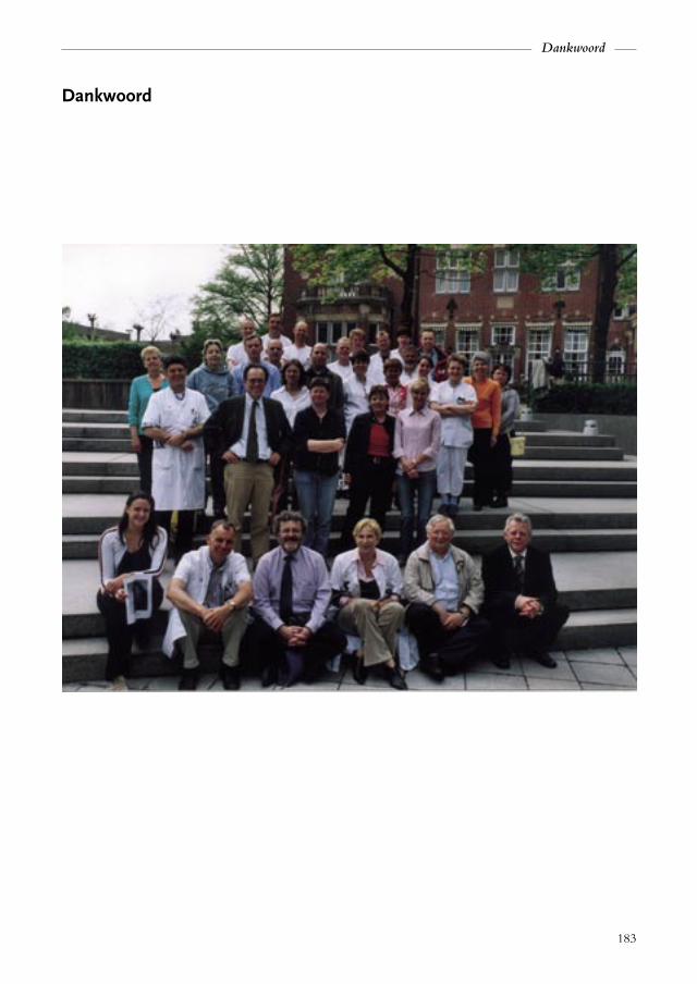
182
_____ Chapter 16 ____________________________________________________________________________________________________________ _____________________________________________________________________________________________________________ Dankwoord _____
183
Dankwoord
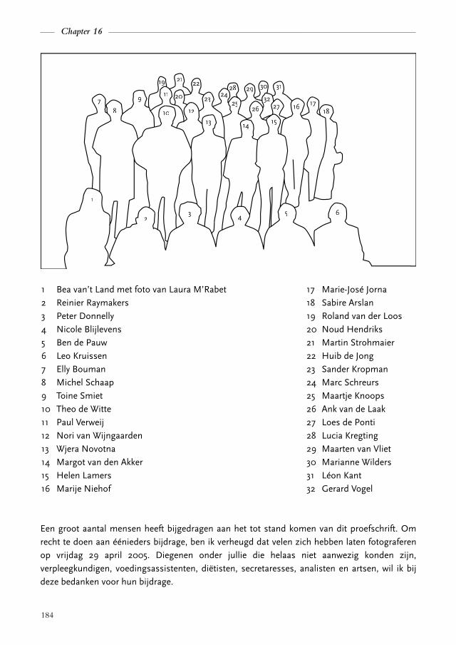
184
_____ Chapter 16 ____________________________________________________________________________________________________________ _____________________________________________________________________________________________________________ Dankwoord _____
185
Een groot aantal mensen heeft bijgedragen aan het tot stand komen van dit proefschrift. Om recht te doen aan éénieders bijdrage, ben ik verheugd dat velen zich hebben laten fotograferen op vrijdag 29 april 2005. Diegenen onder jullie die helaas niet aanwezig konden zijn, verpleegkundigen, voedingsassistenten, diëtisten, secretaresses, analisten en artsen, wil ik bij deze bedanken voor hun bijdrage.
1 Bea van’t Land met foto van Laura M’Rabet2 Reinier Raymakers3 Peter Donnelly 4 Nicole Blijlevens5 Ben de Pauw 6 Leo Kruissen7 Elly Bouman8 Michel Schaap9 Toine Smiet 10 Theo de Witte11 Paul Verweij 12 Nori van Wijngaarden 13 Wjera Novotna14 Margot van den Akker15 Helen Lamers16 Marije Niehof
17 Marie-José Jorna 18 Sabire Arslan19 Roland van der Loos20 Noud Hendriks21 Martin Strohmaier22 Huib de Jong23 Sander Kropman24 Marc Schreurs 25 Maartje Knoops26 Ank van de Laak27 Loes de Ponti28 Lucia Kregting29 Maarten van Vliet30 Marianne Wilders31 Léon Kant32 Gerard Vogel
1
23 4
5 6
1210
9
87
11 20
13 14 15
16 1718
19 21 22
2324
25
28
26
2932
30
27
31
184
_____ Chapter 16 ____________________________________________________________________________________________________________ _____________________________________________________________________________________________________________ Dankwoord _____
185
Maar mijn grootste dank gaat uit naar al die patiënten die geparticipeerd hebben in de supportive care studies van de afdeling Hematologie, zonder hun inzet en enthousiasme zou ik niet zulke mooie resultaten hebben gekregen.
Verder wil ik graag een aantal personen nog speciaal bedanken die een bijzondere rol in mijn promotie traject hebben gespeeld.
Ten eerste, mijn promoter, Ben de Pauw, voor het simpele feit dat hij geloofde in mijn capaciteiten als klinisch onderzoekster en de weg vrij maakte om mijn onderzoek in huis op poten en internationaal op de kaart te zetten. Jouw scherpe geest, jouw betrokkenheid bij patiënten en uiteraard de dirty jokes zijn de ideale mix voor mij geweest om door te gaan ondanks allerlei obstakels. Dank.
Peter, it was a privilege to be trained by you in the field of science. You challenged me to look at the raw data without stretching them to fit the truth. Your criticism was never unjustified although it did hurt, only occasionally, however. You taught me how to make attractive power point presentations and, in addition, showed me how to perform and how to present my results and contemplations; cool, calm and collected. When I experienced a turbulent change in my personal life, you were always there to support me. I feel much obliged. I admit that I am proud to share a room with you, a former Red-handed Scotsman. We are not only a perfect team in supportive care management; we also share our humour that makes life pleasant.
Ton, het is maar goed dat wij géén kamer samen delen want jouw gezelschap is uitermate aangenaam. Jouw enthousiasme voor het vak en met name voor de behandeling van patiënten met een stamceltransplantatie is altijd zeer motiverend geweest voor mij. Tezamen met John heb jij ervoor gezorgd dat mijn interventie studie op de afdeling kon worden uitgevoerd.
Toine, je weet het, je bent niet voor niets mijn paranimf, zonder jouw motivatie van het verpleegkundig team en patiënten zou mijn boekje er zeker anders hebben uitgezien.
Theo, dank dat je de “supportive care business” hebt laten bloeien binnen de afdeling Hematologie.
Marie-José, jouw doortastendheid in de verpleegkundige leiding van de verpleegafdeling werpt haar vruchten af, samen maken we de zorg nog beter.
Staf Hematologie, dank voor het door jullie in mij gestelde vertrouwen.
Ralph, writing is a family asset after all.
17 Marie-José Jorna 18 Sabire Arslan19 Roland van der Loos20 Noud Hendriks21 Martin Strohmaier22 Huib de Jong23 Sander Kropman24 Marc Schreurs 25 Maartje Knoops26 Ank van de Laak27 Loes de Ponti28 Lucia Kregting29 Maarten van Vliet30 Marianne Wilders31 Léon Kant32 Gerard Vogel
186
_____ Chapter 16 ____________________________________________________________________________________________________________
Pa en Ma, bedankt dat jullie mij altijd gestimuleerd hebben om door te leren en ik ben blij dat jullie beiden dit mogen meemaken.
Mariëtte, jouw liefde en hartverwarmende zorgzaamheid laten mij leven, elke dag opnieuw.