METABOLISM, FLOW AND CONTRACTILITY OF THE LANGENDORFF Hans.pdf · metabolism, flow and...
176
METABOLISM, FLOW AND CONTRACTILITY OF THE LANGENDORFF HEART PROEFSCHRIFT TER VERKRIJGING VAN DE GRAAD VAN DOCTOR IN DE GENEESKUNDE AAN DE ERASHUS UNIVERSITEIT TE ROTTERDAM OP GEZAG VAN DE RECTOR PROF.DR. B. LEIJNSE EN VOLGENS BESLUIT VAN HET COLLEGE VAN DEKANEN. DE OPENBARE VERDEDIGING ZAL PLAATS VINDEN OP WOENSDAG 18 OKTOBER 1978 DES NAl'!IDDAGS TE 16.15 UUR PRECIES DOOR HANS STAH GEBOREN TE ABBEKERK
Transcript of METABOLISM, FLOW AND CONTRACTILITY OF THE LANGENDORFF Hans.pdf · metabolism, flow and...
METABOLISM, FLOW AND CONTRACTILITY
OF THE LANGENDORFF HEART
PROEFSCHRIFT
TER VERKRIJGING VAN DE GRAAD VAN
DOCTOR IN DE GENEESKUNDE
AAN DE ERASHUS UNIVERSITEIT TE ROTTERDAM
OP GEZAG VAN DE RECTOR ~~GNIFICUS PROF.DR. B. LEIJNSE
EN VOLGENS BESLUIT VAN HET COLLEGE VAN DEKANEN.
DE OPENBARE VERDEDIGING ZAL PLAATS VINDEN OP
WOENSDAG 18 OKTOBER 1978
DES NAl'!IDDAGS TE 16.15 UUR PRECIES
DOOR
HANS STAH
GEBOREN TE ABBEKERK
PROMOTOR: PROF.DR. W.C. HULSMANN
COREFERENTEN: PROF. P.G. HUGENHOLTZ
PROF.DR. P. HARRIS (LONDON)
Dit proefschrift werd bewerkt binnen de afdelingen
Biochernie I en Cardiovasculaire Research (Thoraxcentrum)
van d~ Faculteit der Geneeskunde, Erasmus Universiteit
Rotterdam.
Het onderzoek en het verschijnen van dit proefschrift
werd mogelijk gemaakt door financiele steun van de
Nederlandse Hartstichting.
1978 Drukkerij van der Poel - Bolnes
Ye mariners all as you pass by
call in and drink if you are dry
come spend me lads your money brisk
and pop your nose in a jug of this
(Traditioneel Engels volksliedje)

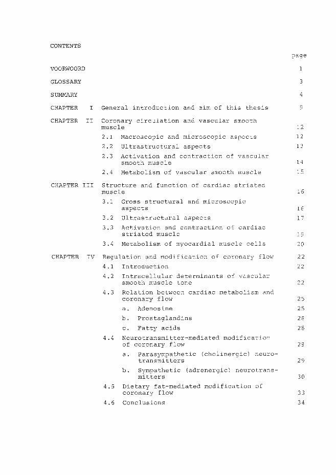
CONTENTS
VOORWOORD
GLOSSARY
SUMMARY
CHAPTER I General introduction and aim of this thesis
CHAPTER II Coronary circulation and vascular smooth muscle
2.1 Macroscopic and microscopic aspects
2.2 Ultrastructural aspects
2.3 Activation and contraction of vascular smooth muscle
2.4 Metabolism of vascular smooth muscle
CHAPTER III Structure and function of cardiac striated muscle
3.1 Gross structural and microscopic aspects
3.2 Ultrastructural aspects
3.3 Activation and contraction of cardiac striated muscle
3.4 Metabolism of myocardial muscle cells
page
1
3
4
9
12
12
13
14
15
16
16
17
19
20
CHAPTER IV Regulation and modification of coronary flow 22
4.1 Introduction
4.2 Intracellular determinants of vascular smooth muscle tone
4.3 Relation between cardiac metabolism and coronary flow
a. Adenosine
b. Prostaglandins
c. Fatty acids
4.4 Neurotransmitter-mediated modification of coronary flow
a. Parasympathetic (cholinergic) neuro-
22
22
25
25
26
28
29
transmitters 29
b. Sympathetic (adrenergic) neurotrans-mitters 30
4.5 Dietary fat-mediated modification of coronary flow
4.6 Conclusions
33
34
page
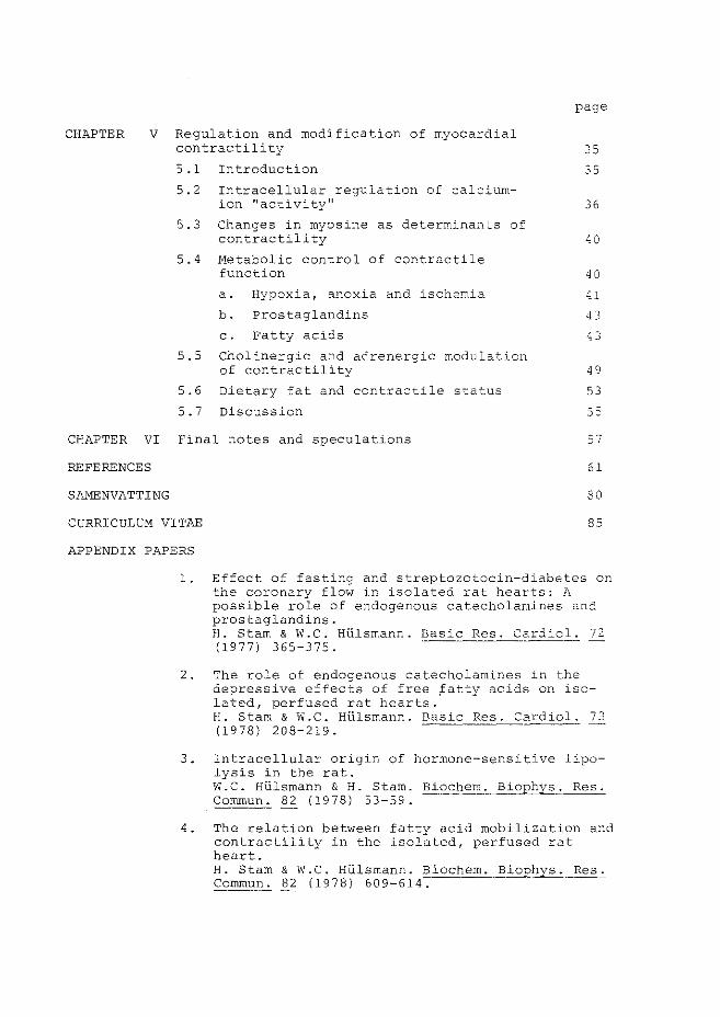
CHAPTER V Regulation and modification of myocardial contractility 35
35 5.1 Introduction
5.2 Intracellular regulation of calciumion "activity n
5.3 Changes in myosine as determinants of contractility
5.4 Hetabolic control of contractile function
a. Hypoxia, anoxia and ischemia
b. Prostaglandins
c. Fatty acids
5.5 Cholinergic and adrenergic modulation of contractility
5.6 Dietary fat and contractile status
5.7 Discussion
36
40
40
41
0
43
49
53
55
CHAPTER VI Final notes and speculations 57
REFERENCES 61
SAMENVATTING 80
CURRICULUM VITAE 85
APPENDIX PAPERS
1. Effect of fasting and streptozotocin-diabetes on the coronary flow in isolated rat hearts: A possible role of endogenous catecholamines and prostaglandins. H. Starn & W.C. Hlilsmann. Basic Res. Cardiol. 72 (1977) 365-375.
2. The role of endogenous catecholamines in the depressive effects of free ~atty acids on isolated, perfused rat hearts. H. Starn & W.C. Hlilsrnann. Basic Res. Cardiol. 73 (1978) 208-219.
3. Intracellular origin of hormone-sensitive lipolysis in the rat. W.C. Hlilsmann & H. Starn. Biochern. B~ophys. Res. Commun. 133. (1978) 53-59.
4. The relation between fatty acid mobilization and contractility in the isolated, perfused rat heart. H. Starn & W.C. Hlilsmann. Biochem. Biophys. Res. Commun. 82 ( 1978) 609-614.
5. Sephadex-induced reduction of coronary flow in the isolated rat heart: A model for ischemic heart disease. H. Starn & J.W. de Jong. J. Mol. Cell. Cardiol . .'l (1977) 633-650.
6. Effect of glucose on AMP-catabolite release during fatty acid perfusion in normal and ischemic rat hearts. H. Starn & w. Breeman. Life Sciences, accepted for publication.

VOORWOORD
Gaarne wil ik deze gelegenheid aangrijpen om een ieder die
in rneer of mindere mate zijn steentje heeft bijgedragen aan de
totstandkoming van dit proefschrift te bedanken. Mijn gedachten
gaan daarbij vooral uit naar:
* Mijn ouders, wier stimulans en belangstelling ik altijd als
onmisbaar heb ervaren,
* Willem HUlsmann, mijn promotor, voor de enthousiaste, ideeen
rijke en niet van Amsterdams humor gespeende wijze waarqp hij
het onderzoek heeft begeleid en mijn interesse in de lipiden
biochemie en fysiologie heeft opgewekt,
* Prof. Peter Harris from the Cardiothoracic Institute (London)
whom I wish to thank for refereeing this thesis and for his
generous hospitality during my visit to his laboratory,
*Prof. Paul Hugenholtz, die als coreferent en cardioloog altijd
de klinische relevantie van dit onderzoek voor ogen heeft gehad
hetgeen mijn belangstelling voor de kliniek heeft doen groeien,
* Jan \··Hllem de Jong, voor de prettige en leerzame samenwerking
met name in de eerste fase van het onderzoek en de wijze waarop
hij een Amsterdammer gedurende de lunchwandelingen wegwijs heeft
gemaakt in dorstlessend Rotterdam,
* Piet Verdouw, die mijn uiterst geringe kennis omtrent de hemo
dynamica van het cardiovasculaire systeem wat heeft aangevuld en
nooit karig was met italiaans ijs,
* Jasper Scholte, Hugo de Jonge, Jos Lamers en Wim Ruitenbeek,
wier opbouwende kritiek en nuttige wenken tijdens de wekelijkse
werkbesprekingen (en borrels) ik ten zeerste heb gewaardeerd,
* Hans Jansen, die altijd klaar stand om de streptozotocine
injecties minsten zo goed te verzorgen als hij kon tafeltennissen,
* Alle overige medewerkers van de afdelingen Biochemie I en Cardio
vasculaire Research {Thoraxcentrurn) voor de 11 hart"elijke werk
atmosfeer en hulpvaardigheid,
* Michael Parnham from the department of Pharmacology for his
spontaneous help in the prostaglandin-(bio)assay and stimulatory
discussions,
1
* Dr. F. ten Hoor en Dr. U.M.T. Houtsmuller van Unilever Research
(Vlaardingen) , die ons rijkelijk voorzagen van prostaglandine
E 1 en de raapolie dieten,
* Liesbeth Eysbroek-Sipman, Mariys Stewart, Tiny Geelhoed-Mieras,
Liz Keijzer en vooral Wout Breeman, voor de "analytische" hand
die zij regelmatig hebben toegestoken,
* Rob van Bremen, die altijd daar was wanneer mijn onkunde van de
electronica fatale vormen dreigde aan te nemen,
* Clarie Viool-Kegge, Coby de Mik-v.d. Kleyn, voor de genoten
redactionele hulp in een deel van de artikelen,
* Cecile Hanson, die mijn (welhaast onleesbare) handgeschreven
versie van dit proefschrift (en enkele artikelen) vaardig,
accuraat en onwaarschijnlijk snel wist om te zetten in een
leesbaar geheel,
* Arie Roodnat, die als keuze-practikant een nuttige bijdrage
heeft geleverd aan de perfusie-experimenten en die mij beloofd
heeft te leren wind-surfen,
*Jan Ekas en overige medewerkers van de glasblazerij, uit wier
handen heel wat uitbreidingen en vervolmakingen van de perfusie
opstelling zijn voortgekomen,
* Mijn teamgenoten van de Thoraxkrakers (zaalvoetbal) en v & A
Zevenklappers (volleybal), voor een ontspannen, sportief en vaak
geestverruimend einde van menige werkdag,
* Bart Bender, voor de fraaie omslag.
* ''Last'' maar zeker niet ''least'', Mariel, als dank voor het einde
loze geduld dat je de laatste jaren voor een halve "vakidioot"
wist op te brengen en voor een altijd gezellig thuis, wil ik
dit proefschrift aan jou opdragen.
2
GLOSSARY
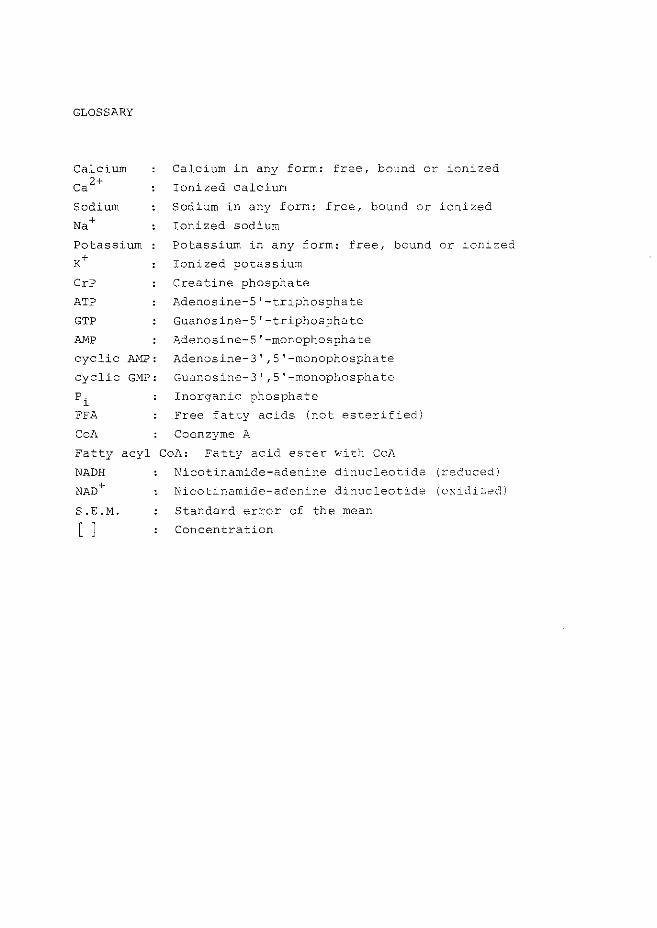
Calcium
ca 2+
Sodium
Na+
Potassium K+
CrP
ATP
GTP
AMP
cyclic AMP:
cyclic GMP:
pi
FFA
CoA
Calcium in any form: free, bound or ionized
Ionized calcium
Sodium in any form: free, bound or ionized
Ionized sodium
Potassium in any form: free, bound or ionized
Ionized potassium
Creatine phosphate
Adenosine-5 1 -triphosphate
Guanosine-5 1 -triphosphate
Adenosine-5'-monophosphate
Adenosine-3' ,5'-monophosphate
Guanosine-3' ,5'-monophosphate
Inorganic phosphate
Free fatty acids (not esterified)
Coenzyme A
Fatty acyl CoA: Fatty acid ester with CoA
NADH Nicotinamide-adenine dinucleotide (reduced)
NAD+
S.E.H.
[ J
Nicotinamide-adenine dinucleotide (oxidized)
Standard error of the mean
Concentration
SUMMARY
This thesis reviews current literature and describes
experimental studies on the regulation and modification of
coronary flow and contractility in isolated rat hearts. In
chapter I and introduction is given to the problems of fatty
acid toxicity and myocardial function. Coronary flow rate and
pump function of the myocardium are mainly determined by the
contractile status of vascular smooth muscle cells and cardiac
striated muscle cells, respectively. Therefore in chapters II
and III morphological and (ultra)structural aspects of both
types of cells have been described.
In chapters IV and V functional and metabolic aspects of coronary
circulation and contractility are illustrated. In both vascular
smooth and cardiac striated muscle cells:
(i) the intracellular calcium concentration is the
main determinant of the contractile status of
actomyosin,
(ii) contraction takes place after the action potential
induced calcium-influx through the plasmamembrane
and calcium release from intracellular stores
(sarcoplasmic reticulum, mitochondria),
(iii) relaxation is achieved after reduction of the cyto
plasmic calcium level by calcium-pump systems in
the plasmamembrane, sarcoplasmic reticulum and
mitochondria,
(iv) calcium-ions trigger the coupling between the
contraction-relaxation cycle with energy metabolism
since glycogenolysis and lipolysis are both
stimulated by calcium.
Cardiac striated muscle contraction and relaxation are energy
dependent processes. Under conditions of limited ATP production
(anoxia, hypoxia, ischemia) contractile function impairs.
Pharmacological, neurohumoral and metabolic control of intra
cellular calcium levels in smooth and striated muscle cells takes
place via two mechanisms:
4
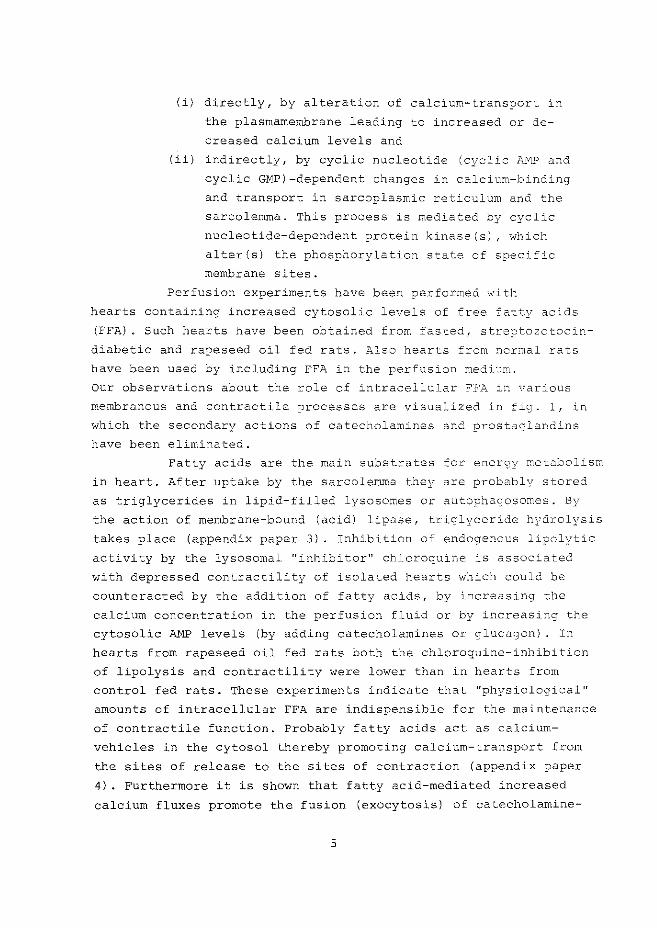
(i) directly, by alteration of calcium-transport in
the plasmamembrane leading to increased or de
creased calcium levels and
Iii) indirectly, by cyclic nucleotide (cyclic &~ and
cyclic GMP)-dependent changes in calcium-binding
and transport in sarcoplasmic reticulum and the
sarcolemma. This process is mediated by cyclic
nucleotide-dependent protein kinase(s), which
alter(s) the phosphorylation state of specific
membrane sites.
Perfusion experiments have been performed wi tl-:
hearts containing increased cytosolic levels of free ::atty acids
(FFA). Such hearts have been obtained from fasted, streptozotoci::-!
diabetic and rapeseed oil fed rats. Also hearts from normal rats
have been used by including FFA in the perfusion medL;m.
Our observations about the role of intracellular FFA ir:. various
membranous and contractile processes are visualized in =ig. 1, in
which the secondary actions of catecholamines and prostaglandins
have been eliminated.
Fatty acids are the main substrates for energy metabolism
in heart. After uptake by the sarcolernna they are probably stored
as triglycerides in lipid-filled lysosomes or autophagosomes. By
the action of membrane-bound (acid) lipase, triglyceride h:rrdrolysis
takes place (appendix paper 3). Inhibition of endogenous lipolytic
activity by the lysosomal "inhibitor" chloroquine is associated
with depressed contractility of isolated hearts which could be
counteracted by the addition of fatty acids, by increasing the
calcium concentration in the perfusion fluid or by increasing the
cytosolic AMP levels (by adding catecholamines or glucagon) . In
hearts from rapeseed oil fed rats both the chloroquine-inhibition
of lipolysis and contractility were lower than in hearts from
control fed rats. These experiments indicate that "physiological"
amounts of intracellular FFA are indispensible for the maintenance
of contractile function. Probably fatty acids act as calcium
vehicles in the cytosol thereby promoting calcium-transport from
the sites of release to the sites of contraction (appendix paper
4). Furthermore it is shown that fatty acid-mediated increased
calcium fluxes promote the fusion (exocytosis) of catecholamine-
5
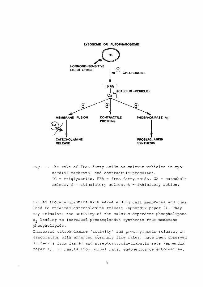
LYSOSOME OR AUTOPHAGOSOME
~8 (ACID) LIPASE 8
~CHLOROQUINE
HORMONE-SENSITIVE J
MEMBRANE FUSION
~l CATECHOLAMINE RELEASE
I (CALCIUM-VEHICLE) lFFA] Ca2+
CONTRACTILE PROTEINS
PHOSPHOLIPASE A2
l PROSTAGLANDIN SYNTHESIS
Fig. 1. The role of free fatty acids as calcium-vehicles in myo
cardial membrane and contractile processes.
TG = triglyceride, FFA = free fatty acids, CA = catechol
amines. $ = stimulatory action, e = inhibitory action.
filled storage granules with nerve-ending cell membranes and thus
lead to enhanced catecholamine release (appendix paper 2) . They
may stimulate the activity of the calcium-dependent phospholipase
A2 leading to increased prostaglandin synthesis from membrane
phospholipids.
Increased catecholamine ''activity'' and prostaglandin release, in
association with enhanced coronary flow rates, have been observed
in hearts from fasted and streptozotocin-diabetic rats (appendix
paper 1). In hearts from normal rats, endogenous catecholarnines,
6
released spontaneously during perfusion, proved to be involved
in the maintenance of the coronary flow rate under normoxic
conditions (appendix papers 1 and 2). The vasodilatory actions
of (exogenous and endogenous) catecholamines and of prostaglandin
like substances are likely to be mediated by a rise in vascular
smooth muscle cytosolic cyclic ANP levels. In contrast to
vascular smooth muscle cells, which relax under conditions of
enhanced cytoplasmic cyclic AMP levels, cardiac striated muscle
cells show increased contractility following addition o_.;'" nor
epinephrine, glucagon and prostaglandin E1 (appendix paper 4).
The calcium-ionophoric properties of fatty acids and
of certain prostaglandins are involved in coronary vasodilation,
probably by stimulating calcium sequestration in coronary vascular
smooth muscle cells. Therefore, the increased coronary flo'.'~ rates
in hearts from rapeseed oil fed rats may be related to the observed
increased rates of basal (and hormone-sensitive) lipolytic activity
The expected increase of fatty acid release from the lipid infil
trated striated muscle cells may be responsible £or vascular
smooth muscle relaxation by the Calcium-removing action of fatty
acids as well.
Experiments were carried out to study the effects cf
fatty acid perfusion during normoxic and ischemic conditions.
Embolization (Sephadex polysaccharide microspheres)-induced reduc
tion of coronary flow was developed and characterized as an experi
mental model for myocardial ischemia (appendix paper 5) . Reduced
concentrations of creatine phosphate and ATP as well as cellular
acidosis during anoxia, hypoxia and ischemia, are responsible for
impairment of the energy-dependent calcium pumps (Ca 2+-ATPases)
of sarcolemma, sarcoplasmic reticulum and possibly also of mito
chondria, so that a hypercontracted "stone" 'heart may result. It
is proposed that the release of ATP catabolites (adenosine,
inosine and hypoxanthine) during oxygen (and/or substrate)-limited
conditions may be used as a marker for coronary artery and ischemic
heart diseases in man.
Perfusion with a fatty acid-albumin complex (at a
relatively high fatty acid:albumin molar ratio, which is the main
determinant of FFA uptake) in the perfusion buffer resulted in a
depression of contractile function. The deleterious role of increa-
7
seo intcacel:ular le~els of fattv acids and t~eir CoA and carni-
102r ncrmoxic and ischemic conditions have teen
·~Jescribcc'i. en "-''co basis of their chaotrcpic act:'.-on on membrans.s
as .c1eterc;ents (see fig, and appendix ~apers L ana 6}. Catechol-
aT;lnes, liber2t:eC f:com endogen.ous stores upon fa-tty acid. :;:;erfusicn
nay also participate in the decline of myocardia- function ~here-
fore, mention is made o~ observations on detrimental actions of
catecholamines upon cellular membranes and metabolism< j_ c. "'1as
f,-Jund that £:atty acid-induced i;r,pairrnent of contractility could
be Drever_ ted by ca techolamine-deiJletion indeed< E'er chermore, t:'Je
.:o,ddi tion of glucose, by supnlying glycerol-3-phosphate for tri-
glyceride synthesis, which probably reduces the intracellular
levels of (and of the CoA- and carnitine ester derivatives)
',•.ras found to
(i) decrease catecholamine release
(ii) counteract the depression of myocardial energetics
and
(iii) Lncrease contractile function,
all occurring d.1ring fatty acid-perfusion under fully oxygenated
a~d oxygen-limj_ted conditions.
o·~1r result.s suggest an important role of fatty acids in
myocardial ~unction under normal and pathoJ.ogical conditions, a
role ir:. ~ich their iono0horic properties are of central signifi-
c 2.r·,ce.
CHAPTER I
GENERAL INTRODUCTION AND AIM OF THIS THESIS
In all tissues blood flow is responsible for the
supply of substrates and oxygen, and for the removal of
catabolites. The maintenance and regulation of the circulation
is dependent on the contractile activity of the myocardium.
For its own energy demand and waste product removal the heart
has a coronary circulatory system which perfuses the cardiac
muscle tissue. It is obvious that myocardial contractility must
be adaptable to the variation of energy demands of the body and
that, in turn, there must be a close relationship between its
own performance and the actual coronary drainage rate. ror that
reason myocardial contractile function as well as coronary
circulation are most likely subject to a complex control syste~.
Exogenous as well as endogenous metabolic, neural and humoral
influences are involved in regulation. Knowledge of their pro
perties is essential to understand the physiological mechanisms
of control as well as for the evaluation of preventive and thera-' ..
peutic measures in (coronary) heart disease. Since the main
elements of which the coronary circulation and cardiac muscle
consist are vascular smooth and cardiac striated muscJ.e cells,
detailed knowledge of the properties of these tvm types oi
muscle is mandatory.
Therefore attention is firstly paid to morphological, structural,
and functional aspects of both the coronary circulatiorl and the
myocardial striated musculature.
In the last decades numerous reports about the effects,
and in particular the toxic effects of free fatty acids (FFA)
upon the myocardium under normal and pathological conditions have
appeared. Yet, the biochemical mechanisms underlying these
detrimental effects are still not fully understood. hTe therefore
studied FFA-induced alterations of metabolism, coronary flow and
contractility in a rather simple heart-preparation, the isolated
rat heart, perfused retrogradely as first described by Lanoendorff
in 1895. In this preparation the aorta is cannulated and the
9
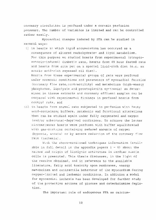
coronary circulation is perfused under a certain perfusion
pressure. The number of variables is limited and can be controlled
rather easily.
Myocardial changes induced by FFA can !Je studied in
several ways:
i) in hearts in which lipid accumulation has occurred as a
consequence of altered carbohydrate- and lipid metabolism.
For this purpose we studied hearts from experimental (strepto
zotocin-induced) diabetic rats, hearts from 48 hour fasted rats
and hearts from rats put on a special lipid-rich diet (e.g. an
erucic acid-rich rapeseed oil diet) .
Hearts from these experimental groups of rats were perfused
under normoxic conditions and parameters of myocardial function
(coronary flow rate, contractility) and metabolism (high-energy
phosphates, lipolysis and prostaglandin synthesis) as deter
mined in tissue extracts and coronary effluent samples can be
compared with experimental findings in perfused hearts from
control rats, and
ii) in hearts from normal rats subjected to perfusion with fatty
acid-containi.ng buffers . .f:'-1etabolic and functional alterations
then can be studied again under fully oxygenated and oxygen
(and/or substrate)-deprived conditions. To achieve the latter
circumstances hearts were perfused with buffer equilibrated
'itli th gas-mixtures containing reduced amounts of oxygen
(hypoxia, anoxia) or by severe reduction of the coronary flov.7
rate (ischemia).
~Vi t.h the above-mentioned techniques information (avail
able in full detail in the appendix papers l - 6) about the
nature and origin of lipolytic activities in cardiac muscle
cells is presented. This thesis discusses, in the light of
the results obtained, and in reference to the available
literature, fatty acid toxicity upon membranes, qnergy
metabolism and contractile behaviour of the myocardium during
oxygen-limited and ischemic conditions. In addition a model
for myocardial ischemia has been developed for further study
of the protective actions of glucose and catecholamine deple
tion.
The important role of endogenous FFA as calcium-
10
ionophores and intracellular calcium-vehicles appears throughout
the present work. Free fatty acids induce the release of endogenous
catecholarnines and prostaglandin-like substances and play an impor
tant role in the regulation of myocardial function.
ll
CHAPTER II
CORONARY CIRCULATION AND VASCULAR SHOOTH HUSCLE
2. l. Macroscopic and microscopic aspects
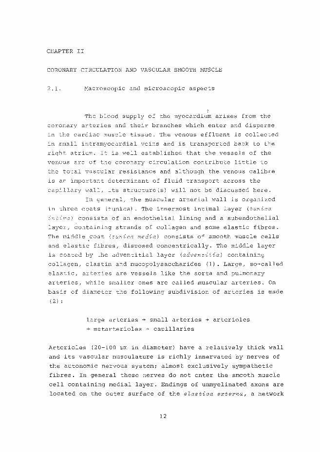
The blood supply of the myocardium arises from the
coronary arteries and their branches which enter and disperse
in the cardiac muscle tissue. The venous effluent is collected
in small intramyocardial veins and is transported back to the
right atrium. It is well established that the vessels of the
venous arc of the coronary circulation contribute little to
the total vascular resistance and although the venous calibre
is an important determinant of fluid transport across the
capillary wall, its structure{s) will not be discussed here.
In general, the muscular arterial wall is organized
in three coats (tunics) . The innermost intimal layer (tunica
c:v,' t-in;.;c) consists of an endothelial lining and a subendothelial
layer, containing strands of collagen and some elastic fibres.
The middle .coat (t!AWica media) consists of smooth muscle cells
and elastic fibres, disposed concentrically. The middle layer
is coated by the adventitial layer (adventitia) containing
collagen, elastin and mucopolysaccharides (1). Large, so-called
elastic, arteries are vessels like the aorta and pulmonary
arteries, while smaller ones are called muscular arteries. On
basis of diameter the following subdivision of arteries is made
( 2 I :
large arteries ~ small arteries ~ arterioles
~ metarterioles ~ capillaries
Arterioles (20-100 wm in diameter) have a relatively thick wall
and its vascular musculature is richly innervated by nerves of
the autonomic nervous system; almost exclusively sympathet~c
fibres. In general these nerves do not enter the smooth muscle
cell containing medial layer. Endings of unmyelinated axons are
located on the outer surface of the elastica externa, a network
12
of elastic fibres around the layers of smooth muscle cells. Frorr
the finest arterioles or metarterioles (10-20 wm) numerous capjJ.
laries (7-9 pm) arise,At the origin of the capillary netv.wrk so
called precapillary sphincters are located. Anatomically we can
say that the precapillary sphincter is the .final srEoot~ muscle
cell of the (met) arteriolar distribution (3). The preca~illary
sphincters possess contractile activity and they are conpletely
devoid of control by the autonomic nervous systerr_, b'.Jt their
muscle tone is sensitive to local chemical in:"'l:.J.ences (4).
The capillaries are narrow tubes, ·w·it~ walls corr.posed
of a single layer of endothelial cells. Outside the endothelial
layer a basement-membrane is found, which plays a role as
physical barrier to penetrating materials or as a supportive
structure affording apparent rigidity to the capillaries. In the
capillaries pericytes were demonstrated in clcse 3sscci~tio:: ':l~l~
the capillary basement-membrane. Their branched c:-ltoplasmic
processes {side-arms) form a web in the endothelinv. and may i1avc
elastic capacity (5). Capillaries possess no known effector
mechanism to control their calibre and hence undergo flo'd changes
passively. The capillary wall permits water and solutes to pass
through (permeability) either via interendothelial ga?S or trans
cellularly {5) . Capillary permeability can be in~luenced by
several substances (histamine, bradykinin, see ref. 6).
2. 2. Ultrastructural aspects
The contractile elements within the vascular smooth
muscle cells are arranged in a multidirecti0nal way which
enables the cell to have local contraction. The plasman1er1bre1 1,~
consists of two types of alternating areas. The first one is tr.c
plasrnamembrane dense body with \Vhich bundles of myofilaments
appear to merge. The other one contains numerous micro;)inocyto
tic vesicles (7) . The surface vesicles are supposed to play a
role in the calcium homeostatis during t:he contraction-relaxatiol
cycle {see later). The presence of receptors for various natu1·a1
and pharmacologica~ substances has been observed (7) .
13
In the cell several contractile proteins have been
visualized (8) and ultrastructure of the contractile apparatus is
compatible with a conventional sliding filament mechanism of
contraction in which thick (myosin) and thin (actin) filaments
slide relative to each other. The thin filaments are anchored
to the above mentioned p-lasmamembrane dense bodies. The parallel
distribution of myosin filaments and their length relative to
the filaments of striated muscle may contribute, in addition to
their lower myosin ATPase activity, to the relative high tension
bearing capacity of smooth muscle. Intracellular organelles, in
volved in the contraction process and in energy metabolism, are
characterized. Both the sarcoplasmic reticulum and the mito
chondria are in close proximity to the plasmamembrane and the
surfnce vesicles.
2. 3. Activation and contraction of vascular smooth muscle
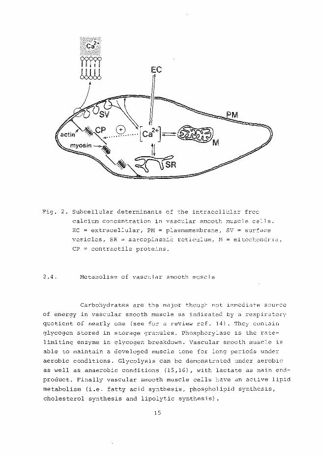
Electro-mechanical and pharmaco-mechanical coupling {9)
activate a contractile mechanism based on a calcium-sensitive
actomysin ATPase system in vascular smooth muscle (10). The
primary event in smooth muscle contraction is a rise in the intra
cellular free calcium concentration (10,11). The participation of
intracellular structures in the determination of the cytoplasmic
calcium-ion concentration is presented in Fig. 2. Transport of
extracellular calcium through the plasmamembrane and their
vesicular excavates (surface vesicles), mobilization of membrane
bound calcium and release of calcium from internal storage sites
(sarcoplasmic reticulum and mitochondria) determine the cyto
solic calcium concentration which induces an interaction between
actin and myosin leading to contraction (12). The regulation of
filament interaction does probably not involve a troponin-like
protein (such as has been described for cardiac striated muscle)
but a myosin-linked calcium binding component (13). Agents which
are known to modify calcium transpo1:t in membranes may influence
the contractile state of vascular smooth muscle.
14
EC
PM
Fig. 2. Subcellular determinants of the intracellular free
calcium concentration in vascular smooth muscle cells.
EC = extracellular, Pl:'-1 = plasmamembrane, SV = surface
vesicles, SR = sarcoplasmiC reticulum, N =mitochondria,
CP = contractile proteins.
2. 4. Metabolism of vascular smooth muscle
Carbohydrates are the major though not immediate source
of energy in vascular smooth muscle as indit:::ated by a respiratory
quotient of nearly one (see for a review ref. 14). They contain
glycogen stored in storage granules. Phosphorylase is the rate
limiting enzyme in glycogen breakdown. Vascular smooth muscle is
able to maintain a developed muscle tone for long periods under
aerobic conditions. Glycolysis can be demonstrated under aerobic
as well as anaerobic conditions (15,16), with lactate as main end
product. Finally vascular smooth muscle cells have an active lipid
metabolism (i.e. fatty acid synthesis, phospholipid synthesis,
cholesterol synthesis and lipolytic synthesis) .
15
CHAPTER III
STRUCTURE AND FUNCTION OF CARDIAC STBIATED MUSCLE
3 . l Gross structural and microscopic aspects
The heart consists of pacemaker and conducting cells,
striated contractile cells and a fibro-elastic matrix. Macro
scopically the heart is divided in four pumping enteties: the
right and left atria and left and right ventricles. Valves are
situated between the cavities of the atria and ventricles, and
between the ventricles and the pulmonary and aort.ic outfl0w
tracts.
The experimental studies presented in this thesis
were performed with rat hearts perfused according to rangendcr>ff
(17). In this preparation the heart is perfused retrogradely via
the aorta at a certain perfusion pressure. Due to this pressure
the valves in the aortic "outflow" tract close and the perfusion
buffer is ''for~ed'' into the coronary circulation originating
from sinuses above the mentioned mitral valves. Atrial tissue is
removed and an atrio-ventricular block is made by cutting a
bundle of stimulus conducting cells situated in the atrial
septum: the H;:s bundle (see appendix paper l). The coronary
effluent flows over the surface of the heart and can easily be
collected. The structural aspects of pacemaker and conducting
cells will not be discussed because in all experiments the hearts
were electrically paced via electrodes placed on the right ven
tricle.
The left ventricle is the main determinant of the con-
tractile activity of the Langen heart. Its wall consists of
three layers. The inner layer is the endocardium, the outer the
epicardium. Both layers consist mainly of connective tissue. In
between the ventricular myocardium is situated, consisting of
series of overlapping sheets of muscle bundles containing the
myofibrils.
The heart is innervated by :Oath sympathetic and parasympathetic
nerve fibres. No specialized nerve endings have been identified
} s
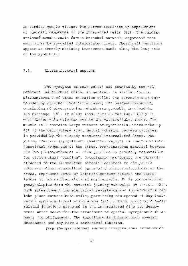
in cardiac muscle tissue. The nerves terminate in depressions
of the cell membranes of the innervated cells (18) . The cardiac
striated muscle cells form a branched networkr separated from
each other by so-called intercalated discs. These cell junctions
appear as densely staining transverse bands along the long axis
of the myofibril.
3.2. Ultrastructural aspects
The myocytes (muscle cells) are bounded by the cell
membrane (sarcolemma) which, in general, is similar to the
plasmamembrane of other mammalian cells. The sarcolemma is sur
rounded by a rather indefinite layer, the basement-meTLibrane,
consisting of glycoproteins, which are probably involved in
ion-exchange (19). It holds ions, such as calcium, likely in
equilibrium with calcium-ions in the extracellular space. The
muscle cell contains large numbers of myofibrils, which make up
47% of the cell volume (20). Mutual cohesion between myocytes
is provided by the already mentioned intercalated discs. The
fascia adherens (myofilament insertion region) is the predominant
junctional component of the discs. Proteinaceous mat12rial between
the two plasmamembranes at this junction is probably responsible
for tight mutual "binding". Cytoplasmic myofibrils are strongly
attached to the filamentous material adjacent to the _,:\;2 ,,,·
adherens. Other specialized parts of the intercalated discs, the
nexus, represent sites of intimate contact between the sarco
lemmas of two cardiac striated muscle cells. It is proposed that
phospholipids form the material joining two ·cells at a llc';<:_,s (21)
Such sites have a low electrical resistance and ion-movements can
take place between both cells, permitting the spread of depolari
zation upon electrical stimulation (22) . A third group of closely
related junctions situated in the intercalated disc are desmo
somes which serve for the attachment of special cytoplasmic fila
ments (tonofilaments). The tonofilaments interconnect several
desmosomes and may have a mechanical function.
From the sarcolemmal surface invaginations arise which
17
run mainly transversely across the cell. These transverse tubules
(T-tubules or T-system) represent portions of the extracellular
space carried into the interior of the cell. Sarcolemma and T
system act as a barrier permitting an intracellular environment
which differs from that of the extracellular space. The mainte
nance of ·the chemical and charge differences between the intra
and extracellular space is mediated by ion-pumps and channels in
these membranes. Inside the cardiac striate6 muscle cell there is
a three dimensional network of fine tubules, the sarcoplasmic
reticulum (or sarcotubular system). It forms cisternae with the
transverse tubules 1 Ylhere they meet.
"Rough-surfaced" endoplasmic reticulum is present in
small quantities, its tubules being continues with the "smooth
surfaced" sarcotubular system. It is considered to be involved
in protein synthesis (23) .
The contractile elements are subdivided into contractile
units (sarcomeres) consisting of thick filaments (myosin) and thin
filaments (actin). Situated at certain intervals along the actin
are the regulatory proteins: troponin and tropomyosin.
Contraction and relaxation of the myocardium has been visualized
in terms of binding and release of calcium-ions from troponin
(i.e. troponin C, which is the "calcium-receptor"), whereas tropo
myosin transmits the calcium-induced conformational changes from
troponin to the contractile proteins, leading to a sliding move
ment of actin and myosin and so to shortening of the sarcomere.
The energy for the contraction-relaxation cycle is provided by
hydrolysis of ATP by the (acto)myosin ATPase.
For the supply of energy for contraction and ion
transport the cell contains numerous mitochondria and storage
granules for glycogen and lipid containing vesicles. Other
organelles found in the myocardial muscle cell are lysosomes
(24,25) 1 Golgi-apparatus and specific granules which contain
catecholamines (mainly norepinephrine, see refs. 26,27).
18
3 0 3 0 Activation and contraction of cardiac striated muscle
Four cellular organelles are involved in the process
of calcium-mediated contraction: sarcolemma, sarcotubular system,
mitochondria and the myofibrils. Pacemaker-induced electrical
depolarization of the sarcolemmal T-system complex is associated
with a calcium influx from superficial sites in the sarcolemma
(the slow inward current of the action potential) and from intra
cellular organelles (sarcoplasmic reticulum and mitochondria),
which is followed by contraction. This proces of excitation
contraction coupling has been subject to many excellent reviews
(28-37) 0
EC
N
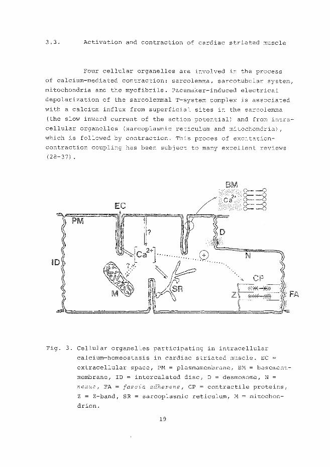
Fig. 3. Cellular organelles participating in intracellular
calcium-homeostasis in cardiac striated muscle. EC
extracellular space, PM= plasmamembrane, BM = basement
membrane, ID = intercalated disc, D = desmosome, N =
nexus, FA fascia adherens, CP =contractile proteins,
Z = Z-band, SR = sarcoplasmic reticulum, M = mitochon
drion.
19
Fig. 3 illustrates the involvement of the already mentioned
cellular components participating in the regulation of intra
cellular calcium concentration. The intracellular free calcium
concentration is lowered by the activation of different
"calcium pumping" mechanisms located at the sarcolemma, sarco
plasmic reticulum and probably also mitochondria ( 38) . The
"calcium pumps" are ATP-dependent and their activity leads to
uptake and accumulation of calcium in sarcoplasmic reticulum
and possibly in mitochondria, and to calcium extrusion into the
intracellular space. Sarcolemmal calcium extrusion may, however, + 2+ also take place through a carrier involving a Na -Ca exchange
+ + mechanism (39). Cardiac sarcolemma contains Na ,K -ATPase and
adenylate cyclase (40,41). Both enzymes are involved in the
regulation and modification of calcium movements across the
plasmamernbrane. Sarcoplasmic reticular membranes can bind and 2+ . l d 2+ take up calcium, and a ca -stlmu ate , Mg -dependent ATPase,
possibly involved in calcium transport has recently been isolated
and purified by Levitsky et al. (42). Like the sarcolemma, sarco
plasmic reticulum might contain Na+,K+-ATPase and adenylate
cyclase activities (43), and these enzymes are also involved in
the calcium transport properties of the sarcotubular membranes.
The contribution of mitochondrial calcium uptake in the
contraction-relaxation process is still controversial.
Metabolism of myocardial muscle cells
Under normal (physiological) conditions myocardial
metabolism is entirely aerobic and oxygen utilization is closely
related to ATP synthesis. In 'iJiVo and under normoxic circum
stances fatty acids and lactate are the main substrates for
cardiac muscle cells (44,45). When the fatty acids are abundant
ly available, insulin is generally limited and glucose utiliza
tion inhibited by depressed glucose uptake and inhibition of
several steps in the glycolytic pathway. The fatty acid:albumin
molar ratio is the main determinant of fatty acid uptake. During
20
-, ·nc;: --·--~---
e:ciC:
stcrage are ~ncreased i~S)
rat:..o is higl1 ''3)
limi t.ing fatt.v
acety~-CoA, acetyl-ca~ni~l~e, fatt•;
endogenous tri;lycerides, store6 in lipid-dropLets. ca~ b0 ~sed
for energy ~etabollsm, ~spending on the availabilit)' nf cxo-
genous fatt\i ;:,_-:=::::Lds,. hormones (cat.echol&IT_ir:es,. insulin··, c-1nd ,~n
the level of cardiac work.
use many substrates to provide in t.heir ener?y dercta!cds (ke-::one
bodiesf fatty aclds, pyruvate, glu_cose, see refs, .:c_;J--o•::J' alL~.c·d~~,
an important source of energy may be tl1e oxidation o~ e~dc9enous-
ly stored triglycerides (49). Tl'.e tsr:dogercolls ~ricjlyccridc.:> J:i.-:_:-:ascc
activity is horc'.'One-sensi"cive (50-53:: and ~na--- :Oe l::::>cc~lizecl j_rJ -r:::l1c
lysosomal or au-tophagic vacuolar r:1embranes (see app-c'n:5i.:-~ ;a per --))
Direct hormonal control of metabolism is not possible in isol2-r:::e6
heart. However, endogenous catecholamines, stored j11 ti1c ~erve-
endings of adrenergic (orthosympa.Jche-::icl ner,_,-e-.::nc1_L'0S 11'-J.y l~~::e_t_---
fere under conditions -I hen they are rel-ease·.:':
activity,by determining the reguireme~t or hl0h-en01r,,. :JI10S]J~3::es
and therefore the redox-state of the tissue, s
of energy metabolism,
Under oxygen-deprived conditio~s 1~ .e. l1'f'OY~3. 2nO~l3
and ischemia) fatty acid ::,xida tio'."l :~s rec;-LJ_c:·2c': slJ.a"-. n :__-,- \<n L,, ".:.he
utilization of glucose and glycogen (fror' s-corag-e gran·_llesl
accelerates markedly (54), These processes •,iill lead t_o enhE',nced
tissue levels of lactate, fatty acids, fatty acyl-CoA esters and
fatty acyl-carnitine, and of ATP depletion since glycol~·tic ATP
synthesis cannot meet ATP demands for contraction and csrnotic
work, when oxidative phosphorylation is inhibitedc
CHAPTER IV
REGULATION AND MODIFICATION OF CORONARY FLOW
4 .l. INTRODUCTION
The circulation of the myocardium is influenced by
mechanical, neural and metabolic factors. Coronary flow is
dependent on the (''driving") perfusion pressure and on the
vascular resistance. The latter is mainly determined by the
muscle tone of coronary resistance vessels (diameter <100 wrn)
but also by a cyclic extravascular compression of the coronary
arteries by the contracting myocardial muscle during systole
(55,56). Early observations of Driscoll et al. (57) indicated
a correlation between coronary flow and perfusion pressure in
the isolated, nonworking and fibrillating heart. However, an
autoregulatory mechanism which implies that steady-state flow
remains constant despite changes in perfusion pressure has
been observed (55,58). Isolated, perfused rat hearts do not
show this phenomenon {appendix paper l): changes in perfusion
pressure were always followed by changes in coronary flow rate.
Before discussing the metabolic and neural influences on coro
nary circulation we will review current knowledge about the
determination of smooth muscle contractility.
4. 2 0 Intracellular determinants of vascular smooth muscle
tone
As already mentioned in chapter III, the muscle tone
of vascular smooth muscle is responsible for coronary resistance
and the contractile state of vascular smooth muscle is primary
determined by the intracellular concentration of calcium-ions
{see fig. 1). Smooth muscle depends largely upon intracellular
supplies of calcium, since they can function for a long time
in the absence of extracellular calcium (59). Since smooth
22
muscle cells are e~citable, the action potential, by increasing
calcium permeability may play a direct role in mediating contrac
tion. It will be clear that pharmacological modification of
calcium-influx during the action potential directly. will deter
mine coronary tone (e.g. acetylcholine and other parasympathico
mimetics) . The energy metabolism is coupled to contraction since
enhanced calcium levels provoke to conversion of phosphorylase b
+phosphorylase~ and thus initiate glycogenolysis. Relaxation
of vascular smooth muscle is achieved by the calcium-pump
activity of the plasmamembrane, sarcoplasmic reticulum and mito
chondria. The calcium-pumps can be stimulated by a mechanism
involving cyclic AMP which is formed from ATP through the action
of membrane bound or soluble adenylate cyclase (60,61). It is
proposed that cyclic AMP, by facilitating phosphorylation of
protein or lipoprotein (by a cyclic AMP-dependent protein kinase)
increases the calcium binding to "binding-sites 11 (and subsequent
transport) in smooth muscle cells (60,62,63). The resulting de
crease in free intracellular calcium levels causes relaxation.
The involvement of a cyclic AHP stimulation of microsomal and + + + 2+ plasmamembrane Na ,K -ATPase, followed by a decreased Na -ca
exchange as relaxatory mechanism has been proposed by Limas and
Cohn (64). A rise in intracellular cyclic AMP is not only
mediated by increased adenylate cyclase activity but can also be
achieved by inhibition of cyclic AI'1P phosphodiesterase (e.g. by
methylxanthines).
If relaxation is caused by an increased level of cyclic
AMP it seems reasonable to assume that decreased levels may be
associated with contraction enhancement (65). Indeed it was
noticed that drug-induced aortic vasoconstriction was associated
with a decreased cyclic A}W level (66) . Since also changes in
vascular smooth muscle cyclic GNP levels during the contraction
relaxation cycle are observed, this nucleotide may also be
involved in the regulation of the .intracellular calcium level.
Moreover cyclic GMP has been proposed to stimulate calcium
release from rnicrosomes in various types of smooth muscle (67)
Drug-induced activation of membrane-bound or cytosolic (soluble)
guanylate cyclase, followed by rises in cyclic GMP were associated
with smooth muscle contraction (68). However, (i) the fact that
23
guanylate cyclase activity is stimulated by calcium-ions, (ii)
the observation that cytoplasmic cyclic GMP increases under
conditions when smooth muscle cells do neither contract nor
relax and, (iii) the finding of drug-induced smooth muscle
contraction without a change in the level of cyclic GMP {69)
seem to rule out any direct regulatory action of cyclic GMP in
the smooth muscle contraction-relaxation cycle. It may be con
ceivable that cyclic GMP plays a role as feedback signal to
speed up the calcium removal by decreasing the influx of calcium
( 6 8) •
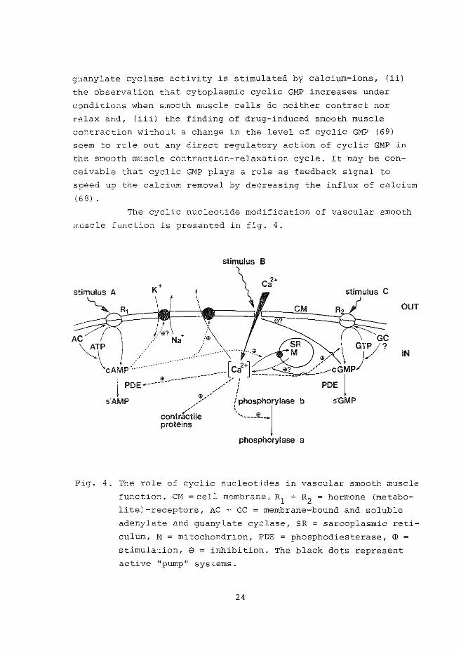
The cyclic nucleotide modification of vascular smooth
muscle function is presented in fig. 4.
OUT
IN
Fig. 4. The role of cyclic nucleotides in vascular smooth muscle
function. CM =cell membrane, R1
+ R2
= hormone (metabo
lite)-receptors, AC + GC =membrane-bound and soluble
adenylate and guanylate cyclase, SR = sarcoplasmic reti
culum, M = mitochondrion, PDE = phosphodiesterase, $ =
stimulation, 9 = inhibition. The black dots represent
active "pump 11 systems.
24
In the next paragraphs a number of metabolic and neural influen
ces on coronary flow of isolated hearts will be discussed on the
basis of this (hypothetical) scheme.
4. 3. Relation between cardiac metabolism and coronary flow
Although extra-cardiac mechanisms play a role in the
modification of coronary flow, the primary regulatory mechanism(s)
responsible for the adjustment of coronary resistance reside{s)
within the heart. The metabolic demands of vascular smooth muscle
cells are negligible when compared to the high energy demands of
contracting cardiac striated muscle cells. Therefore, smooth
muscle can maintain contractile function under long periods of
hypoxia, while striated muscle will loose its contracting and
relaxing capacity. Alterations in cardiac striated muscle
metabolism, however, modulate smooth muscle 11 activity" and nume
rous intrinsic factors have been suggested as mediators {for a
review see ref. 55). Lactate, co 2 , K+, Pi, osmolality, low po 2 and adenine nucleotides have all been proposed to play a role in
the modification of coronary flow under various experimental con
ditions, but most fail to fulfil physiological criteria. Substan
tial evidence has accumulated to show that increased cardiac
activity leads to enhanced coronary flow (70,71) and the relation
between these two processes has been attributed to increased
cardiac levels of cyclic AMP {72), since phosphodiesterase inhibi
tors potentiate this metabolically induced coronary dilatation
during hyperactivity (72). In the next sections some metabolites
which may be involved in coronary flow regu'lation are discussed.
a. Adenosine
During cardiac hyperactivity (associated with increased
oxygen demand) , whether induced by pacing at high rates or due to
catecholamines, adenosine is released from the isolated rat heart
25
(73-75). Furthermore adenosine formation is enhanced in associa
tion with vascular dilatation following hypoxia and anoxia (73,
appendix paper 5) , reactive hyperemia (coronary vasodilation
occurring after short periods of interruption of flow; see
refs. 76,77) and ischemia (appendix papers 5 and 6, 78). Adeno
sine is form.ed by enzymatic hydrolysis of 5 '-AMP through the
action of 5'-nucleotidase which is a plasmamernbrane-bound enzyme
( 79) . A rise in intracellular AMP levels, indeed, is likely to
occur during the conditions mentioned above, while changes in
ATP and ADF levels (inhibitory) and AMP (stimulatory) determine
3'-nucleotidase activity (80).
The mechanism by which adenosine exerts its vasodila
tory action is still unknown. Adenosine-receptors have been
proposed to mediate the vasodilatory capacity of adenosine (81-
83). Olsson dt JZ. (81,82) first observed adenosine receptors on
the coronary myocyte surface while Schrader et aZ. (83) presented
evidence for adenosine-receptors both on coronary myocytes and
atrial muscle cells. Adenosine-stimulation of adenylate cyclase
has been reported for ventricular muscle (84,85), and if the same
mechanism occurs in vascular smooth muscle it may well be that
the increased cytoplasmic cyclic AMP levels are the mediators of
adenosine-induced coronary vasodilation (adenosine may then act
as stimulus A, see ref. 3). Only limited experimental evidence is
available which does not support a role for cyclic M1P in the
adenosine-induced coronary relaxation (86). Schrader et aL (87)
observed adenosine-inhibition of action potential-linked calcium
ion influx in atrial muscle, also leading to decreased intra
cellular calcium levels ("negative" stimulus B in fig. 4).
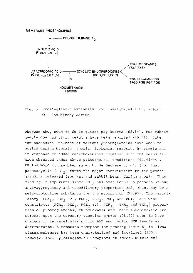
b. Prostaglandins
Endogenous prostaglandins, synthetized from essential
fatty acids in phospholipids (as indicated in fig. 5) have been
implicated in the regulation of coronary flow (appendix paper l)
In the isolated dog and rat heart they are probably not involved
in the maintenance of coronary flow during normoxic conditions
26
MEMBRANE PHOSPHOLIPIDS r--- PHOSPHOLIPASE A2
LINOLEIC ACID (CI8,2,L>9,12)
THROMBOXANES
<(TXA,TXB) ARACHIDONIC ACID T(CYCLIC) ENDOPEROXIDES (C2Q, 4,L>5,8,11,14) (PGG,PGH, PGR)
9 PROSTAGLANDINS . (PGD.PGE.PGF,PGI)
INDOMETHACIN ASPIRIN
Fig. 5. Prostaglandin synthesis from unsaturated fatty acids.
e : inhibitory action.
whereas they seem to do in guinea pig hearts (88,89). For rabbit
hearts contradictory results have been reported (90,91). Like
for adenosine, release of various prostaglandins have been re
ported during hypoxia, anoxia, ischemia, reactive hyperemia and
in response to added catecholamines together with the vasodila
tion observed under these pathological conditions (90, 92-94).
Furthermore it has been shown by De Deckere c ~- (I,' ( 9 S) that
prostacyclin (PGI 2 ) forms the major contribution to the prosta
glandins released from rat and rabbit heart during anoxia. This
finding is important since PGr 2 has been found to possess strong
anti-aggregatory and vasodilatory properties and, thus, may be a
self-protective substance for the myocardium (96,97). The vasodi
latory [PGE 1 , PGE 2 (?), PGG 2 , PGH 2 , PGH 3 and PGI 2 ] and vaso
constrictor [PGA2 , PGD2 , PGE 2 (?), PGF 2a' TXA2 and TXA3 ] proper
ties of prostaglandins, thromboxanes and their endoperoxide pre
cursors upon the coronary vascular system (98,99) seem to have
changes in intracellular cyclic AMP and cyclic GMP levels as
determinants. A membrane receptor for prostaglandin E1 in liver
plasrnamernbranes has been characterized and localized (100).
However, about prostaglandin-receptors in smooth muscle and
27
cardiac striated muscle plasmamembranes no information is
available. In smooth muscle cells of various origin a vasodila
tor prostaglandin-induced increase in cyclic A~W levels has
been reported (72,101-104), while vasoconstrictor-prostaglandin
induced incr~ase in cyclic GMP has been mentioned (102). Al
though these findings indeed may infer a role of cyclic nucleo
tides in the vasoactive actions of prostaglandins other explana
tions can be opposed.
Firstly, prostaglandins, particularly PGE 1 , can bind
calcium-ions and may act as calcium-ionophores (105,106) thereby
directly stimulating the efflux of calcium from smooth muscle
cells and secondly by inhibition of Na+-activated ATPase as a
result of increased calcium binding. The latter is observed for
rat brain synaptosomal Na+-stimulated ATPase (107).
During fasting and experimental (streptozotocin)
diabetes the release of prostaglandin-like substances is
enhanced in association with increased coronary flow rates
(aJ?pendix paper 1). However, endogenous catecholamines are
involved in this process (see later).
c. Fatty acids
When ra·t hearts are perfused with medium and long
chain fatty acids vasodilation is observed (104, appendix papers
2 and 6). As shown by Hi.ilsmann (104) this fatty acid-mediated
relaxation of coronary smooth vasculature is neither mediated by
a release of adenosine from the hearts, nor by stimulation of
adenylate cyclase (increase in cyclic AMP) nor by activation of
the adrenergic system although under some conditions fatty acid
i::l.duced release of endogenous catecholamines is observed (appen
dix paper 2) .
Lipophilic fatty acids (and their CoA and carnitine
esters), like for example prostaglandin E 1 (105,106), may possess
ionophoric properties (104;108) concerning calcium binding. and
transport through membranes. This could lead to calcium sequestra
tion from vascular smooth muscle cells and result in vasodilation.
28
On the other hand, fatty acid-induced inhibition of myocardial
and plasmamembrane
Hiilsmann (109)
+ + Na ,K -ATPase, as reported by Lamers
might be involved, since it may lead to decrea-
sed extra(myocardial)cellular and smooth muscle calcium levels.
4. 4. Neurotransmitter-mediated modification of coronary
flow
Nerve endings from both divisions of the autonorr,ic
nervous system (sympathetic and parasympathetic) innervate
coronary vessels. Stimulation of these autonomic nerves in
fluences myocardial performance and metabolism. In isolated
hearts a direct neural influence is absent. However, under
various experimental conditions release of neurotransmitter sub
stances, stored in nerve-ending vesicles can take place thereby
mimicking the effects of direct nerve stimulation. Sympathetic
neurotransmitters (catecholamines) are also synthesized in the
adrenal glands and can be released in the circulation. Circula
ting neurotransmitters have the same influence as transmitters
released upo~ nerve stimulation.
a. Parasympathetic (cholinergic) neurotransmitters
Acetylcholine is the neurotransmitter in parasympathe
tic nerve endings. Acetylcholine is capable to "activate"
several types of membrane-receptors: nicotinic-receptors present
in skeletal muscle and muscarinic-receptors present in myocar
dial striated and smooth muscles. It has been shown that stimula
tion of parasympathetic (vagal) nerves results in coronary vaso
dilation in the in situ dog heart (110). This observation might
be related to the acetylcholine-induced increase in smooth
muscle cyclic GM~ levels (111). For isolated hearts little is
known about cholinergic control of coronary flow. However,
isolated, perfused mammalian hearts (cat, rabbit, guinea-pig)
29
do not release acetylcholine, neither spontaneously, nor during
vagal stimulation (112) and for this reason it seems reasonable
to state that cholinergic neurotransmitters do not participate
in the maintenance of coronary flow in isolated hearts under
normoxic conditions. About the role of acetylcholine on coro
nary flow during pathological conditions (anoxia, hypoxia,
ischemia) no information is available.
b. Sympathetic (adrenergic) neurotransmitters
The myocardium and coronary vasculature receive a
plentiful sympathetic nerve supply. Epinephrine and in particu
lar norepinephrine are neurotransmitter substances in the nerve
endings of the adrenergic system. Together they are called
catecholamines, while other synthetic agents like isoproterenol,
phenylephrine and oxyphedrine behave like catecholamine-agonists.
Their effects are mediated via "activation" of several receptors
(113). These receptors are designated a, s1 ,and s2 (114, see for a
review ref. 115). a-Receptors are not present in arterioles. They
are mainly present in the larger coronary arteries (116).
o:-Receptors are specifically activated by phenylephrine and can
be blocked by phentolamine, phenoxy-benzamine and dibozane. s2-Receptors exist in large and small arteries of the coronary
circulation although some disagreement concerning the latter is
present. Isoproterenol is a specific S-receptor agonist while
propranolol is a specific P-blocking agent. The relative
potencies of some catecholamines for a-receptor stimulation are:
epinephrine ~ norepinephrine >> isoproterenol, while for S-recep
tors this is: isoproterenol> epinephrine> norepinephrine (113)
The overall coronary vascular response to catechol
amines is vasodilation (117), and comprised of direct vasocon
striction via a-adrenoceptors (118) and vasodilation mediated by
S-adrenoceptors, probably of the s2-type (116,118). Mohrman and
Feigl (56), however, oppose that vasodilation after sympathetic
stimulation is a result of an a-receptor mediated constrictor
mechanism overruled by a metabolic vasodilation, which may be
30
related to chatecholamine-induced release of adenosine (74) .
Catecholamines exert their actions via the cyclic nucleotide
system. This would imply that a-receptor stimulation leads to
a decrease in intracellular levels of cyclic AMP (65) while
the opposite occurs during S-receptor stimulation. However,
the contraction of coronary arterioles associated with
a-receptor stimulation appeared not to be mediated by changes
in cyclic AMP levels while, indeed, relaxation associated with
S-receptor stimulation was mediated by an increase in cyto
solic cyclic AMP levels (65,119).
In isolated, perfused rat hearts released endogenous
catecholamines are involved in the maintenance of coronary
flow under normoxic conditions (appendix papers 1 and 2) ,
since after catecholamine-depletion by reserpin pretreatment
or preperfusion with tyramine, a lower coronary flow rate is
observed. During fasting and streptozotocin-diabetes the in
creased flow rates are probably determined by a direct vasodi
latory action of released catecholamines and by a catecholamine
induced release of vasodilatory prostaglandin-like substances
(appendix paper 1). Increased catecholamine contents in tissue
slices of hearts from fasted rats have been reported (120) while
Chaudurni and Shipp (121) observed increased cyclic ANP levels
in hearts of diabetic rats. Furthermore, catecholamine-induced
release of prostaglandins from the heart is well established
(93' 122) .
A large part of nerve-ending catecholamines is stored
in so-called adrenergic vesicles. The lipid composition of this
vesicle membrane reveals a relatively high concentration of
the membrane-fusion promoting lyso-phosphatidylcholine (123,124)
Calcium-ions are the trigger for fusion of adrenergic vesicles
and plasmamembranes resulting in exocytosis and catecholamine
release while removal of calcium from the nerve endings will
be responsible for the budding of, to form granules (124).
Catecholamine-secretion from isolated hearts, indeed, proved to
be stimulated by perfusion with ca2+,Mg 2+-ionophore X-537A,
long chain fatty acids and high concentrations of prostaglandin
E1 (appendix paper 2) . The calcium-ionophoric properties of
fatty acids and prostaglandins may have triggered catecholamine-
31
w
"'
Table I
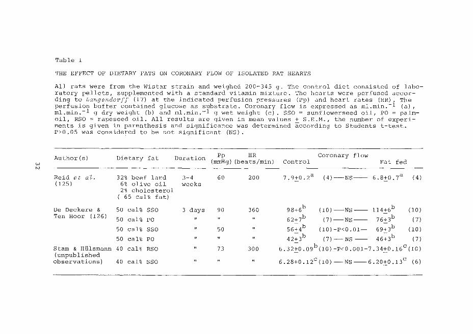
THE EFFECT OF DIETARY FATS ON CORONARY l'LO\V OF ISOLATED RAT HEARTS
All rats were from the Wistar strain and weighed 200-345 g. The control diet consisted of laboratory pellets, supplemented with a standard vitamin mixture. The hearts were perfused according to Langendorf! (17) at the indicated perfusion pressures (Pp) and heart rates (HR). The perfusion buffer contained glucose as substrate. Coronary flow is expressed as rnl.min.-1 (a), ml.rnin.-1 g dry weight (b) and rnl.rnin.-1 g wet weight (c). SSO = sunflowerseed oil, PO= palmoil, RSO =rapeseed oil. All results are given in mean values+ S.E.H., the number of experiments is given in parenthesis and significance was determined according to Students t-test. P>O.OS was considered to be not significant (NS).
Author (s) Dietary fat Duration Pp HR Coronary flow (mmHg) (beats/min) Control Fat fed
Reid et aZ. 32% beef lard 3-4 60 200 7.9+0.2a (4)-NS-- 6.8+0.7a ( 4) (125) 6% olive oil weeks
2% cholesterol ( 65 cal% fat)
De Deckere & 50 cal% SSO 3 days 90 360 98:J:6b (10)-NS-- 114:J:6b (10) Ten Hoar ( 126)
50 cal% PO " " " 62+7b (7)-NS-- 76+3b (7)
50 cal% SSO " 50 " 56~4b (l0)-P<0.01- 69~3b (10)
50 cal% PO " " " 42~3b (7)-NS -- 46~3b (7)
Starn & Hlilsrnann 40 cal% RSO " 73 300 6.32±0.09b(10)-P<0.001-7.34±0.16c(10) (unpublished
6. 28:J:O .12c (10)- NS --6. 20:J:O .13c observations) 40 cal% SSO " " " ( 6)
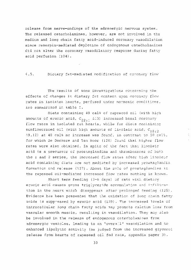
release from nerve-endings of the adrenergic nervous system.
The released catecholamines, however, are not involved in the
medium and long chain fatty acid-induced coronary vasodilation
since reserpin-mediated depletion of endogenous cateCholamines
did not alter the coronary vasodilatory response during fatty
acid perfusion (104).
4. 5. Dietary fat-mediated modification of coronary flow
The results of some investigations concerning the
effects of changes in dietary fat content upon coronar::-· flow
rates in isolated hearts, perfused under normoxic conditions,
are summarized in table I.
Diets containing 40 cal% of rapeseed oil (with high
amounts of erucic acid, c 22 : 1 ~13) increased basal coronary
flow rates in isolated rat hearts, while for diets containing
sunflowerseed oil (with high amounts of linoleic acid, c 18 : 2 ~9,12) at 40 cal% no increase was found, in contrast to 50 cal%,
for which De Deckere and Ten Boor (126) found that higher flow
rates were also obtained. In spite of the fact that linoleic
acid is a precursor o£ prostaglandins and thromboxanes of both
the 1 and 2 series, the increased flow rates after high linoleic
acid containing diets are not mediated by increased prostaglandi!1
formation and release (127). About the role of prostaglandins in
the rapeseed oil-mediated increased flow rates nothing is known.
Short term feeding (3-6 days) of rats with dietary
erucic acid causes gross triglyceride accumulation and infiltra
tion in the heart which disappears after prolonged feeding (128)
Evidence has been presented that the oxidation of long chain fatty
acids is suppressed by erucic acid {129) . The increased levels of
intracellular long chain fatty acids may promote calcium loss from
vascular smooth muscle, resulting in vasodilation. They may also
be involved in the release of endogenous catecholamines from
adrenergic vesicles, leading to an "overall" vasodilation and to
enhanced lipolytic activity (as judged from the increased glycerol
release from hearts of rapeseed oil fed rats, appendix paper 3).
33
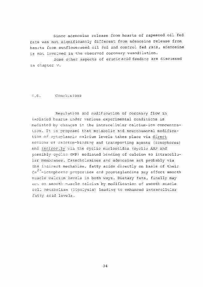
Since adenosine release from hearts of rapeseed oil fed
rats was not significantly different from adenosine release from
hearts from sunflowerseed oil fed and control fed rats, adenosine
is not involved in the observed coronary vasodilation.
Some other aspects of erucic acid feeding are discussed
in chapter V.
4. 6. Conclusions
Regulation and modification of coronary flow in
isolated hearts under various experimental conditions is
mediated by changes in the intracellular calcium-ion concentra
tion. It is ?roposed that metabolic and neurohumoral modifica
tion of cytoplasmic calcium levels takes place via direct
actions of calcium-binding and transporting agents (ionophores)
and indirectly via the cyclic nucleotides (cyclic AMP and
possibly cyclic GMP) mediated binding of calcium to intracellu
lar membranes. Catecholamines and adenosine act probably via
the indirect mechanism, fatty acids directly on basis of their
ca 2+-ionophoric properties and prostaglandins may affect smooth
muscle calcium levels in both ways. Dietary fats, finally may
act on smooth muscle calcium by modification of smooth muscle
cell metabolism (li~olysis) leading to enhanced intracellular
f~tty acid levels.
34
CHAPTER V
REGULATION AND MODIFICATION OF MYOCARDIAL CONTRACTILITY
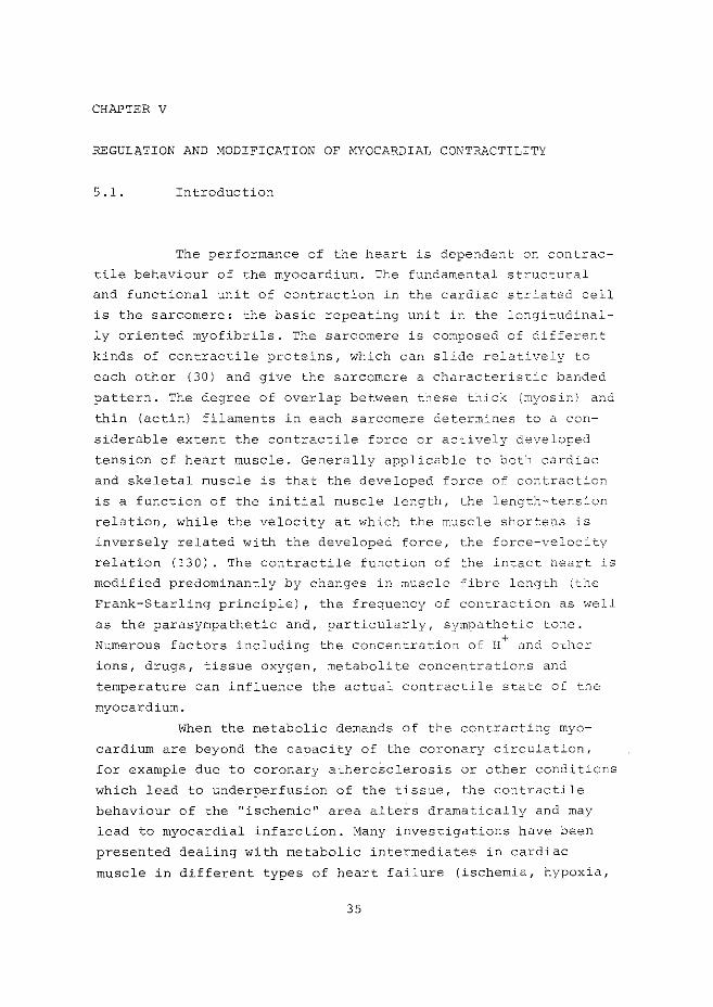
5 .1. Introduction
The performance of the heart is dependent on contrac
tile behaviour of the myocardium. The fundamental structural
and functional unit of contraction in the cardiac striated cell
is the sarcomere: the basic repeating unit in the longitudinal
ly oriented myofibrils. The sarcomere is composed of di£ferent
kinds of contractile proteins, which can slide relatively to
each other (30) and give the sarcomere a characteristic banded
pattern. The degree of overlap between these thick (myosin) and
thin (actin) filaments in each sarcomere determines to a con
siderable extent the contractile force or actively developed
tension of heart muscle. Generally applicable to both cardiac
and skeletal muscle is that the developed force of contraction
is a function of the initial muscle length, the length-tension
relation, while the velocity at which the muscle shortens is
inversely related with the developed force, the force-velocity
relation (130). The contractile function of the intact heart is
modified predominantly by changes in muscle fibre length (the
Frank-Starling principle) , the frequency of contraction as well
as the parasympathetic and, particularly, sympathetic tone.
Numerous factors including the concentration of H+ and other
ions, drugs, tissue oxygen, metabolite concentrations and
temperature can influence the actual contractile state of the
myocardium.
When the metabolic demands of the contracting myo
cardium are beyond the capacity of the coronary circulation,
for example due to coronary atheroSclerosis or other conditions
which lead to underperfusion of the tissue, the contractile
behaviour of the "ischemic" area alters dramatically and may
lead to myocardial infarction. Many investigations have been
presented dealing with metabolic intermediates in cardiac
muscle in different types of heart failure (ischemia, hypoxia,
35
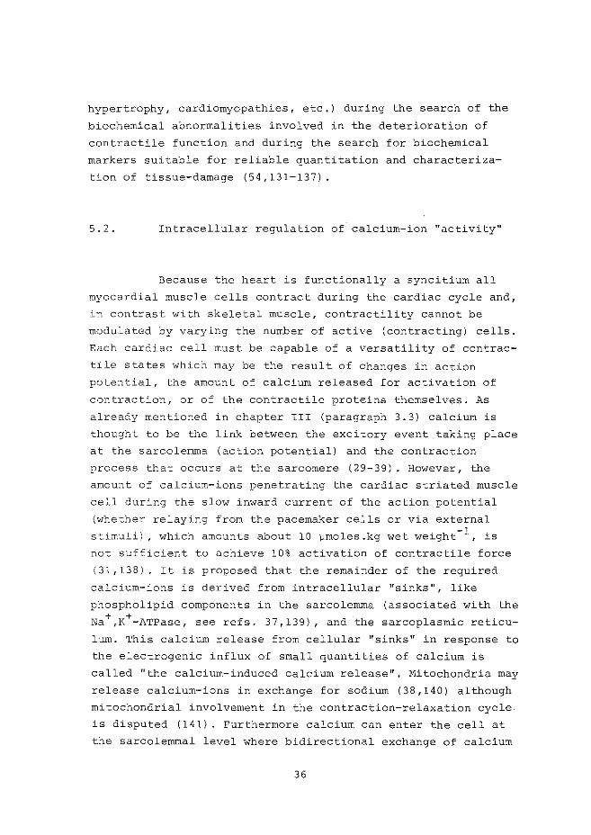
hypertrophy, cardiomyopathies, etc.) during the search of the
biochemical abnormalities involved in the deterioration of
contractile function and during the search for biochemical
markers suitable for reliable quantitation and characteriza
tion of tissue-damage (54,131-137).
5. 2. Intracellular regulation of calcium-ion "activity"
Because the heart is functionally a syncitium all
myocardial muscle cells contract during the cardiac cycle and 1
in contrast with skeletal muscle, contractility cannot be
modulated by varying the number of active (contracting) cells.
Each cardiac cell must be capable of a versatility of contrac
tile states which may be the result of changes in action
potential, the amount of calcium released for activation of
contraction, or of the contractile proteins themselves. As
already mentioned in chapter III (paragraph 3.3) calcium is
thought to be the link between the excitory event taking place
at the sarcolemma (action potential) and the contraction
process that occurs at the sarcomere (29-39). However, the
amount of calcium-ions penetrating the cardiac striated muscle
cell during the slow inward current of the action potential
(whether relaying from the pacemaker cells or via external
stimuli), which amounts about 10 vmoles.kg wet weight- 1 , is
not sufficient to achieve 10% activation of contractile force
(31,138). It is proposed that the remainder of the required
calcium-ions is derived from intracellular "sinks", like
phospholipid components in the sarcolemma (associated with the
Na+,K+-ATPase, see refs. 37,139), and the sarcoplasmic reticu
lum. This calcium release from cellular "sinks" in response to
the electrogenic influx of small quantities of calcium is
called "the calcium-induced calcium release 11, Mitochondria may
release calcium-ions in exchange for sodium (38,140) although
mitochondrial involvement in the contraction-relaxation cycle
is disputed (141) . Furthermore calcium can enter the cell at
the sarcolemmal level where bidirectional exchange of calcium
36
with cations such as Na+, K+, H+ and possibly Mg 2+ is proposed
although the exact mechanisms are not understood (36).
Relaxation is a consequence of decreased cytosolic
calcium levels. A number of mechanisms are involved· in the
sequestration of calcium-ions from the cytoplasm. Firstly,
sarcolemma and sarcoplasmic reticulum possess a stimulated
Ng 2+-dependent ATPase which may be responsible for calcium
binding and transport (uptake, efflux). secondly, mitochondria
can accumulate calcium linked to respiration (142,143). Thirdly,
the already mentioned bidirectional exchange systems may be
involved. The Na+,K+-ATPase, mainly localized at the sarcolemma,
plays a central role in some of the calcium-exchange systems.
For instance, inhibition of the enzyme by glycosides or,possibly,
cyclic AMP (144) gives rise to increased intracellular sodium
concentration and may be followed by a Na+-ca 2+ exchange at the
sarcolemma (145), or calcium release from the mitochondria. The
resulted increased cytoplasmic calcium levels alter the contrac
tile state of the myocardium.
Other enzymes, involved in the regulation of myocar
dial contractility, are adenylate and guanylate cyclase, since
oscillations of cyclic Ar>1P and cyclic GI--1P during one cardiac
contraction cycle have been observed (146,147). It indicates a
participiation of cyclic nucleotides under normal physiological
conditions. It is suggested by Brooker (147) that oscillations
in cyclic AMP are caused by transient stimulation of adenylate
cyclase during the action potential while it also may reflect
fluctuating calcium levels through activation of cyclic M1P
phosphodiesterase (148) or as mentioned by Tada et at. (41)
through feedback inhibition of adenylate cxclase.
Adenylate cyclase is localized in sarcolemma and in
microsomal fractions (43,149) and its activity is responsible
for cyclic AMP formation. Together with cyclic AMP phospho
diesterase activity it determines· the actual intracellular
concentration of cyclic AMP (150). This cyclic nucleotide in
turn can stimulate specific cyclic AMP-dependent protein
kinases involved in phosphorylation of specific sites of the
sarcoplasmic reticulum leading to augmented calcium uptake
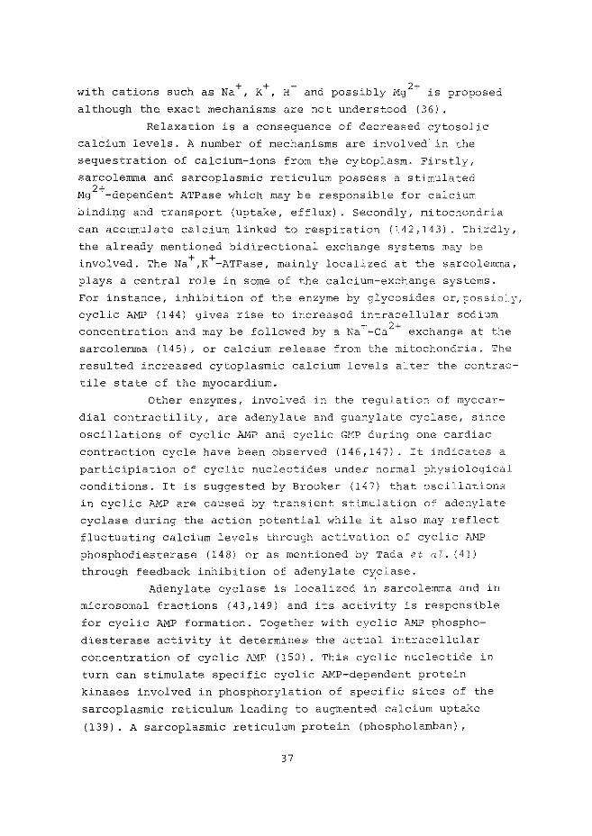
(139). A sarcoplasmic reticulum protein (phospholamban)
37
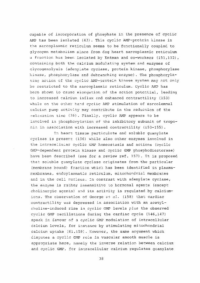
capable of incorporation of phosphate in the presence of cyclic
fu~P has been isolated (43) . This cyclic AMP-protein kinase in
the sarcoplasmic reticulum seems to be functionally coupled to
glycogen metabolism since from dog heart sarcoplasmic reticulum
a fraction has been isolated by Entman and co-workers (151,152)
containing both the calcium modulating system and enzymes of
glycogenolysis (adenylate cyclase, protein kinase, phosphorylase
kinase, phosphorylase and debranching enzyme). The phosphoryla
ting action of the cyclic P...11P-protein kinase system may not only
be restricted to the sarcoplasmic reticulum. Cyclic AMP has
been shown to cause elongation of the action potential, leading
to increased calcium influx and enhanced contractility (153)
while on the other hand cyclic AMP stimulation of sarcolemmal
calcium pump activity may contribute in the reduction of the
relaxation time (36). Finally, cyclic AMP appears to be
involved in phosphorylation of the inhibitory subunit of tropo
nin in association with increased contractility (153-155) .
In heart tissue particulate and soluble guanylate
cyclase is present (156) while also other enzymes involved in
the intracellular cyclic GMP homeostasis and actions (cyclic
GMP-dependent protein kinase and cyclic GMP phosphodiesterase)
have been described (see for a review ref. 157). It is proposed
that soluble guanylate cyclase originates from the particular
(membrane bound) fraction which has been identified in plasma
membranes, endoplasmatic reticulum, mitochondrial membranes
and in the cell nucleus. In contrast with adenylate cyclase,
the enzyme is rather insensitive to hormonal agents (except
cholinergic agents) and its activity is regulated by calcium
ions. The observation of George et al. (158) that cardiac
contractility was depressed in association with an acetyl
choline-induced rise in cyclic GMP levels plus the observed
cyclic GMP oscillations during the cardiac cycle (146,147)
speak in favour of a cyclic GMP modulation of intracellular
calcium levels, for instance by stimulating mitochondrial
calcium uptake (61,159). However, the same argument which
disputes a cyclic GMP role in vascular smooth muscle is
appropriate here, namely the inverse relation between calcium
and cyclic GMP, for intracellular calcium regulates guanylate
38
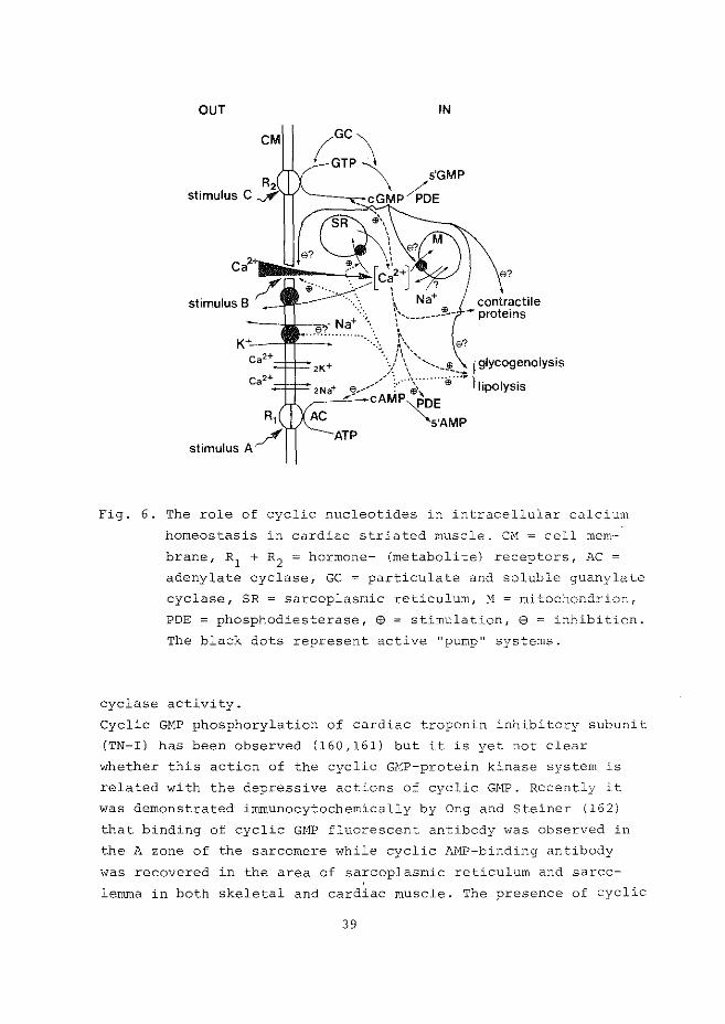
OUT IN
Fig. 6. The role of cyclic nucleotides in intracellular calcium
homeostasis in cardiac striated muscle. CM = cell mem-
brane, R1 + R2 = hormone- (metabolite) receptors, AC =
adenylate cyclase, GC = particulate and soluble guanylate
cyclase, SR = sarcoplasmic reticulum, M mitochondrion,
POE = phosphodiesterase, @ = stimulation, 8 = inhibition.
The black dots represent active "pump" systems.
cyclase activity.
Cyclic GMP phosphorylation of cardiac troponin inhibitory subunit
(TN-I) has been observed (160,161) but it is yet not clear
whether this action of the cyclic GMP-protein kinase system is
related with the depressive actions of cyclic GMP. Recently it
was demonstrated immunocytochemically by Ong and Steiner (162)
that binding of cyclic GHP fluorescent antibody was observed in
the A zone of the sarcomere while cyclic M1P-binding antibody
was recovered in the area of sarcoplasmic reticulum and sarco
lemma in both skeletal and cardiac muscle. The presence of cyclic
39
GMP-binding antibody in the A band suggests a role for cyclic
GHP in the regulation of myosin function.
The various above mentioned mechanisms which may be
involved in the regulation and modification of the intra
cellular calcium-ion concentration in cardiac muscle, in
relation with contractile function and metabolism, are summa
rized in fig. 6. In the subsequent paragraphs, neurohumoral,
metabolic and pharmacological effects on the calcium-ion
activity are discussed, while reference is made to this figure.
s. 3. Changes in myosin as determinant of contractility
Hydrolysis of ATP by myosin ATPase activity determines
the velocity of the contractile machinery of cardiac muscle, and
it has been suggested by Katz (133) that changes in contractility
in the failing heart are associated with changes in the rate of
ATP hydrolysis by the myofibrils. Decreased myosin ATPase and
altered myocardial contractility have been observed in endo
crinop~thies (hyper- and hypothyreoidism, adrenalectomy) , during
ageing and in heart failure due to chronic hemodynamic overload
(hypertension, aortic constriction, pulmonary stenosis, see refs.
133,135,163). Furthermore, significant changes in myosin and its
ATPase activity have been observed after administration of
catecholamines and during myocardial ischemia (164,165).
5. 4. Metabolic control of contractile function
Intracellular energy metabolism in myocardial muscle
may be divided in three stages:
(i) energy production by oxidation of substrates
coupled to ATP synthesis,
(iii "storage" of high-energy phosphate-bonds in
creatinephosphate and
(iii) utilization of chemical energy for mechanical
work and ion-pumps (ATPases) .
40
It has been long apparent that ATP is necessary for the chemi
cal reactions which form the basis of cardiac contractile
function. During contraction myosin cross-bridges interact
with the thin actin filaments which are pulled towa.rds the
center of the sarcomere. Relaxation takes place during dis
sociation of the actomyosin complex. However, it is still not
clear whether ATP is involved in the contractory events or in
the relaxation process. It is proposed that dissociation of the
actomyosin complex only proceeds when ATP is present and, if
not, muscle stiffness and inextensibili ty (rigor, "stone heart 11)
may ensue (166). Nevertheless, it is obvious that under condi
tions when energy production is limited, contractile function
is impaired. However, since decline in contractility and tissue
high-energy phosphates were not found to be associated, other
factors, directly or indirectly related with cellular energy
metabolism, have to be involved, while the imbalance between
intracellular compartments of adenine-nucleotides needs further
study ( 167-169) .
a. Hypoxia, anoxia and ischemia.
The delivery of oxygen to myocardial cells is crucial
for normal function of the heart. With increased cardiac work,
oxygen demand .increases proportionally and it is met by an auto
regulatory increase in coronary flow rate and increased oxygen
extraction. ATP synthesis during oxidative metabolism appears
to be strictly controlled by ATP utilization, while mitochon
drial respiration and citric acid cycle activity are controlled
by the cytoplasmic phosphate potential ( [ATP] I ( [ADP] x [Pi])
and the mitochondrial NAD oxidation-reduction state (170).
During acute ischemia, hypoxia and anoxia energy metabolism
alters drastically. Some of the alterations occurring in
isolated rat hearts are discussed in appendix papers 5 and 6.
In that work reduction of coronary flow was achieved by micro
sphere-embolization of the coronary circulation. In this model
for ischemia the vascular response (vasodilation of the non-
41
obstructed vessels) is retained. Reduced ATP levels observed
during ischemia, hypoxia (and anoxia) were correlated with
depressed contractility although "factors other than tissue
levels of creatine phosphate and ATP were rate-limiting". An
important factor may be acidosis, produced by metabolic altera
tions during substrate- and oxygen limited conditions (171).
Both proton generation and reduced levels of high-energy phos
phates may influence several calcium-mediated events in the
contraction-relaxation cycle:
(i) inactivation of the slow calcium-ion channels
(slow inward calcium influx during the action
potential) by reduced membrane phosphorylation
when ATP levels are decreased (172),
(ii) reduced interaction between the contractile
proteins or failure of actin and myosin filaments
to dissociate when ATP is lacking (166),
(iii) proton competition with calcium for activation
sites at the troponin molecules leading to inter
ruption (or reduction) of actin-myosin interaction
( 17 3 I ,
(iv) reduced calcium efflux due to inactivation of
calcium "pumps, in sarcolemma, sarcoplasmic reti
culum and mitochondria at low ATP levels,
(v) proton inhibition of calcium accumulation in
sarcoplasmic reticulum as shown for skeletal
muscle (174) and
(vi) proton inhibition of actomyosin ATPase (Prof.
P. Harris, personal communication).
The observed decrease in Na+,K+-ATPase activity (175)
and increased lysosomal fragility (176} during ischemia may also
be hazardous to the myocardial tissue.
In early stages, loss of contractility due to the above
mentioned events is probably fully reversible. However, sustained
ischemia will lead to irreversible damage of myocardial cells
(infarction} . During the onset of ischemia catecholamine release
from endogenous stores takes place leading to enhanced cytosolic
cyclic AMP levels (177). It is proposed that cyclic AMP is
involved in arrhythmogenic action occurring in response to
42
administered catecholamines (178) and during ischemia (179,
180). The metabolic response to increased intracellular cyclic
AMP during ischemia are (i) stimulation of prostaglandin syn
thesis and release (93) (ii) enhanced lipolysis, which in com
bination with decreased carnitine levels and res9iratory-chain
activity, lead to increased levels of long chain fatty acids
and their CoA and carnitine esters (181) and (iii) enhanced
glyco(geno)lysis resulting in depletion of glycogen stores
(182,183).
b. Prostaglandins
Besides the already mentioned effect on coronary flow,
prostaglandins have marked effects on cardiac contractility and
on lipid and carbohydrate metabolism (see for a review ref. 184)
while they are supposed to play a modulatory role in neuro
(muscular)transmission (94,122). Generally spoken, most prosta
glandins increase cardiac contractility in a dose dependent
manner. The positive inotropic effects are mediated by cyclic
AMP since prostaglandin-induced increased in adenylate cyclase
activity (185) and enhanced cyclic AMP levels have been observed
(186). On the other hand prostaglandin-induced increased cellular
membrane permeability to calcium has been observed in frog
·~earts (187). It has been proposed by Vergroesen and De Boer
(188) that prostaglandins were antagonizing the depressive
actions of potassium-ions rather than facilitating calcium uptake.
Prostaglandin-mediated increase in contractile function is
coupled to increased glucose and fatty acid oxidation (189) cmd
increased phosphorylase activity has been reported (187). Further
more, prostaglandin E1
proved to inhibit myocardial lipolysis at
high doses while increased lipolysis occurred at low doses (190,
191) .
The modulatory action of prostaglandins (especially the
E, A and F series) upon adrenergic neurotransmission and/or
response to exogenous catecholarnines has recently been reviewed
by Westfall (122). In numerous investigations in various species
43
and preparations, prostaglandins have been reported to inhibit,
to stimulate or to possess no influence upon the overflow of
catecholarnines and/or effector cell response to nerve stimula
tion. Our studies proved that, in isolated, perfused norepine
phrine-loaded rat hearts, high concentrations of prostaglandin
E 1 induced a sudden release of catecholamines (+ metabolites)
in the coronary effluent (appendix paper 2) . This observation
can be explained by the proposed calcium-ionophoric properties
of prostaglandins (104,105,124,187, see also chapter IV, para
graph 4.4.b).
Prostaglandin E1 and r 2 possess membrane stabilizing properties
(lysosomes, red blood cells) and inhibit platelet aggregation
(192-194). Together with the already mentioned vasodilatory
properties of prostaglandins, these effects may be involved in
the preservation of ischemic tissue as judged from reduced myo
cardial enzyme release (195), rhythm disturbances (196) while
it has been proposed by Riemersma et al. ( 197) that prostaglan
din-mediated inhibition of lipolysis and concomittantly reduced
myocardial free fatty acid extraction and oxygen consumption,
are involved in the beneficial role of prostaglandins during
ischemia.
c. Fatty acids
Endogenous triglycerides form a major source of ATP
in vivo as well as in isolated, perfused hearts during normoxia.
Even with exogenous glucose present the perfused heart still
metabolizes fatty acids (derived from endogenous triglycerides)
to a large extent (49,148). Some aspects of fatty acid metabolism
in the myocardium are presented in fig. 7. Lipoprotein lipase
localized in the endothelial cell membrane, is involved in the
removal (hydrolysis) of exogenous triglycerides from lipoproteins
(chylomicrons and very low density lipoproteins). Subsequently,
fatty acids penetrate the cell, are bound to a fatty acid
~inding protein (Z-protein) or as observed by Gloster and Harris
(199) to myoglobin. They are activated to fatty acyl-CoA esters
44
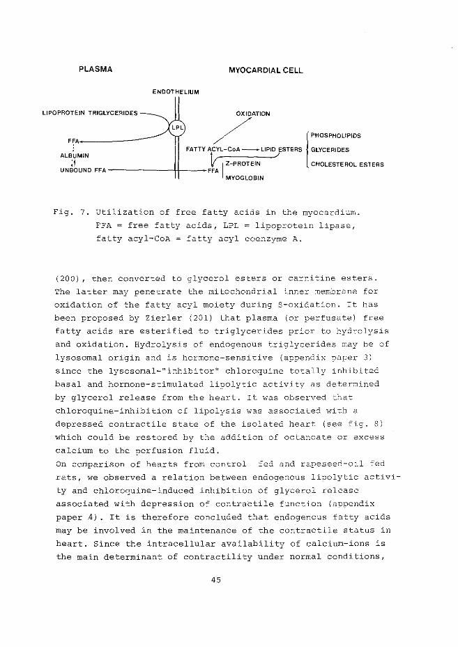
PLASMA MYOCARDIAL CELL
ENDOTHELIUM
LIPOPROTEIN TRIGLYCEAIDES
F~A-------
FATTYACYL-CoA--LtPID ESTERS GLYCERIDES
LPL /ATION l PHOSPHOLIPIDS
ALBUMIN t,.. ./ It v I Z-PROTEIN CHOLESTEROL ESTERS
UNBOUND FFA -------f+----FFA MYOGLOBIN
Fig. 7. Utilization of free fatty acids in the myocardium.
FFA = free fatty acids, LPL = lipoprotein lipase,
fatty acyl-CoA = fatty acyl coenzyme A.
(200), then converted to glycerol esters or carnitine esters.
The latter may penetrate the mitochondrial inner membrane for
oxidation of the fatty acyl moiety during S-oxidation. It has
been proposed by Zierler (201) that plasma (or perfusate) free
fatty acids are esterified to triglycerides prior to hydrolysis
and oxidation. Hydrolysis of endogenous triglycerides may be of
lysosomal origin and is hormone-sensitive (appendix paper 3)
since the lysosomal-"inhibitor 11 chloroquine totally inhibited
basal and hormone-stimulated lipolytic activity as determined
by glycerol release from the heart. It was observed that
chloroquine-inhibition of lipolysis was associated with a
depressed contractile state of the isolated heart (see fig. 8)
which could be restored by the addition of octanoate or excess
calcium to the perfusion fluid.
On comparison of hearts from control fed and rapeseed-oil fed
rats, we observed a relation between endogenous lipolytic activi
ty and chloroquine-induced inhibition of glycerol release
associated with depression of contraCtile function (appendix
paper 4) . It is therefore concluded that endogenous fatty acids
may be involved in the maintenance of the contractile status in
heart. Since the intracellular availability of calcium-ions is
the main determinant of contractility under normal conditions,
45
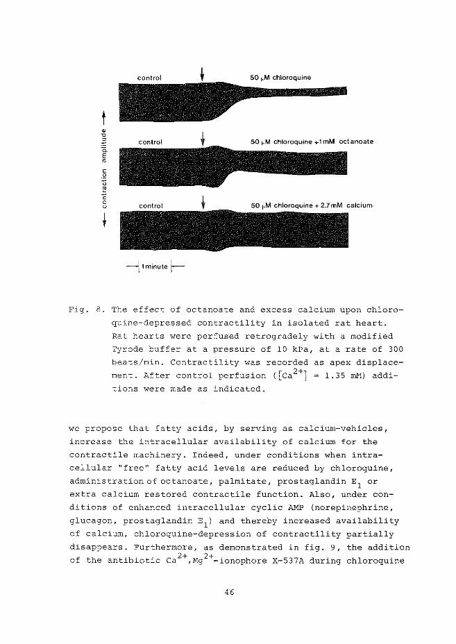
t • "0
-~ a_ E • c .2 u ~ c 0 u
50 J.lM chloroquine
--j 1 minute f---
Fig. 8. The effect of octanoate and excess calcium upon chloro
quine-depressed contractility in isolated rat heart.
Rat hearts were perfused retrogradely with a modified
Tyrode buffer at a pressure of 10 kPa, at a rate of 300
beats/min. Contractility was recorded as apex displace
ment. After control perfusion ([ca 2+] = 1.35 rnN) addi
tions were made as indicated.
we propose that fatty acids, by serving as calcium-vehicles,
increase the intracellular availability of calcium for the
contractile machinery. Indeed, under conditions when intra
cellular "free" fatty acid levels are reduced by chloroquine,
administration of octanoate, palmitate, prostaglandin E1
or
extra calcium restored contractile function. Also, under con
ditions of enhanced intracellular cyclic AMP (norepinephrine,
glucagon, prostaglandin E1
) and thereby increased availability
of calcium, chloroquine-depression of contractility partially
disappears. Furthermore, as demonstrated in fig. 9, the addition 2+ 2+ . of the antibiotic Ca ,Mg -1onophore X-537A during chloroquine
46
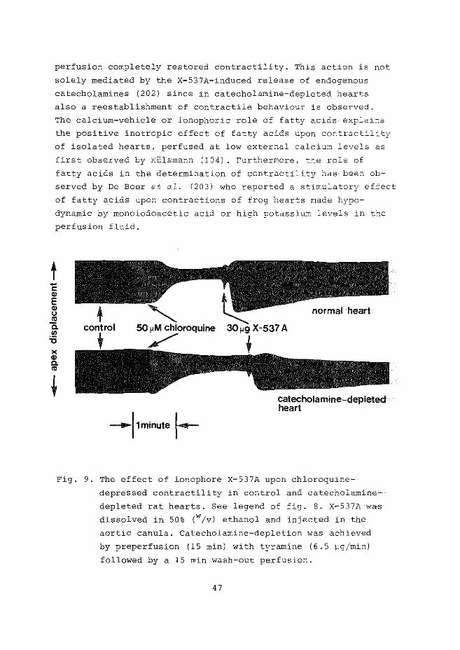
perfusion completely restored contractility. This action is not
solely mediated by the X-537A-induced release of endogenous
catecholamines (202) since in catecholamine-depleted hearts
also a reestablishment of contractile behaviour is observed.
The calcium-vehicle or ionophoric role of fatty acids explains
the positive inotropic effect of fatty acids upon contractility
of isolated hearts, perfused at low external calcium levels as
first observed by Hi.i.lsmann (104). rurthermore, the role of
fatty acids in the determination of contractility has been ob
served by De Boer et al. (203) who reported a stimulatory effec.t
of fatty acids upon contractions of frog hearts made hypo
dynamic by rnonoiodoacetic acid or high potassium levels in- the
perfusion fluid.
t -c: ., E ~ "' c. .!!! '0 )(
~
-11 minute 1--
normal heart
catecholamine-depleted heart
Fig. 9. The effect of ionophore X-537A upon chloroquine
depressed contractility in control and catecholamine
depleted rat hearts. See legend of fig. 8. x-537A wa"s
dissolved in 50% (w/v) ethanol and injected in the
aortic canula. Catecholamine-depletion was achieved
by preperfusion (15 min) with tyramine (6 .5 JJg/rnin)
followed by a 15 min wash-out perfusion.
47
In contrast to "physiological" levels of fatty acids
which as we propose are necessary for the maintenance of con
tractile function, high intracellular levels of fatty acids
(and their CoA and carnitine ester derivatives) are harmful
for the myocardium. The intracellular concentration of fatty
acids (+ esters) is determined by the uptake (which is mainly
dependent on their molar ratio with serum or perfusate albumin;
it is stimulated by catecholamines, see ref. 204) by the rate
of mitochondrial S-oxidation and by the balance between esteri
fication and (hormone-sensitive) lipolysis. Under oxygen
limited conditions (anoxia, hypoxia and ischemia) indeed eleva
ted endogenous free fatty acid levels (and their CoA and carni
tine esters) is observed (205) . The deleterious effects of high
free fatty acid levels are mediated by their membrane-detergent
properties, by their calcium-ionophoric properties and their
inhibiting capacity of various enzymes involved in energy meta
bolism (see table II). The actions of a high molar fatty acid:
albumin ratio in the perfusion buffer upon metabolism and
function in normal and ischemic rat hearts are discussed in
appendix papers 2 and 6. It was noticed that both depletion of
endogenous catecholamines and the addition of 11 mM glucose
during fatty acid perfusion partially restored the depressive
actions upon contractility and energy metabolism (as determined
by release of A.l:-1P-cataboli tes from the heart) . The protective
action of glucose can be explained by its ability to supply
glycerol-3-phosphate for triglyceride formation and/or by an
increased glycolytic ATP synthesis. These protective actions
may form at least part of the basis of the glucose-insulin
potassium therapy, advocated by Sodi-Pallares et al. (218) and
Opie (219) in the treatment of ischemia and other forms of
coronary heart disease. Besides glucose, anti-lipophilic drugs
(220,221) have been proposed to protect the myocardium from the
free fatty acid "toxicity", while also carnitine receives much
attention in this field since it is hypothesized by Shug et al.
(181) to reduce the adenine nucleotide translocator inhibition
by long chain fatty acids.
48
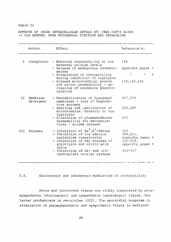
Table II
EFFECTS OF (HIGH INTRACELLULAR LEVELS OF) FREE FATTY ACIDS (+ CoA ESTERS) UPON ~!YOCARDIAL FUNCTION AND HETABOLISM
Action Effect Reference(s)
I Ionophoric - Enhanced contractility at low 104
II Membrane detergent
III Enzymes
external calcium levels - Release of endogenous catechol- appendix paper 2
amines - Stimulation of contractility
during inhibition of lipolysis - Altered mitochondrial proton
and cation permeability ~ uncoupling of oxidative phosphorylation
- Destabilization of lysosomal membranes + loss of degradative enzymes
- Swelling and labilization of mitochondria, directly or via lipolysis
- Alteration of plasmarnembrane permeability for macromolecules + enzyme release
+ + - Inhibition of Na ,K -ATPase - Inhibition of the adenine
nucleotide translocator - Inhibition of key enzymes of
glycolysis and citric acid cycle
- Inhibition of di- and tricarboxylate carrier systems
4
108,143,206
207,208
206,209
210
109 206,211, appendix paper 6 212-214, appendix paper 6
215-217
5.5. Cholinergic and adrenergic modulation of contractility
Atria and junctional tissue are richly innervated by para
sympathetic (cholinergic) and sympathetic (adrenergic) fibres. The
latter predominate in ventricles {222) . The myocardial response on
stimulation of parasympathetic and sympathetic fibres is mediated
49
by acetylcholine and (nor)epinephrine respectively and can be
mimicked by exogenous perfusion with these transmitters. The sup
uression of cardiac contractility by acetylcholine was associated
with the rise in ventricular muscle cyclic GMP and with decrea
sed cyclic AMP levels (111,158) and could be imita~ed by the
exogenous addition of cyclic GHP-derivatives (223) while di
butyryl-cyclic GMP could reverse both the positive ionotropic
and glycogen phosphorylase activating actions of isoproterenol
(224).
Adrenergic stimulation of cardiac contractility is
associated with increased myocardial cyclic AMP levels (153)
and concomittant decrease in cyclic GMP content (225). Together
with the observed phasic changes of both cyclic AMP and cyclic
GMP during the contraction-relaxation cycle (146,147) it has
led to the "Yin Yang" hypothesis for both cyclic nucleotides in
heart and other organs (226). However, the correlation between
inotropic actions of acetylcholine (+ derivatives) and catechol
amines and myocardial tissue levels of cyclic GMP and cyclic
AMP, respectively, has been disputed (227,228). Without
neglecting the importance of cholinergic actions we will not
further discuss acetylcholine-mediated affection of myocardial
metabolism and function.
As hypothesized by Robinson et al. (65) catecholamine
actions via a- and S-adrenergic receptors can modify the
activity of adenylate cyclase and result in changes in the rat~
of cyclic kMP formation. Indeed, in addition to the well
established positive inotropic effects evoked by B-adrenoceptor
stimulation, inotropic effects (although contradictory) have
been reported for myocardial a-adrenoceptor stimulation (229,
230) . Since, however, B-receptor density is larger than
a-receptor density in the myocardium, S-adrenergic effects are
predominant ( 2 2 9) .
Both cardiac smooth muscle and striated muscle 'cells
possess specific catecholamine-binding sites which are mainly
located in plasmamembranes. They show (i) reversible catechol
amine-binding, (ii) high affinity and saturation kinetics and
(iii) stereo specificity and thus fulfil the criteria for true
receptors (for a review see ref. 231). S-Receptor stimulation
50
and adenylate cyclase activity seem to be related although Hu
and Venter (232) did not observe changes in cardiac cellular
cyclic AMP levels upon stimulation of the S-receptors with
isoproterenol which was bound to polymers whereas stimulation
of cat papillary muscle contractility continued. The catalytic
unit of adenylate cyclase may be coupled not only to P-recep
tors but also to receptors for glucagon and histamine. In
other wordS, probably two or more different receptors are
coupled to the same adenyl cyclase (233,234).
The receptor-adenylate cyclase interaction starts
after release ofcatecholamines from endogenous stores
(adrenergic vesicles) in the postsynaptic space by exocytosis
of the storage vesicles; a process probably mediated by increa
sed calcium influx (124), while acetylcholine, nicotinic acid,
ionophore X-537A, cyclic nucleotides, phosphodiesterase inhibi
tors and inhibition of oxidative phosphorylation, which causes
loss of Ca++ from mitochondria, also lead to increased catechol
amine liberation (124,140,202). As discussed in appendix paper
2 and chapter IV, paragraphs 4b, 4c and 4d, fatty acids and
prostaglandin E1 may also promote catecholamine release from
nerve-ending adrenergic vesicles. Receptor-adenylate cyclase
interactions at the adrenergic neuroeffector junction cease
after catecholamine inactivation which occurs by at least three
different mechanisms:
(i) enzymatic destruction by catechol-a-methyl
transferase (COMT) located in the post-synaptic
membranes,
(ii) overflow in the circulation and
(iii) reuptake by the nerve-terminal.
Reuptake of secreted catecholamines is an aCtive transport, and
believed to be the chief mechanism of neurotransmitter inactiva
tion (235). After reuptake, catecholarnines are restored in
vesicles or metabolized by mitochondrial monoamine oxidase (MAO)
The inotropic and metabolic response upon S-adreno
ceptor stimulation are mediated by cyclic AMP-dependent protein
kinases (see paragraph 5.2). Increased contractility is coupled
to increased metabolic rates. Catecholarnines induce glycogeno
lysis and stimulate endogenous lipolysis (see appendix papers
51
3 and 6). However, the elevation of cyclic AMP in the myocar
dial muscle cell may not be necessarily sufficient for com
plete control of physiological and metabolic responses to
catecholamines. Besides cyclic AMP, allosteric effectors and
inhibitors may influence the overall response to catecholamines,
while calcium-ions are also involved in the determination of
the rate of glycogenolysis and lipolysis (236) both in the ab
sence or presence of cyclic AMP (237)-. It is possible that
fatty acids by serving as calcium-vehicles may be involved in
these actions since ca2+-stirnulation of protein kinase is well
established (238), as well as ca2+-stirnulation of various lipo
lytic enzymes. The close relation between enhanced contractility
and metabolism is possibly accounted for by phosphorylase
kinase, a calcium-dependent kinase involved in glycogenolysis
and in phosphorylation of the inhibitory subunit of troponin
(TN-I, see ref. 239).
Since the observations of Moore and Ruska (240) and
Parker (241) concerning budding, vacuolization and expansion of
vascular smooth muscle cell membranes which are situated in the
vicinity of adrenergic nerve terminals, many reports appeared
about catecholamine-induced damage of myocardial plasmamernbra
nes, resulting in increased permeability for macromolecules
(horse-radish peroxidase, see ref. 242), enzyme release {243-
245) and platelet aggregation (246). The detrimental effects of
circulating and/or released endogenous catecholamines may be
responsible for the induction of myocardial necrosis as
reported by Rona et al. (247). Explanations for these catechol
amine effects are still hard to give, but some interesting
observations have been done:
(i) catecholamines may alter membrane fluidity
(248) and hereby the activity of membrane
bound enzymes (144,249)
(ii) catecholamines may affect membrane phospho
lipid metabolism (250) and
(iii) oxidation products of catecholamines interfere
with "calcium pump"-systems of the microsomal
membranes and may lead to calcium overload
and cellular disfunction (251)
52
(iv) catecholamine, by stimulating endogenous lipo
lysis, may increase the detergent (soaps and
activated fatty acids) concentration.
Moreover, studies of WaldenstrOm et a2. (252) indicated that
massive release of endogenous catecholamines induced by per
fusion with tyramine, caused a many-fold increase of the loss
of creatine kinase from isolated perfused rat hearts. Hence,
catecholamine stores in isolated, denervated hearts are large -6 enough (a value of ~ 10 N in heart homogenates can be
estimated, see ref. 253) to affect membranous and metabolic
processes when they are released. Catecholamine-induced altera
tion of membrane structure and function may be amplified by the
observation that the fatty acid products of lipolysis will in
its turn again stimulate catecholamine release (appendix paper
2). Catecholamines (and other hormones, such as glucagon) may,
however, not only modify sarcolemma but also intracellular
membranes via phosphorylation by the cyclic AMP-protein kinase
system and thereby affect enzyme activities associated with
these membranes. Some actions of the cyclic N1P-protein kinase
system upon microsomal and lysosomal membranes are discussed in
appendix paper 3.
5. 6. Dietary fat and contractile status
Since cardiac membranes are highly involved in funda
mental cellular processes like contractility, osmotic regulation
and various energy producing metabolic pathways, the fatty acid
composition of cardiac phospholipids may be very important in
the determination of myocardial function. Membrane-phospholipid
fatty acid composition is influenced by nutritional (dietary)
hormonal, pharmacological and environmental means and since
dietary fat and serum lipids are considered as risk factors in
cardiovascular diseases in man, attention has been paid to the
relation between dietary fat and cardiac contractility.
In studies of Gudbjarnason and Hallgrimson (254) it
53
was shown that increasing amounts of various polyunsaturated
fatty acids in cardiac lipids were able to protect development
of myocardial necrosis in rats following overstimulation with
isoproterenol while, on the other hand, rapeseed-oil feeding is
associated with cardiopathogenic lesions (255) . Erucic acid
rich diets i·nduce gross fat accumulation in the myocardium ( 128,
255) after 3-6 days of feeding, which may be caused by (i) in
creased uptake of erucic acid (256, see however ref. 257), (ii)
erucyl carnitine inhibition of palmitate oxidation (129,258)
and (iii) decreased B-oxidation of erucate (259). Intracellular
levels of free fatty acids are increased (appendix paper 3),
while also changes in overall myocardial but, in particular,
mitochondrial phospholipid composition and cholesterol content
have been observed (260-262). As a result of these changes in
membrane phospholipids mitochondrial function may be impaired
(259,263,264) and the activity of adenylate cyclase reduced
(265). In table III the effects of a four day diet of rats with
40 cal% rapeseed oil upon the energy charge of their isolated,
perfused hearts is presented. The results are compared with
values from perfused hearts o£ rats fed with 40 cal% sunflower
seed oil and hearts ·from rats fed control laboratory pellets.
The reduced energy charge in hearts from rapeseed oil fed rats
may be a consequence of moderate uncoupling by intracellular
fatty acids of mitochondrial oxidative phosphorylation (209,215,
266) or inhibition of the adenine nucleotide translocator (211).
'I'he inhibition of mitochondrial function may be responsible for
the decrease in contractility of hearts from rapeseed oil (267 I
a."1d beef lard fed rats ( 125) . Another contribution may be the
hormonal imbalance of animals fed diets rich in (very) long chain
fatty acids, which could lead to myocardial lesions. Enlargement
of thyreoid and adrenal glands have been observed (268,269) while
HUlsmann (270) recently demonstrated enhanced levels of cortico
sterone and testosterone and concluded that long chain fatty
acid rich diets provoke abnormal stress reactions.
Decreased contractility, probably associated with impaired
mitochondrial function has also been observed in hearts from
essential fatty acid-deficient rats (271, 272).
54
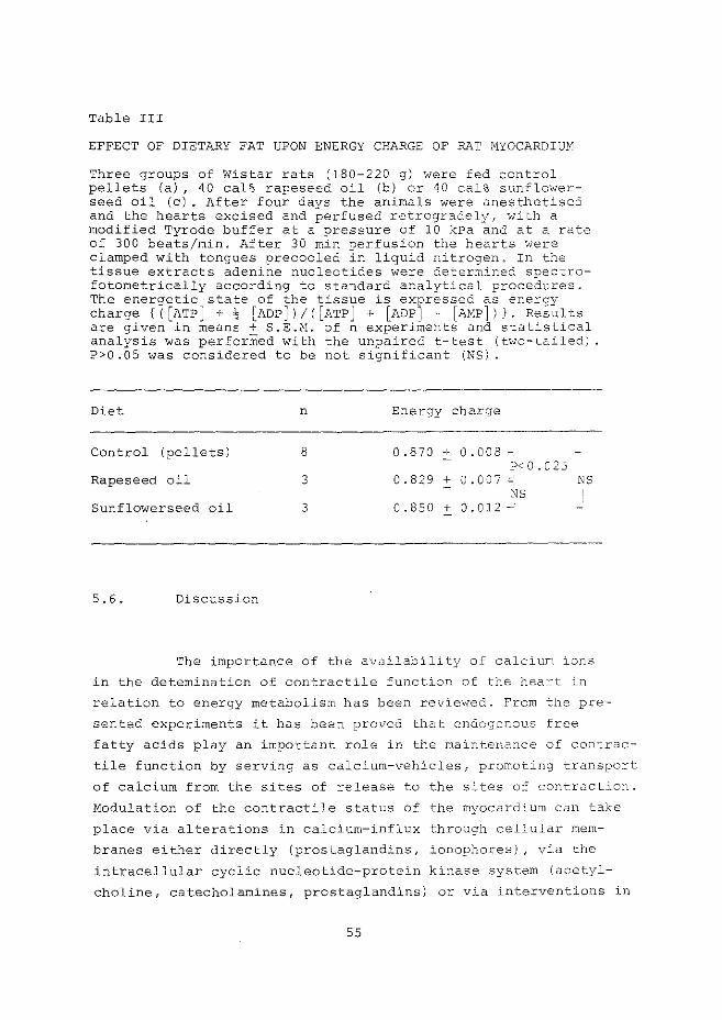
Table III
EFFECT OF DIETARY FAT UPON ENERGY CHARGE OF RAT MYOCARDIUH
Three groups of Wistar rats (180-220 g) were fed control pellets (a) , 40 cal% rapeseed oil (b) or 40 cal% sunflowerseed oil (c). After four days the animals were anesthetised and the hearts excised and perfused retrogradely, with a modified Tyrode buffer at a pressure of 10 kPa and at a rate of 300 beats/min. After 30 min perfusion the hearts were clamped with tongues precooled in liquid nitrogen. In the tissue extracts adenine nucleotides were determined spectrofotometrically according to standard analytical procedures. The energetic state of the tissue is exl?ressed as energy charge {([ATP] + l; [ADP])/i[ATP] + [ADPJ + [ANPj)). Results are given in means + S.E.M. of n experiments and statistical analysis was perforffied with the unpaired t-test (two-tailed) P>O.OS was considered to be not significant (NS).
Diet n Energy charge
Control (pellets) 8 0.870 + 0.008-P<0.025
Rapeseed oil 3 0.829 + 0. 00 7 ,;:.: NS NS
Sunflowerseed oil 3 0.850 + 0.012 ~
5. 6. Discussion
The importance of the availability of calcium ions
in the detemination of contractile function of the heart in
relation to energy metabolism has been reviewed. From the pre
sented experiments it has been proved that endogenous free
fatty acids play an important role in the maintenance of contrac
tile function by serving as calcium-vehicles, promoting transport
of calcium from the sites of release to the sites of contraction.
Modulation of the contractile status of the myocardium can take
place via alterations in calcium-influx through cellular mem
branes either directly (prostaglandins, ionophores), vi.a the
intracellular cyclic nucleotide-protein kinase system (acetyl
choline, catecholamines, prostaglandins) or via interventions in
55
energy metabolism (anoxia, hypoxia, ischemia and dietary fats).
Intracellular free fatty acids (and their CoA and/or carnitine
esters) may be involved in all three mentioned mechanisms;
(i) by acting as calcium-ionophores and by
increasing calcium transport through the sarco
lemma,
(ii) by releasing endogenously stored catecholamines
and
(iii) by affecting mitochondrial oxidative phosphoryla
tion (uncoupling and inhibition of the adenine
translocator).
The importance of free fatty acids in cell function implies
their strict metabolic control. It is generally believed now
(201) that plasma free fatty acids, taken up from the circulation,
are esterified, probably by sarcoplasmic reticulum, transported
as triglycerides and stored in "lipid droplets". Similar "drop
lets" have been isolated from beef heart, while electronmicrosco
PY revealed that these particles were surrounded by a unit mem
brane (273). This also holds for the lipid containing droplets
from hearts of rats fed rapeseed oil, which when stained for acid
phosphatase were found to contain reaction product in their peri
phery (N.C. Hlilsmann, unpublished). Therefore it is likely that
the lipid particles are lipid-filled lysosomes or autophagic
vacuoles. Indeed, both basal and norepinephrine-stimulated
lipolysis are inhibited when lysosomal-"activity" was eliminated
by the lysosomal inhibitor chloroquine. Acid lipase can therefore
be considered as the enzyme responsible for intracellular tri
glyceride hydrolysis and be involved in the supply of fatty acids
for energy metabolism and in intracellular calcium-transport.
56
CHAPTER VI
FINAL NOTES AND SPECULATIONS
* Isolated rat hearts, perfused retrogradely under
controlled conditions are useful objects in studies of myo
cardial metabolism, and of the regulation and modification of
coronary circulation, while an estimation of contractile
function from isotonic contractions of the heart can be made
by registration of the apex displacement in the vertical
direction.
The question arises now whether control of coronary flow rates
and contractile behaviour in isolated, hemoglobin-free perfused
hearts are comparable with the in vivo regulation?
Hyocardial capillary density is rather uniform and
capillary perfusion is maintained by the diameter of resistance
vessels and the tonus of precapillary sphincters. When we com
pare various systems which control the contractile state of
arterial (and arteriolar) and sphincter smooth muscle cells, it
appears that local metabolic influences overrule the neuro
hormonal control mechanisms. Metabolic control appears to opera
te in isolated hearts although autoregulation during pressure
flow studies cannot be observed. In non-working L:znaendcJ'ff
hearts mechanical expansion of coronary vessels probably over
rules any other mechanism while metabolic demands of the hearts
are low and also saturated at low perfusion pressure. The in
tactness of intrinsic control (hypoxia, anoxia),however, makes
it more reliable to extrapolate findings concerning flow regula
tion in isolated hearts to the in vivo heart.
Coronary circulation appears to be not homogenous, A
close examination of studies from Steenbergen tot: a!. (274) with
NADH surface fluorescence of hemoglobin-free, perfused, ";~~orking
rat hearts, reveals that even under normoxic condi tioJ.1S
(p02
>600 rnm Hg) a heterogenous tissue perfusic- pattern exists
which aggravates during hypoxia and ischemia. Thus, not all
capillaries are perfused under control conditions. This may
explain the apparent latency of myocardial lipoprotein lipase
57
release from endothelial plasmamembranes upon perfusion with
heparin. In fact, under conditions when it is likely to have
a more homogenous perfusion of myocardial tissue (high per
fusion pressure and heart rate, and norepinephrine in the
perfusion fluid) , heparin-perfusion could provoke almost
complete release of lipoprotein lipase from the isolated
heart (appendix paper 3).
In contrast to apparent similarity in control of
coronary circulation in isolated, perfused hearts and the in
o;-ivo beating heart, the regulation and modification of
contractility in the in vivo myocardium is much more complex.
Especially, the absence of direct neurohumoral regulation in
isolated hearts (although endogenous neurotransmitter sub
stances may still be of interest) makes it hard to extrapolate
findings in the isolated, non-working heart to the in vivo
cardiovascular system with its mutual interrelationships, is
absent in isolated hearts. However, the function of isolated
as well as in vi~o hearts is directly dependent on proper
energy metabolism; therefore findings, in this field, in iso
lated hearts may also be important in the in vivo heart.
** Coronary vascular and cardiac striated muscle cells
are the functional "units" in circulatory and contractile be
haviour. A review of both types of cells leads to the impression
that they possess opposite properties. For instance:
(i) fatty acids relax vascular smooth muscle and
increase contractile status of striated muscle
at low calcium levels (104) ,
(ii) agents which increase intracellular cyclic AMP
levels (catecholarnines, prostaglandins) induce
increased contraction of cardiac striated muscle
cells while they relax vascular smooth muscle
and
(iii) adenosine induces substantial coronary vasodila
tion, while no or little effect is observed upon
contractile behaviour of the striated muscle cell.
Since the contractile state of both types of muscle cells is
58
mediated by the intracellular calcium-ion concentration, the
regulation of calcium-homeostasis may be different in both
types of cells. Apparently, like in the first example, fatty
acids induce a one-way shift of calcium from the smooth muscle
to the cardiac striated muscle and this process fits well in
the proposed calcium-vehicle function of fatty acids in
contracting myocardial muscle cells. Fatty acid-induced libera
tion of endogenous catecholamines and subsequent elevation of
cyclic AlviP is not involved in the fatty acid-mediated coronary
vasodilation since the same observation was done in catec~ol-
amine depleted hearts (appendix paper 2) .
Cyclic AMP is involved in microsomal calcium-binding i~ both
types of cells. However, nothing is known about a cyclic A:,lP
stimulated calcium-influx in smooth muscle as is generally
accepted for striated muscle. A suitable hypothesis may be
that in smooth muscle only relaxing (calcium-binding) actions
of cyclic AMP are present while in striated muscle both
contracting (increased calcium-influx) and relaxing actions of
cyclic AMP are involved. This assumption explains the relaxing
action of cyclic AMP in skinned cardiac muscle cells (275).
*** The role of endogenous catecholamines in the function
of hearts, must not be neglected. During perfusion experiments
with electrically paced hearts, there will be a continuous over
flow of catecholamines which is, at least, involved in the
maintenance of coronary flow. Their role in merrbrane damage,
enzyme release and metabolic "disorders", especially under
stressful conditions (diabetes, starvation, long chain fatty
acid-rich feeding) warrant further investigations.
**** Increased intracellular levels of free fatty acids
(their CoA and/or carnitine esters} may be involved in the onset
of arrhythmias via inhibition of Na+,K+-ATPase as shown by
Lamers and Hlilsmann (109)" Since chloroquine bo·th inhibits basal
and catecholamine-increased lipolysis, and reveals anti
arrhythmic properties, we hypothesize that inhibition of endo-
59
genous (overstimulated) lipolysis is the mechanism of action
of certain anti-arrhythmic drugs. Indeed lidocaine, a well
established anti-arrhythmic drug, proved to inhibit basal and
norepinephrine-stimulated glycerol release from the perfused
rat heart (unpublished observations) .
60
REFERENCES
1. P.D.M.V. Turlapaty, R.K. Hesler & 0. Carrier Jr.: The role of calcium in different layers of vascular smooth muscle in norepinephrine contraction. Blood Vessels 13, 1976, 19 3-209.
2. R.M. Berne & H.N. Levy: In: "Cardiovascular Physiology", 1972, C.V. Mosby, St. Louis, U.S.A.
3. M.P. Wiedeman, R.F. Turna & M.N. Mayrovitz: Defining the precapillary sphincter. Microvasc. Res. 12, 1976, 71-75.
4. V. Navaratnam: In: "The human heart and circulation". Eds. R .J. Harrison & A .W. Asscher r Academic Press I Lonc'ccn' New York, San Francisco, 1975, pp. 98-109.
5. S. Baez: Microcirculation. Ann. Rev. Physiol. 39, 1977, 391-415.
6. R.D. Carter, W.L. Joyner & E.M. Reuhin: Effects of hista~ine and some other substances on molecular specificity c·f the capillary wall to plasma-proteins and dextran. Micro\·asc. Res. 7, 1974, 31-48.
7. F.S. Fay, P.H. Cooke & P.G. Canaday: Contractile properties of isolated smooth muscle cells. In: "Physiology of sn.ooth muscle''. Eds. E. Blilbring & M.F. Shuba, Raven Press, New York, 1976, pp. 249-264.
8. A.P. Somlyo, C.E. Devine, A.V. Somlyo & R.V. Rice: Filament organization in vertebrate smooth muscle. Phil. Transact. Royal Soc. Biol. 26, 1973, 223-229.
9. A.V. Somlyo & A.P. Somlyo: Electromechanical and pharr ~o
rnechanical coupling in vascular smooth muscle. J. Phc . .:.·macol. Exp. Ther. 159, 1968, 129-145.
10. M.P. Sparrow, L.C. r.1axwell, J.C. Ruegg & D.F. Bohr: Preparations and properties of a calcium ion-sensitive actomyosine from arteries. Am. J. Physiol. 219, 1970, 1366-1371.
11. D.F. Bohr: Vascular smooth muscle updated. Circ. Res. 32, 1973, 665-672.
12. K. Yamashita, T. Takagi & K. Hotta: Mobilization of cellular calcium and contraction-relaxation of vascular smooth muscle. Jap. J. Physiol. 27, 1977, 551-564.
13. R.A. Murphy: Structural proteins in the myofilaments and regulation of contraction in vertebrate smooth muscle. Fed. Proc. 35, 1976, 1302-1306.
14. A.P. Somlyo & A.V. Somlyo: Vascular smooth muscle. Pharm. Rev. 20, 1968, 197-272.
15. A.L. Lehninger: The metabolism of the arterial wall. ln: "The arterial wall". Ed. A.L. Lansing, Williams & Wilkins, Baltimore, 1959, pp. 200-246.
16. F. Takenaka, M. Sakunashi & M. Higuchi: High-energy phosphate metabolism of isolated coronary arteries in the dog. Blood Vessels 15, 1978, 190-198.
61
17. 0. Langendorf£: Untersuchungen am liberlebenden saugetierherzen. Pflligers Archiv Ges. Physiol. 61, 1895, 225-241.
18. N.S. McNutt & D.W. Fawcett: Myocardial ultrastructure. In: "Themamrnalianmyocardium", Eds. G.A. Langer & A.S. Brady. John Wiley & Sons, New York, London, Sydney, 1974, pp. 1-49.
19. H.D. Howse, V.J. Ferrans & R.G. Hibbs: A comparative histochemical and electronmicroscopic study of the surface coatings of cardiac muscle cells. J. Mol. Cell. Cardiel. 1, 1970, 157-168.
20. E. Page & L.P. McCallister: Quantitative electronmicroscopic description of heart muscle cells. Application to normal 1
hypertrophied and thyroxine stimulated hearts. Am. J. Cardiel. 31, 1978, 172-181.
21. D.A. Goodenouah & J.P. Revel: A fine structural analysis of intracellular junctions in the mouse liver. J. Cell Biol. 45, 1970, 272-290.
22. F.O. Simpson, D.G. Ryan & J.M. Ledingham: Fine structure of mammalian myocardial cells. In: 11 The Myocardium", Adv. Cardiel. 12, 1974, 15-33 (Karger, Basel).
23. D.W. Fawcett & N.S. l·1cNutt: The ultrastructure of cat myocardium. I. Ventricular papillary muscle. J. Cell Biol. 42, 1969, 1-45.
24. A.L. Smith & J.W.C. Bird: Distribution and particle properties of the vacuolar apparatus of cardiac muscle tissue. I. Biochemical characterization of cardiac muscle lysosomes and the isolation of acid, neutral and alkaline proteases. J. Mol. Cell. Cardiol. 7, 1975, 39-61.
25. K. Wildenthal: Lysosomes and lysosomal enzymes in the heart. In: "Lysosomes in Biology and Pathology", val. 4. Eds. J.T. Dingle & R.T. Dean, North-Holland, Amsterdam, 1975, pp. 167-190.
26. C.T. Potter & J. Axelrod: Properties of norepinephrine storage particles of the rat heart. J. Pharmacal. Exp. Ther. 142, 1963, 299-315.
27. J.C. Sosa, F. de la Iglesia, G. Lurnb, J.M. Berger & S. Bencosme: Subcellular distribution of catecholamines and specific granules in rat heart. Lab. Invest. 21, 1969, 19-26.
28. S. Hajdu & E.J. Leonard: A calcium transport system for mammalian cells. Life Sciences 17, 1976, 1527-1534.
29. S. Ebashi & M. Endo: Calcium and muscle contraction. Progr. Biophys. Mol. Biol. 18, 1968, 123-183.
30. H.E. Huxley: The mechanism of muscular contraction. Science 164, 1969, 1356-1366.
31. G.A. Langer: Excitation-contraction coupling. Ann. Rev. Physiol. 35, 1973, 708-757.
32. W.G. Nayler & J. Dunnett: Regulation of myocardial contraction. In: "The Myocardium", Adv. Cardiel. 12, 1974, 45-58 (Karger, Basel) .
62
33. J. Gergely: Excitation-contraction coupling- cardiac muscle events in the myofilament. Fed. Proc. 35, 1976, 1283-1287.
34. S. Ebashi: Excitation-contraction coupling. Ann. Rev. Physio1. 38, 1976, 293-313.
35. A.M. Katz: Physiology of the heart. Raven Press, New York, 1977, pp. 137-160.
36. N.S. Dhalla, A. Ziegelhoffer & J.A.C. Harrow: Regulatory role of membrane systems in heart function. Can. J. Physiol. Pharmacal. 55, 1977, 1211-1234.
37. H. LUllman & Th. Peters: Plasmalemma! calcium in cardiac excitation-contraction coupling. Clin. Exp. Pharmacal. Physiol. 4, 1977, 49-57.
38. E. Carafoli; Mitochondria, ca2+ transport and the regulation of heart contraction and metabolism. J. Mol. Cell. Cardiol. 7, 1975, 83-89.
39. H. Reuter: Exchange of calcium ions in the mammalian myocardium. Circ. Res. 34, 1974, 599-605.
40. P.V. Sulakhe, D.B .. HcNamara & N.S. Dhalla: Characterization of partially purified heart sarcolemmal Na+-K+-stimulated ATPase. In: 11 The Sarcolemma", Rec. Adv. Stud. Card. Struct. Metab.,vol. 9. Eds. P.E. Roy & N.S. Dhalla, Univ. Park Press, Baltimore, London, Tokyo, 1976, pp. 229-302.
41. M. Tada, N.A. Kirchberger, J.M. Iorio & A.M. Katz: Control of cardiac sarcolemmal adenylate cyclase and sodiumpotassium-activated adenosine-triphosphatase activities. Circ. Res. 36, 1975, 8-17.
42. D.O. Levitsky, M.K. Aliev, A.V. Kuzmin, T.S. Levchenko, V.N. Smirnov & E.I. Chazov: Isolation of calcium pump system and purification of calcium ion-dependent ATPase from heart muscle. Biochim. Biophys. Acta 443, 1976, 468-484.
43. M.L. Entman, G.L. Levey & S.E. Epstein: Demonstration of adenyl cyclase activity in canine cardiac sarcoplasmic reticulum. Biochem. Biophys. Res. Cornmun. 35, 1969, 728-733.
44. R.J. Bing: Cardiac metabolism. Physiol. Rev. 45, 1965, 171-213.
45. J.R. Neely & H.E. Morgan: Relationshi.p between carbohydrate and lipid metabolism and the energy balance of heart muscle. Ann. Rev. Physiol. 36, 1974, 413-459.
46. J.R. Williamson & H.A. Krebs: Acetoacetate as fuel of respiration in perfused rat heart. Biochem. J. 80, 1961, 540-547.
47. J.F. Oram, S.L. Benneth & J.R. Neely: Regulation of fatty acid utilization in isolated perfused rat hearts. J. Biol. Chern. 248, 1973, 5299-5309.
48. L.H. Opie & P. Owen: Effects of increased mechanical work by isolated perfused rat heart during production or uptake of ketone bodies. Biochem. J. 148, 1975, 403-415.
63
49. R.E. Olson & R.J. Hoeschen: Utilization ot endogenous lipid by the isolated perfused rat heart. Biochern. J. 103, 1967, 796-801.
50. J. R. ~IJ'illiarnson: Metabolic effects of epinephrine in isolated perfused rat heart. I. Dissociation of the glycogenolytic from the metabolic stimulatory effect. J. Biol. Chern. 239, 1964, 2721-2729.
51. R.A. ·Kreisberg: Effect of epinephrine on myocardial triglyceride and free fatty acid utilization. Am. J. Physiol. 210, 1966, 385-389.
52. M.P. Crass III: Regulation of triglyceride metabolism in the isotopically prelabeled perfused heart. Fed. Proc. 36, 1977, 1995-1999.
53. J.J. Lech, G.J. Jesmok & D.N. Calvert: Effect of drugs and hormones on lipolysis in heart. Fed. Proc. 36, 1977, 2000-2008.
54. L.H. Opie: Metabolism of the heart in health and disease. Am. Heart J. 76, 1968, 685-698.
55. R. Rubio & R.M. Berne: Regulation of coronary blood flow. Progr. Cardiovasc. Dis. 18, 1975, 105-122.
56. D.E. Mohrman & E.O. Feigl: Competition between sympathetic vasoconstriction and metabolic vasodilation in the canine coronary circulation. Circ. Res. 42, 1978, 79-86.
57. T.E. Driscoll, T.W. Hoir & R.W. Eckstein: Vascular effects of changes in perfusion pressure in the nonischemic and ischemic heart. Circ. Res. 14-15 (Suppl I), 1964, I-94 -I-102.
58. R. BUnger, F.J. Haddy, A. Querengasser & E. Gerlach: An isolated guinea pig heart preparation with in vivo like features. Pflligers Archiv 353, 1975, 317-326.
59. S. Greenberg, J.P. Long & F.P.J. Diecke: Differentiation of calcium pools utilized in the contractile response of canine arterial and venous smooth muscle to norepinephrine. J. Pharmacal. Exp. Ther. 185, 1973, 1059-1067.
60. H.P. Bar: Cyclic nucleotides and smooth muscle. Adv. Cycl. Nucl. Res. 4, 1974, 195-237.
61. M.J. Berridge: The interaction of cyclic nucleotides and calcium in the control of cellular activity. Adv. Cycl. Nucl. Res. 6, 1975, 1-98.
62. R. Andersson: Cyclic AMP as a mediator of the relaxing action of papaverine, nitroglycerine, diazoxide and hydralazide in intestinal and vascular smooth muscle. Acta Pharmacal. Toxicol. 32, 1973, 321-336.
63. W.R. Kukovetz, G. POch & A. Wurm: Role of cyclic AMP as cellular transmitter of positive inotropic and coronary dilator effects of drugs. In: "Symposium on drugs and heart metabolism''. Eds. L. Szekeres, J. Knoll & J.G. Papp, Akademiai Kiado, Budapest, 1973~ pp. 37-59.
64. C.J. Limas & J .N. Cohn: s-timulation of vascular smooth muscle sodium, potassium-adenosine triphosphatase by vasodilators.
64
Circ. Res. 35, 1974, 601-607.
65. G.A. Robinson, R.W. Butcher & E.W. Sutherland: Adenyl cyclase as an adrenergic receptor. Ann. NY Acad. Sci. 139, 1967, 703-723.
66. L. Volicier & S. Hynie: Effect of catecholarnines and angiotensin on cyclic AMP in rat aorta and tail artery. Eur. J. Pharmacal. 15, 1974, 214-220.
67. R. Andersson, K. Nilsson, J. Wikberg, S. Johansson, E. Mohme-Lundholm & L. Lundholm: Cyclic nucleotides and the contraction of smooth muscle. Adv. Cycl. Nucl. Res. 5' 1975' 491-518.
68. G. Schultz, J.G. Hardman, K. Schultz, C.E. Baird & E.W. Sutherland: The importance of calcium ions for the regulation of guanosine 3',5'-cyclic monophosphate levels. Proc. Natl. Acad. Sci. U.S.A. 70, 1973, 3819-3893.
69. I. Takayanagi, H. Ohkubo & T. Takagi: Drug-induced smooth muscle contraction with no change in the level of cyclic GMP. Jap. J. Pharmacal. 26, 1976, 501-504.
70. K.J. Broadley: The release of a coronary vasodilator metabolite from the guinea-pig isolated perfused heart stimulated by catecholamines, histamine and electrical pacing and by exposure to anoxia. Br. J. Pharmacal. 58, 1976, 89-100.
71. A.K. Sen, F.A. Sunahara & J. Talesnik: Coronary reactions to cardiac hyperactivity and to hypoxia in is'olated perfused heart of rat. Br. J. Pharmacal. 61, 1977, 381-393.
72. A.K. Sen, F.A. Sunahara & J. Talesnik: Prostaglandin E2 and cyclic AMP in the coronary vasodilatation due to cardiac hyperactivity. Can. J. Physiol. Pharmacal. 54, 1976, 128-139.
73. M. Katori & R.M. Berne: Release of adenosine from anoxic hearts. Relationship to coronary flow. Circ. Res. 19, 1966, 420-425.
74. J. Schrader & E. Gerlach: Effect of beta-adrenergic stimulation on 14c-adenosine release from the heart. Pflligers Archiv 355, 1975, R17 (Abstract).
75. A.C. Fox, G.E. Reed, E. Glassman, A.J. Kaltman & B.B. Silk: Release of adenosine from human hea~ts during angina induced by rapid atrial pacing. J. Clin. Invest. 53, 1974, 1447-1457.
76. R. Rubio, R.M. Berne & M. Katori: Release of adenosine in reactive hyperemia of the dog heart. Am. J. Physiol. 216, 1969, 56-62.
77. J. Schrader, F.J. Haddy & E. Gerlach: Release of adenosine, inosine and hypoxanthine from isolated guinea pig hearts during hypoxia, flow autoregulation and reactive hyperemia. Pflligers Archiv 269, 1977, 1-6.
78. H. Starn, J.W. de Jong & H.L. van der Wiel: Metabolic consequences of Sephadex-induced reduction of coronary flow
65
in isolated rat hearts. In: 11 Cardiac Adaptation", Rec. Adv. Stud. Card. Struct. Metab., vol. 12. Eds. T. Kobayashi, Y. Ito & G. Rona, 1978, pp. 253-258.
79. R. Rubio, R.M. Berne & K.G. Dobson Jr.: Sites of adenosine production in cardiac and skeletal muscle. Am. J. Physiol. 225, 1973, 938-953.
80. D.E. Atkinson: Energy charge of the adenylate pool as a regulatory parameter: Interaction with feedback modifiers. Biochemistry 7, 1968, 4030-4034.
81. R.A. Olsson, C.J. Davis, E.M. Khouri & R.E. Patterson: Evidence for an adenosine receptor on the surface of dog coronary myocyte. Circ. Res. 39, 1976, 93-98.
82. R.A. Olsson, C.J. Davis & E.M. Khouri: Coronary activity of adenosine covalently linked to polylysine. Life Sciences 21' 1977' 1343-1356.
83. J. Schrader, s. Nees & E. Gerlach: Evidence for a cell surface adenosine receptor on coronary myocytes and atrial muscle cells. Pflligers Archiv 369, 1977, 251-257.
84. M. Huang & G.I. DruiTUTLond: Effect of adenosine on cyclic AMP accumulation in ventricular myocard.ium. Biochem. Pharmacal. 25, 1976, 2713-2719.
85. M. Huang & G.I. DruiTUTLond: Interaction between adenosine and catecholamines on cyclic AMP accumulation in guinea pig ventricular myocardium. Biochem. Pharmacal. 27, 1978, 187-191.
86. J.T. Her-lihy, E.L. Bock:ffian, -R.M. Berne, R. Rubio & H.L. Baxter: Effects of adenosine on the cyclic AMP levels and ca2+-K+ dos.e-re·sponse curves of V?-Scular smooth muscle. Physiologist 17, 1974 ,. 244 (Abstract).
87. J. Schrader, R. Rubio & R.M. Berne·: Inhibition of slow action potentials of guinea pig atrial muscle by adenosine: a possible effect on ca2+-influx. J. Mol. Cell. Cardiol. 7, 1975, 427-433.
88. K. Schr6r, R. Krebs & C. Nookhwunn: Increase in coronary vascular resis.tance by indomethacin in the isolated quinea .pig heart preparation in the absence of changes in mechanical performance and oxygen consumption. Eur. J. Pharmacal. 39, 1976, 161-169.
89. Th.H. Hintze & G. Kaley: Prostaglandins and the control of blood flow in the canine myocardium. Circ. Res. 40, 1977, 313-320.
90. A.Z. Block, H. Feinberg, K. Herbaczynska-Cedro & J.R. Vane: Anoxia induced release of prostaglandins in isolated rabbit hearts. Circ. Res. 36, 1975, 34-42.
91. R.L. Moreti & S. 1iliraham: Stimulation of microsomal prostaglandin synthesis by a blood plasma constituent which augments autoregulation and maintenance of vascular tone in isolated rabbit hearts. Circ. Res. 42, 1978, 317-323.
92. P, Needleman: The synthesis and function of prostaglandins in the heart. Fed. Proc. 35, i976, 2376-2381.
66
93. G. Markelonis & J. Garbus: Alterations of intracellular oxidative metabolism as stimuli evoking prostaglandin biosynthesis. Prostaglandins 10, 1975, 1087-1106.
94. F. ten Hoor & A.J. vergroesen: Prostaglandins and the heart. J. Mol. Cell. Cardiol. 7, 1975, 335-541.
95. E.A.M. de Deckere, D.H. Nugteren & P.. ten Hoor; Prostacyclin is the major prostaglandin released by the isolated rabbit and rat heart. Nature 268, 1977, 160-163.
96. S. Moncada, R. Gryglewski, S. Bunting & J.R. Vane: An enzyme isolated from arteries transforms prostaglc,r.Gi:l. endoperoxides to an unstable substance that inhibits platelet aggregation. Nature 263, 1976, 663-665.
97. G.J. Dusting, S. Moncada & J.R. Vane: Pro~;tacyclin (PGX) is the endogenous metabolite responsible f0r relaxation of coronary arteries induced by arachidonic acid. Prostaglandins 13, 1977, 3-15.
98. P. Needleman, P.S. Kulkarni & A. Raz: Coronary tone nodulation: Formation and actions of prostaglandins, endoperoxides, and thrornboxanes. Science 195, 1977, 409-412.
99. K. SchrOr: Prostaglandin D2 (PGD2) -A potent coronary vasoconstrictor in the guinea pig isolated heart. N.S. Arch. Pharmacal. 302, 1978, 61-62.
100. M. Smigel & S. Fleischer: Characterization and localization of prostaglandin E1 receptors in rat liver plasmamembranes. Biochim. Biophys. Acta 332, 1974, 358-373.
101. A.P. Shepherd, C.C. Hao, E.D. Jacobson & L.L. Shanbour: The role of cyclic AMP in mesenteric vasodilation. !>Ucrovasc. Res. 6, 1973, 332-341.
102. E.W. Dunham, M.K. Haddox & N.D. Goldberg: Alteration of vein cyclic 3',5 1 nucleotide concentrations during changes in contractility. Proc. Natl. Acad. Sci. U.S.A. 71, 1974, 815-819.
103. R. Andersson: Cyclic ArviP, a regulator substance in ',moth muscle function and metabolism. Acta Physiol. Sc,~ ·:d. :9 G, (suppl), 1973, 7-21.
104. W.C. HUlsmann: coronary vasodilation by fatty acids. Basic Res. Cardiol. 71, 1976, 179-181.
105. S.J. Kirtland & H. Baum: Prostaglandin, E1 may act as a ''calcium ionophore''. Nature (New Biol.) 236, 1972, 47-49.
106. E. Carafoli & F. Crovetti: Interaction between prostaglandin E1 and calcium at the level of the mitochondrial membrane. Arch. Biochem. Biophys. 154, 1973, 40-46.
107. J.C. Gilbert & M.G. Willie: Effects of prostaglandins on the ATPase activities of synaptosomes. Biochem. Pharmacal. 24, 1975, 551-556.
108. G.K. Asimakis & L.A. Sordahl: Effects of actractyloside and palmitoyl-coenzyme A on calcium transport in cardiac mitochondria. Arch. Biochem. Biophys. 179, 1977, 200-210.
67
109. J.M.J. Lamers & W.C. Hlilsmann: Inhibition of (Na++K+)stimulated ATPase of heart by fatty acids. J. Mol. Cell. Cardiol. 9, 1977, 343-346.
110. E.O. Feigl: Parasympathetic control of coronary blood flow in dogs. Circ. Res. 25, 1969, 509-519.
111. T.P. Lee, J.F. Kuo & P. Greengard: Role of muscarinic cholinergic receptors in regulation of guanosine 3',5'cyclic monophosphate content in mammalian brain, heart muscle and intestinal smooth muscle. Proc. Natl. Acad. Sci. U.S.A. 69, 1972, 3287-3291.
112. H.A. Dieterich, K. LOffelholz & H. Pompetzki: Acetylcholine overflow from isolated perfused hearts of various species in the absence of cholinesterase inhibition. N.S. Arch. Pharmacal. 296, 1977, 149-152.
113. R.P. Ahlquist: A study of the adrenotropic receptors. Am. J. Physiol. 153, 1948, 586-600.
114. A.M. Lands, A. Arnold, J.P. McAnliff & T.G. Brown: Differentiation of receptor systems activated by sympathomimetic amines. Nature 214, 1967, 597-598.
115. G. Ross: Adrenergic responses of the coronary vessels. Circ. Res. 39, 1976, 461-465.
116. B.L. Bayer, P. Mentz & W. FOrster: Characterization of the adrenoreceptors in coronary arteries of pigs. Eur. J. Pharmacal. 29, 1974, 58-65.
117. P.J. Dempsey & T. Cooper: Pharmacology of the coronary circulation. Ann. Rev. Pharmacal. 12, 1972, 99-110.
118. K.J. Broadley: An analysis of the coronary vascular responses to catecholamines using a modified Langendorf£ heart preparation. Br. J. Pharmacal. 40, 1970, 617-629.
119. C.L. Seidel, R.L. Schnarr & H.V. Sparks: Coronary artery cyclic AI',lP content during adrenergic receptor stimulation. Am. J. Physiol. 229, 1975, 265-269.
120. E.A. Nikkila, P. Torsti & o. Penttila: Effect of fasting, exercise and reserpine on catecholamine content and lipoprotein lipase activity of rat heart and adipose tissue. Life Sciences 4, 1965, 27-35.
121. S.N. Chaudhuri & J.C. Shipp: Cyclic AMP (cAI,1P) in hearts of alloxan-diabetic rats. In: "Myocardial Metabolism", Rec. Adv. Stud. Card. Struct. Hetab.,vol. 3. Ed. N.S. Dhalla, University Park Press, Baltimore, London, Tokyo, 1973, pp. 319-330.
122. Th. Westfall: Local regulation of adrenergic neurotransmission. Physiol. Rev. 57, 1977, 659-728.
123. H. Blaschke, H. Fi~emark, A.D. Smith & H. Winkler: Lipids of the adrenal medulla. Lysolecithine; a characteristic constituent of chromaffin granules. Biochem. J. 104, 1967' 545-549.
124. D. Njus & G.K. Radda: Bioenergetic processes in chromaffin granules. )> ne;.'l perspective on some old problems. Biochim. Biophys. Acta 463, 1978 1 219-244.
68
125. J.V.O. Reid, A. Lochner & A.J. Brink: Effect of a high fat diet on the metabolism and mechanical behaviour of the isolated rat heart. J . .Hol. Cell. Cardiel. 1, 1970, 209-220.
126. E.A.M. de Deckere & F. ten Hoor: Influences of dietary fats on the coronary flow and oxygen consumption of the isolated rat heart. In: 11 Biochemistry and pharmacology of myocardial hypertrophy, hypoxia en infarction", Rec. Adv. Stud. Card. Struct. Metab., vol. 7. Eds. P. Harris, R.J. Bing & A. Fleckenstein. University Park Press, Baltimore, London, Tokyo, 1976, pp. 475-483.
127. E.A.M. de Deckere, D.H. Nugteren & F. ten Hoor: The influence of dietary fats on prostaglandin release of the isolated rabbit and rat heart and rat aorta. Proc. 19th Dutch Fed. Mee·ting, Leiden, 1978, p. 223 (Abstract).
128. R.O. Vles: Nutritional aspects of rapeseed oils. Proc. 4th Internationaler Rapskongress, Giessen, 1974, pp. 17-30.
129. L.HeijkenskjOld & L. Ernster: Studies on the mode of action of erucic acid on heart metabolism. Acta Med. Scand. 585 (supp1), 1975, 75-83.
130. J.R. Blinks & B.R. Jewell: The meaning and measurement of myocardial contractility. In: "Cardiovascular Fluid Dynamics", vol. 1. Ed. D.H. Bergel, Acad. Press, London, New York, 1972, pp. 225-260.
131. L.H. Opie: Metabolism of the heart in health and disease. Am. Heart J. 77, 1969, 100-122 & 383-410.
132. R.J. Kones: Metabolism of the acutely ischemic and hypoxic heart. Crit. Care Med. 1, 1973, 321-330.
133. A.M. Katz: Biochemical ndefect" in the hypertrophied and failing heart: deletereous or compensating. Circulation 47, 1973, 1076-1079.
134. L.D. Hillis & E. Braunwald: Myocardial ischemia. New Engl. J. Med. 296, 1977, 971-978, 1034-1041 & 1093-1096.
135. N.S. Dhalla, P.K. Das & G.P. Sharma: Subcellular basis of cardiac contractile failure. J. Mol. Cell. Cardiol. 10, 1978, 363-385.
136. J.W. de Jong: Biochemistry of acutely ischemic myocardium. In: "Pathophysiology of myocardial p,erfusion", Ed. W. Schaper. Elsevier-North Holland Biomedical Press, 1978, in the press.
137. P.O. Verdouw, H. Starn & W.J. Remme: Fundamental validity and clinical usefulness of myocardial lactate balance during ischemia. A comparison with other biochemical markers. In: "Symposium on lactate and anaerobic metabolism". Eds. J. Weber & P.R. Moret, Springer-verlag, Berlin, 1978, in the press.
138. G.A. Langer: Events at the cardiac sarcolemma: Localization and movement of contractile-dependent calcium. Fed. Proc. 35, 1976, 1274-1278.
69
139. A. Schwartz: Cell membrane Na+,K+-ATPase and sarcoplasmic reticulum: Possible regulators of intracellu:ar ion activity. Fed. Proc. 35, 1976, 1279-1282.
140. E. Carafoli, R. Tiozzo, G. Crovetti & C. Kratzing: The release of calcium from heart mitochondria by sodium. J. Mol. Cell. Cardiel. 6, 1974, 361-371.
141. T. Kitazawa: Physiological significance of Ca uptake by mitochondria in the heart in comparison with that by cardiac sarcoplasmic reticulum. J. Biochem. (Tokyo) 80, 1976, 1139-1147.
142. A.L. Lehninger: Mitochondria and calcium transport. Biochem. J. 119, 1970, 129-138.
143. E.J. Harris: The uptake and release of calcium by heart mitochondria. Biochem. J. 168, 1977, 447-456.
144. C.J. Limas, A.V. Notargiacomo & J.N. Cohn: Effect of c-AMP on the (Na+-K+)-ATPase from myocardial sarcolemma. Cardiovasc. Res. 7, 1973, 477-482.
145. I.R. Wendt & G.A. Langer: The sodium calcium relationship in mammalian myocardium: Effect of sodium deficient perfusion on calcium fluxes. J. Mol. Cell. Cardiol. 9, 1977, 551-564.
146. A. Wollenberger, E. Babskii, E.G. Krause, S. Genz, D. Blohm & E. Bogdanova: Cyclic changes in levels of cyclic M1P and cyclic GMP in frog myocardium during the cardiac cycle. Biochem. Biophys. Res. Cornmun. 55, 1973, 446-452.
147. G. Brooker: Implications of cyclic nucleotide oscillations during the myocardial contraction cycle. Adv. Cycl. Nucl. Res. 5, 1975, 435-452.
148. T.S. Teo & J.H. ~Yang: Mechanism of activation of a cyclic adenosine 3' ,5 1 -rnonophosphate phosphodiesterase from bovine heart by calcium ions. J. Biol. Chern. 248, 1973, 5950-5955.
149. G.I. Drummond & J. Dunham: Adenylate cyclase in cardiac microsomal fractions. J. Mol. Cell. Cardiel. 10, 1978, 317-331.
150. M.M. Appleman & W.L. Terasaki: Regulation of cyclic nucleotide phosphodiesterase. Adv. Cycl. Nucl. Res. 5, 1975, 153-162.
151. M.L. Entman, H.A. Goldstein & A. Schwartz: The cardiac sarcoplasmic reticulum-glycogenolytic complex, an internal betaadrenergic receptor. Life Sciences 19, 1976, 1623-1630.
152. M.L. En~man, E.P. Barnet, A.J. Garber, A. Schwartz, G.S. Leve:-, D.C. Lehotay & L.A. Bricker: The cardiac sarcoplasr:,ic reticulurn-glycogenolytic complex. A possible effector for cyclic AMP. Biochim. Biophys. Acta 499, 1977, 228-237.
153. N.L. ELtrnan: The role of cyclic AMP in the modulation of cardiac contractility. Adv. Cycl. Nucl. Res. 4, 1974, 163-193.
154. R.J. Solaro, A.J.G. I>1oir & S.V. Perry: Phosphorylation of troponine I and the inotropic effect of adrenaline in perfused rabbit heart. Nature 262, 1976, 615-616.
70
155. K.P. Ray & P.J. England: Phosphorylation of the inhibitory subunit of troponin and its effect on the calcium dependence of cardiac myofibril adenosine triphosphatase. FEBS Lett. 70, 1976, 11-16.
156. A.A. White: Guanylate cyclase activity in heart and lung. Adv. Cycl. Nucl. Res. 5, 1975, 353-373.
157. N.D. Goldberg & M.K. Haddox: Cyclic GMP metabolism and involvement in biological regulation. Ann. Rev. Biochem. 46' 1977' 823-896.
158. W.J. George, R.D. Wilkerson & P.J. Kadowitz: Influence of acetylcholine on contractile force and cyclic nucleotide levels in the isolated perfused rat heart. J. Pharmacal. Exp. Ther. 184, 1972, 228-235.
159. H. Rasmussen, P. Jensen, W. Lake, N. Friedman & D.B.P. Goodman: Cyclic nucleotides and cellular calcium metabolism. Adv. Cycl. Nucl. Res. 5, 1974, 375-394.
160. D.K. Blumenthal, J.T. Stull & G.N. Gill: Phosphorylation of cardiac troponin by guanosine 3':5'-rnonophosphatedependent protein kinase. J. Biol. Chern. 253, 1978, 334-337.
161. Th.M. Lincoln & J.D. Corbin: Purified cyclic GMP-dependent protein kinase catalyzes the phosphorylation of cardiac inhibitory subunit (TN-I). J. Biol. Chern. 253, 1978, 338-341.
162. S.H. Ong & A.L. Steiner: Localization of cyclic Gl'-lP and cyclic AMP in cardiac and skeletal muscle: Immunocytochemical demonstration. Science 195, 1977, 183-185.
163. A.M. Katz: "Tonic" and "phasic" mechanisms in the regulation of myocardial contractility. Basic Res. Cardiel. 71, 1976, 447-455.
164. V. Pelouch, Z. Deyl & 0. Poupa: Myosin aggregations in cardiac necrosis induced by isoproterenol in rats. Physiol. Bohemoslov. 20 1 1970, 9-13.
165. V. Pelouch 1 B. OS!adal & T. First: Structural and enzymatic properties of cardiac myosin in ischemic and non-ischemic regions of the rat myocardium. Pflligers Archiv 364, 1976, 1-6.
166. A.M. Katz & M. Tada: The "stone heart". and other challenges to the biochemist. Am. J. Cardiel. 39, 1977, 1073-1077.
167. S. Gudbjarnason, P. Mathes & K.G. Ravens: Functional cornpartrnentation of ATP and creatine phosphate in the heart muscle. J. Mol. Cell. Cardiol. 1, 1970, 325-339.
168. S. Gudbjarnason: Inhibition of energy transfer in ischemic heart muscle. In: "Myocardiology", Rec. Adv. Stud. Card. Struct. Metab., vol. 1. Eds. E. Bajusz & G. Rona, University Park Press, Baltimore 1 London, Tokyo, 1972, pp. 17-26.
169. J. Schrader & E. Gerlach: Compartmentation of cardiac adenine nucleotides and formation of adenosine. Pflligers Archiv 367, 1976, 129-135.
71
170. J.R. Williamson, C. Steenbergen, G. de Leeuw & C. Barlow: Control of production in cardiac muscle: Effects of ischemia and acidosis. In: "Heart Function and Metabolism", Rec. Adv. Stud. Card. Struct. Metab., vel. 11. Eds. T. Kobayashi, T. Sane & N.S. Dhalla, University Park Press, Baltimore, London, Tokyo, 1978, pp. 521-531.
171. W. Gevers: Generation of protons by metabolic processes in heart cells. J. Mol. Cell. Cardiel. 9, 1977, 867-874.
172. N. Sperelakis & J.A. Schneider: A metabolic control mechanism for calcium-ion inf-lux that may protect the ventricular myocardial cell. Am. J. Cardiel. 37, 1976, 1079-1085.
173. R.W. Tsien: Possible effects of hydrogen ions in ischemic myocardium. Circ. Res. 53 (suppl I), 1976; I-14-I-16.
174. r·LC. Berman, D.B. !clcintosh & J.E. Keuch: Proton inactivation of ca2+ transport by sarcoplasmic reticulum. J. Biol. Chern. 252, 1977, 994-1001.
175. G.A. Beller, J. Conroy & Th.W. Smith: Ischemia-induced alterations in myocardial (Na++K+)-ATPase and cardiac glycoside binding. J. Clin. Invest. 57, 1976, 341-350.
176. R.S. Decker, A.R. Poole, E.E. Griffin, J.T. Dingle & K. Wildenthal: Altered distribution of lysosomal cathepsin Din ischemic myocardium. J. Clin. Invest. 59, 1977, 911-921.
177. A. Wollenberger, L.W. Shabab, E.G. Krause, S. Genz, W. Karbonow & s. Nitschkoff: Effects of acute ischemia on myo-cardial cyclic AMP, phosphorylase A and lactate levels in various forms of cardiac hypertrophy. Correlation with cardiac norepinephrine stores. In: "~1yocardial Metabolism", Rec. Adv. Stud. Card. Struct. Metab., vel. 3. Ed. N.S. Dhalla, University Park Press, Baltimore, London, Tokyo, 1973, pp. 551-559.
178. A. Langslet & I. ¢ye: The role of cyclic 3',5'-AMP in- the cardiac response to noradrenaline. Eur. J. Pharmacal. 12, 1970, 137-144.
179. W.F. Lubbe, O.L. Bricknell, T. Podzuweit & L.H. Opie: Cyclic AMP as a determinant of vulnerability to ventricular fibrillation in the isolated rat heart. Cardiovasc. Res. 10, 1976, 697-702.
180. T.· Podzuweit, A.S. Dalby, G.W. Cherry & L.H. Opie: Cyclic AMP levels in ischaemic and non-ischaemic myocardium following coronary artery ligation: Relation to ventricular fibrillation. J. Mol. Cell. Cardiel. 10, 1978, 81-94.
181. A.L. Shug, J.H. Thompson, J.D. Folts, N. Bittar, M.I. Klein, J.R. Koke & P.J. Huth: Changes in tissue levels of carnitine and other metabolites during myocardial ischemia and anoxia. Arch. Biochem. Biophys. 187, 1978, 25-23.
182. W. KUbler & P.G. Spieckerman: Regulation of glycolysis in_ the ischemic and anoxic myocardium. J. Mol. Cell. Cardiel. 1, 1970, 351-377.
183. E.A. Newsholme & C. Start: Regulation of glycogen metabolism.
72
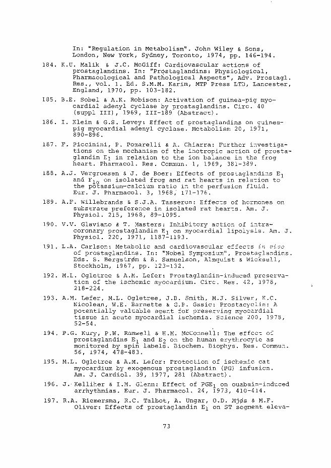
In: 11 Regulation in Metabolism". John Wiley & Sons, London, New York, Sydney, Toronto, 1974, pp. 146-194.
184. K.U. Malik & J .C. McGiff: Cardiovascular actions of prostaglandins. In: "Prqstaglandins: Physiological, Pharmacological and Pathological Aspects", Adv. Prostagl. Res., vel. 1. Ed. S.M.M. Karim, MTP Press LTD, Lancester, England, 1970, pp. 103-182.
185. B.E. Sobel & A.K. Robison: Activation of guinea-pig myocardial adenyl cyclase by prostaglandins. Circ. 40 (suppl III), 1969, III-189 (Abstract).
186. I. Klein & G.S. Levey: Effect of prostaglandins on guineapig myocardial adenyl cyclase. Metabolism 20, 1971, 890-896.
187. F. Piccinini, P. Pomarelli & A. Chiarra: Further investigations on the mechanism of the inotropic action of prostaglandin E1 in relation to the ion balance in the frog heart. Pharmacal. Res. Commun. 1, 1969, 381-389.
188. A.J. Vergroesen & J. de Boer: Effects of prostaglandins E1 and F 1a on isolated frog and rat hearts in relation to the potassium-calcium ratio in the perfusion fluid. Eur. J. Pharmacal. 3, 1968, 171-176.
189. A.F. Willebrands & S.J.A. Tasseron: Effects of hormones on substrate preference in isolated rat hearts. Am. J. Physiol. 215, 1968, 89-1095.
190. V. V. Glaviano & T. r>iasters: Inhibitory action of intracoronary prostaglandin E1 on myocardial lipolysis. Am. J. Physiol. 220, 1971, 1187-1193.
191. L.A. Carlson: Metabolic and cardiovascular effects in vivo of prostaglandins. In: "Nobel Symposium 11
, Prostaglandins. Eds. S. Bergstr¢m & B. Samuelson, Almquist & Wicksell, Stockholm, 1967, pp. 123-132.
192. M.L. Ogletree & A.M. Lefer: Prostaglandin-induced preservation of the ischemic myocardium. Circ. Res. 42, 1978, 218-224.
193. A.M. Lefer, M.L. Ogletree, J.B. Smith, M.J. Silver, K.C. Nicolean, ~V.E. Barnette & G.P. Gasic: Prostacyclin: A potentially valuable agent for preserving myocardial tissue in acute myocardial ischemia. Science 200, 1978, 52-54.
194. P.G. Kury, P.W. Ramwell & H.M. !'1cConnell: The effect of prostaglandins E 1 and E2 on the human erythrocyte as monitored by spin labels. Biochem. Biophys. Res. Commun. 56, 1974, 478-483.
195. M.L. Ogletree & A.M. Lefer: Protection of ischemic cat myocardium by exogenous prostaglandin (PG) infusion. Am. J. Cardiel. 39, 1977, 281 (Abstract) .
196. J. Kelliher & I.M. Glenn: Effect of PGE 1 on ouabain-induced arrhythmias. Eur. J. Pharmacal. 24, 1973, 410-414.
197. R.A. Riemersma, R.C. Talbot, A. Ungar, O.D. Mj¢s & M.F. Oliver: Effects of prostaglandin E1 on ST segment eleva-
73
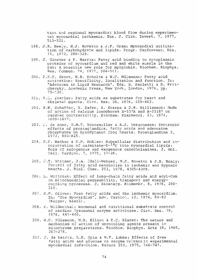
tion and regional myocardial blood flow during experimental myocardial ischaemia. Eur. J. Clin. Invest. 7 1 1977, 515-521.
198. J.R. Neely, H.J. Rovetto & J.F. Oram: Myocardial utilization of carbohydrate and lipids. Progr. Cardiovasc. Dis. 15, 1972, 289-329.
199. J. Gloster & P. Harris: Fatty acid binding to cytoplasmic prot.eins of myocardium and red and white muscle in the rat: A possible new role for myoglobin. Biochem. Biophys. Res. Commun. 74, 1977, 506-511.
200. P.H.E. Groot, H.R. Scholte & w.c. Hlilsmann: Fatty acid activation: Specificity, localization and function. In: "Advances in Lipid Research". Eds. R. Paoletti & D. Kritchevsky, Academic Press, New York, London, 1976 1 pp. 75-126.
201. K.L. Zierler: Fatty acids as substrates for heart and skeletal muscle. Circ. Res. 38, 1976, 459-463.
202. S.W. Schaffer, B. Safer, A. Scarpa & J.R. Williamson: l1ode of action of calcium ionophores X-537A and A-23187 on cardiac contractility. Biochem. Pharmacal. 23, 1974, 1609-1617.
203. J. de Boer, u.M.T. Houtsmuller & A.J. Vergroesen: Inotropic effects of prostaglandins, fatty acids and adenosine phosphates on hypodynamic frog hearts. Prostglandins 3, 1973, 803-825.
204. P.P. Mathur & C.M. Mokler: Subcellular distribution and incorporation of palmitate-u-14c into myocardial lipids: Role of endogenous and exogenous catecholamines. J. Mol. Cell. Cardiol. 7, 1975, 17-26.
205. J.T. Whitmer, J.A. Idell-Wenger, M.J. Rovetto & J.R. Neely: Control of fatty acid metabolism in ischemic and hypoxic hearts. J. Biol. Chern. 253, 1978, 4305-4309.
206. L. Nojtczak: Effect of long-chain fatty acids and acyl-CoA on rnitcchondrial permeability, transport and energycoupling processes. J. Bioenerg. Biomernbr. 8, 1976, 200-210.
207. M.F. Oliver: Free fatty acids and the ischemic myocardium. In: "The Myocardium", Adv. Cardiel. 12, 1974, 84-93 (Karger, Basel).
208. K. Wildenthal: Hormonal and nutritional substrate control of cardiac lysosomal enzyme activities. Circ. Res. 39 1
1976' 441-446.
209. W.C. Hlilsmann, \II.B. Elliot & E.C. Slater: The nature and mechanism of action of uncoupling agents present in mitochrome preparations. Biochim. Biophys. Acta 39, 1960, 267-276.
210. J. de Leiris, L.H. Opie & N.F. Lubbe: Effects of free fatty acids and glucose on enzyme release in experimental myocardial infarction. Nature 253, 1975, 746-747.
74
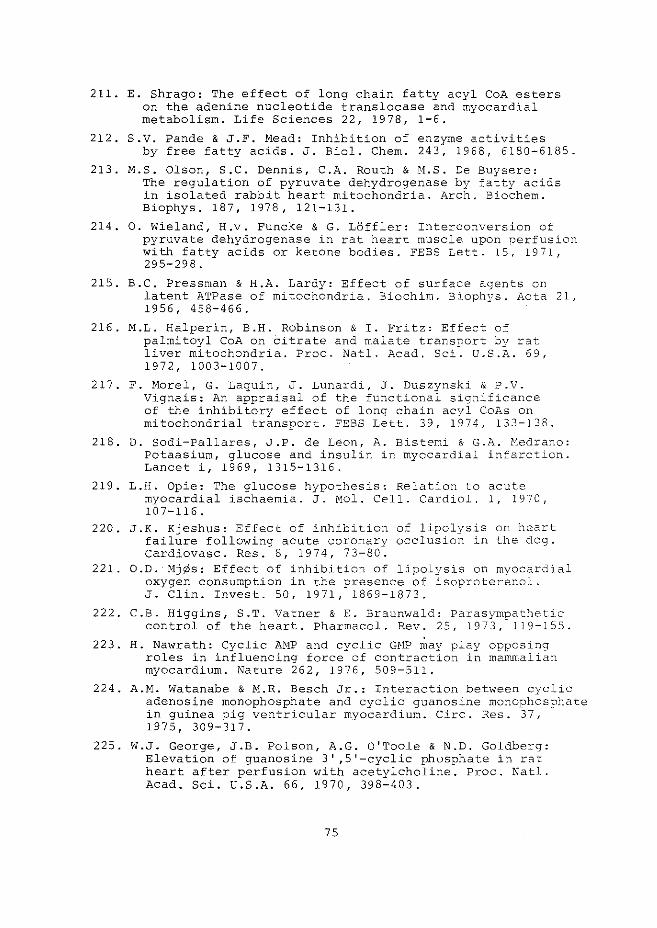
211. E. Shrago: The effect of long chain fatty acyl CoA esters on the adenine nucleotide translocase and myocardial metabolism. Life Sciences 22, 1978, 1-6.
212. S.V. Pande & J.F. Mead: Inhibition of enzyme activities by free fatty acids. J. Biol. Chern. 243, 1968, 6180-6185.
213. M.S. Olson, S.C. Dennis, C.A. Routh & M.S. De Buysere: The regulation of pyruvate dehydrogenase by fatty acids in isolated rabbit heart mitochondria. Arch. Biochem. Biophys. 187, 1978, 121-131.
214. 0. Wieland, H.v. Funcke & G. LOffler: Interconversion of pyruvate dehydrogenase in rat heart muscle. upon perfusion with fatty acids or ketone bodies. FEBS Lett. 15, 1971, 295-298.
215. B.C. Pressman & H.A. Lardy: Effect of surface agents on latent ATPase of mitochondria. Biochim. Biophys. Acta 21, 1956, 458-466.
216. M.L. Halperin, B.H. Robinson & I. Fritz: Effect of palrnitoyl CoA on citrate and malate transport by rat liver mitochondria. Proc. Natl. Acad. Sci. U.S.A. 69, 1972, 1003-1007.
217. F. Morel, G. Laquin, J. Lunardi, J. Duszynski & P.V. Vignais: An appraisal of the functional significance of the inhibitory effect of long chain acyl CoAs on mitochondrial transport. FEES Lett. 39, 1974, 133-138.
218. D. Sodi-Pallares, J.P. de Leon, A. Bistemi & G.A. Medrano: Potaasium, glucose and insulin in myocardial infarction. Lancet i, 1969, 1315-1316.
219. L.H. Opie: The glucose hypothesis: Relation to acute myocardial ischaemia. J. Mol. Cell. Cardiel. 1, 1970, 107-116.
220. J.K. Kjeshus: Effect of inhibition of lipolysis on heart failure following acute coronary occlusion in the dog. Cardiovasc. Res. 8, 1974, 73-80.
221. O.D. Mj¢s: Effect of inhibition of lipolysis on myocardial oxygen consumption in the presence of isoproterenol. J. Clin. Invest. 50, 1971, 1869-1873.
222. C.B. Higgins, S.T. Vatner & E. Braunwald: Parasympathetic control of the heart. Pharmacal. Rev. 25, 1973,-119-155.
223. H. Nawrath: Cyclic A:HP and cyclic GHP may play opposing roles in influencing force of contraction in mammalian myocardium. Nature 262, 1976, 509-511.
224. A.M. Watanabe & M.R. Besch Jr.: Interaction between cyclic adenosine monophosphate and cyclic guanosine monophosphate in guinea pig ventricular myocardium. Circ. Res. 37, 1975' 309-317.
225. W.J. George, J.B. Polson, A.G. O'Toole & N.D. Goldberg: Elevation of guanosine 3' ,5'-cyclic phosphate in rat heart after perfusion with acetylcholine. Proc. Natl. Acad. Sci. U.S.A. 66, 1970, 398-403.
75
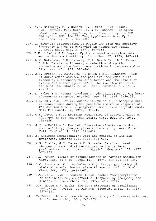
226. N.D. Goldberg, M.K. Haddox, S.E. Nicol, D.B. Glass, C.H. Sanford, F.A. Kuehl Jr. & E. Estenen: Biological regulation through opposing influences of cyclic GMP and cyclic AI~: The Yin Yang hypothesis. Adv. Cycl. Nucl. Res. 5, 1975, 307-330.
227. G. Brooker: Dissociation of cyclic GMP from the negative inotropic action of carbachol in guinea pig atria. J. Cycl. Nucl. Res. 3, 1977, 407-413.
228. B.E. Sobel & S.E. Mayer: Cyclic adenosine monophosphate and cardiac contractility. Circ. Res. 32, 1973, ~07-414.
229. A.M. Watanabe, D.R. Hataway, H.R. Besch Jr., B.B. Farmer & R.A. Harris: a-Adrenergic reduction of cyclic adenosine monophosphate concentrations in rat myocardium. Circ. Res. 40, 1977, 596-602.
230. O.E. Brodde, S. Motomura, M. Endoh & H.J. SchUmann: L"ack of correlation between the positive inotropic effect evoked by a-adrenoceptor stimulation and the levels of cyclic AMP and/or cycli Glv1P in the isolated ventricle strip of the rabbit. J. Hol. Cell. Cardiol. 10, 1978, 207-219.
231. E. Haber & s. Wrenn: Problems in identification of the betaadrenergic receptor. Physiol. Rev. 56, 1976, 317-338.
232. E.H. Hu & J.C. Venter: Adenosine cyclic 3',5'-monophosphate concentrations during the positive inotropic response of rat cardiac muscle to polymeric immobilized isoproterenol. Mol. Pharmacal. 14, 1978, 237-245.
233. G.S. Levey & S.E. Epstein: Activation of adenyl cyclase by g·lucagon in rat and human heart. Circ. Res. 24 1 1969, 151-156.
234. J.K. McNeill & D. Muschek: Histamine effects on cardiac contractility, phosphorylase and adenyl cyclase. J. Mol. Cell. Cardiel. 4, 1972, 611-624.
235. J. Axelrod: Noradrenalin: Fate and control of its biosynthesis. Science 173, 1971, 598-606.
236. N.S. Dhalla, J.C. Yates & V. Proveda: Calcium-linked changes in myocardial metabolism in the isolated perfused rat heart. Can. J. Physiol. Pharmacal. 55, 1977, 925-933.
237. S.E. Nayer: Effect of catecholamines on cardiac metabolism Circ. Res. 34 + 35 (Suppl III), 1974, III-129-III-135.
238. C.O. Brostrom, F.L. Hunkeler & E.G. Krebs: Regulation of skeletal muscle phosphorylase kinase by ca2+. J. Biol. Chern. 246, 1971, 1961-1967.
239. J .T. Stull, C.O. Brostrom & E.G. Krebs: Phosphorylation of the inhibitory component of troponin by phosphorylase kinase. J. Biol. Chern. 247, 1972, 5272-5274.
240. D.N. Hoare & H. Ruska: The fine structure of capillaries and small arteries. J. Biophys. Biochern. Cytol. 3, 1957, 457-462.
241. F. Parker: An electron microscopic study of coronary a~teries. Aln. J. Anat. 103, 1958, 247-273.
76
242. M. Boutet, I. Huttner & G. Rona: Permeability alteration of sarcolemmal membrane in catecholamine-induced cardiac muscle cell injury. Lab. Invest. 34, 1976, 482-488.
243. J. De Leiris, D. Feuvray & C. Corne: Acetylcholineinduced release of lactate dehydrogenase from isolated perfused rat heart. J. Nol. Cell. Cardiol. 4, 1972, 357-365.
244. K. Sakai & P.G. Spieckerrnann: Effects of reserpine and propranolol on anoxia-induced enzyme release from the isolated perfused guinea-pig heart. N.S. Arch. Pharrnacol. 291, 1975, 123-130.
245. W.G. Nayler, A. Grau & C. Yepez: S-Adrenoceptor antagonists and the release of creatine phosphokinase from hypoxic heart muscle. Cardiovasc. Res. 11, 1977, 344-352.
246. J.I. Haft & K. Fani: Stress and the induction of intravascular platelet aggregation in the heart. Circulation 48, 1973, 164-169.
247. G. Rona, M. Boutet, I. Huttner & H. Peters: Pathogenesis of isoproterenol induced myocardial alterations: Fundamental and morphological correlations. In: "Myocardial I''letabolisrn", Rec. Adv. Stud. Card. Struct. Metab., vol. 3. Ed. N.S. Dhalla, Universitv Park Press, Baltimore, London, Tokyo, 1973, pp. 507-525.
248. N.J. Balter, H.C. Cowden & S.L. Schwartz: Induction of membrane alterations by norepinephrine: Studies with macrophages and phospholipid monolayers. J. Pharmacal. Exp. Ther. 201, 1977, 627-635.
249. D.A. Vessey & D. Zahirn: Hernbrane fluidity and the regulation of membrane-bound enzymes. Horiz. Biochem. BioplY~:{S. 1, 1974, 138-174.
250. H.L. Verbeck, E.F. Halewski, L.S. Erhart & A.P. Harti:-1: Membrane phospholipid metabolism in the isoproterenol induced cardiomyopathy of the rat. In: "Pathophysiology and f·1orphology of r.1yocardial Cell Altera_tions", Rec. Adv. Stud. Card. Struct. Metab., vol. 6. Eds. A. Fleckenstein & G. Rona, University Park Press, Baltimore, London, Tokyo, 1975, pp. 175-181.
251. N.S. Dhalla, J.L. Yates, S.L. Lee & A. Singh: Functional and subcellular changes in the isolated rat heart perfused with oxidized isoproterenol. J. Mol.' Cardiel. 19, 1978, 31-41.
252. A.P. WaldenstrOm, A.C. Hjalmarson & L. Thornell: Possible role of noradrenaline in development of myocardial infarction -Experimental study in isolated rat heart. Am. Heart J. 95, 1978, 43-52.
253. S.R. Snidei, 0. Almgren & A. Carlsson: The occurrence and functional sig-nificance of dopamine in some peripheral adrenergic nerves of the rat. N.S. Pharmacal. 278, 1973, 1-12.
254. S. Gudbjarnason & J. Hallgrimson: The role of myocardial lipids in the development of cardiac necrosis. Acta Ned. Scand. 587, 1976, 17-25.
77
255. A.M.r-1. Abdellatif & R.O. Vles: Pathological effects of dietary rapeseed oil in rats. Nutr. Metab. 12, 1Q70, 285-295.
256. S.A. Gumpen & K.R. Narum: The relative amounts of longchain acylcarnitines, short-chain acylcarnitines and carnitine in heart, liver and brown adipose tissue from rats fed on rapeseed oil. Biochirn. Biophys. Acta 316, 1973, 48-55.
257. J. Jaillard, G. Sezille, P. Dewailly, J.C. Fruchhart & r1. Bertrand: Etude experimentale chez l'hornme et chez l' animal du metabolisme de 1' acide erucique: extraction myocardique chez l'homme, apres surcharge en huile de colza et lipides tissulaires chez le rat, apres surcharge en tri8rucine. Nutr. Hetabol. 15, 1973, 336-347.
258. B.O. Christophersen & J. Bremer: Inhibitory effect of erucylcarnitine on the oxidation of palmitate by rat heart mitochondria. FEES Lett. 23, 1972, 230-232.
259. f'LA. Swarttouw: The oxidation of erucic acid by rat heart mitochondria. Biochirn. Biophys. Acta 337, 1974, 13-21.
260. H.W. Hulan, J.K.G. Kramer, S. D-1ahadevan & F.D. Sauer: Relationshin between erucic acid and myocardial changes in male rats. Lipids 11, 1976, 9-15.
261. P. Dewailly, G. Sezille, A. Nouvelot, J.C. Fruchart & J. Jaillard: Changes in rat heart phospholipid composition after ra~eseed-oil feeding. Lipids 12, 1977, 301-306.
262. P. Dewailly, A. Nouvelot, G. Sezille, J.C. Fruchart & J. Jaillard: Changes in fatty acid composition of cardiac mitochondrial phospholipids in rats fed rapeseedoil. Lipids 13, 1978, 225-312.
263. U.M.T. Houtsmuller, C.B. Struyck & A. van der Beek: Decrease in rate of ATP synthesis of isolated rat heart mitochondria induced by dietary erucic acid. Biochim. Biophys. Acta 218, 1970, 564-566.
264. C.M.L. Hsu & F.A. Kummerov: Influence of elaidate and erucate on heart mitochondria. Lipids 12, 1977, 486-494.
265. Th. Cresteil, P. Keteri & D. Lapons: Modulation de l'activite adenyl cyclase du coeur de rat par ingestion d'huile de colza. Cornp. Rend. Acad. Sc. Paris 275, 1972, 1443-1445.
266. W.C. HUlsrnann: Over het mechanisme van adernhalingsketen phosphorylering, 1958, Poortpers, Amsterdam.
267. F. ten Hoar, H .r-1. de Graaf & A.J. Vergroesen: Effects of dietary erucic acid and linoleic acid on myocardial function in rats. In: 11 Myocardial ~letabolism", Rec. Adv. Stud. Card. Struct. Metab., vol. 3. Ed. N.S. Dhalla, University Park Press, Baltimore, London, Tokyo, 1973, pp. 59-72.
268. K.K. Caroll: Erucic acic as the factor in rape oil affecting adrenal cholesterol in the rat. J. Biol. Chern. 200, 1953, 287-292.
269. J.L. Beare-Rogers, E.A. Near & H.A. Heggtveit: Cardiac
78
lipid changes in rats fed oils containing long-chain fatty acids. Can. Inst. Food Technol. 4, 1971, 120-132.
270. W.C. Hlilsrnann: Abnormal stress reactions after feeding diets rich in (very) long-chain fatty acids; high levels of corticosterone and testosterone. Mol. Cell. Endocrinol., in the press.
271. F. ten Hoar & H.M. de Graaf: The influence of a diet deficient in linoleic acid on the force of contraction of the isolated papillary muscle in rats. Circ. 43 + 44 (Supp. II), 1971, II-113 (Abstract).
272. T. Ito & R.M. Johnson: Effects of nutritional deficiency of unsaturated fats on rat liver mitochondria. I. Respiratory control and ATP-Pi exchange activity. J. Biol. Chern. 239, 1964, 3201-3208.
273. K. Christiansen & P.K. Jensen: Membrane-bound lipid particles from beef heart. Chemical composition and structure. Biochim. Biophys. Acta 260, 1972, 449-459.
274. Ch. Steenbergen, G. de Leeuw, C. Barlow, B. Chance & J.R. Williamson: Heterogeneity of the hypoxic state in perfused rat heart. Circ. Res. 41, 1977, 606-615.
275. A. Fabia to & F. Fabia to: Relaxing and inotropic effect of cyclic AMP of skinned cardiac cells. Nature 253, 1975, 556-558.
79
SAMENVATTING
Dit proefschrift geeft een literatuuroverzicht, en
beschrijft experimentele studies met betrekking tot de regulatie
en modificatie van de coronaire doorstroming en contractiliteit
van geisoleerde ratteharten.
Hoofdstuk I geeft een inleiding omtrent het probleern van de vet
zuur-toxiciteit en hartfunktie. De doorstrorningssnelheid van de
coronair vaten en de pornp-funktie van het hart worden hoofdzake
lijk bepaald door de contractie-toestand van, respectievelijk,
de gladde spiercellen in de vaatwand en de dwarsgestreepte cellen
van de hartspier. Hoofdstukken II en III beschrijven dan oak de
morfologische en (ultra)structurele aspecten van beide celtypen.
In hoofdstukken IV en v worden funktionele en stofwisselings
aspecten van de coronaire circulatie en de contractiliteit toe
gelicht. In zowel de gladde spiercellen van de vaatwand als de
dwarsgestreepte spiercellen van het hart:
(i) wordt de contractie-toestand hoofdzakelijk bepaald
door de intracellulaire calcium-concentratie,
(ii) vindt de contractie plaats na calcium-influx door
het plasmamembraan gedurende de actie-potentiaal,
en door het vrijkomen van intracellulair opge
slagen calcium (in het sarcoplasmatisch reticulum
en mitochondrien),
(iii) treedt relaxatie op nadat specifieke calcium-pomp
systemen in het plasmamembraan, het sarcoplasma
tisch reticulum en de mitochondrien de cytoplasma
tische calcium-concentratie hebben verlaagd en,
(iv) vormen calcium-ionen de schakel tussen de contrac
tie-relaxatie cyclus en de energie-stofwisseling
aangezien zowel glycogenolyse (glycogeen-afbraak)
en lipolyse (vet-afbraak) gestirnuleerd worden door
calcium
De contractie en relaxatie van de dwarsgestreepte hart
spiercellen zijn energie-afhankelijke processen. Een verslechte
ring van de contractie-kracht treedt dan ook op wanneer de pro
ductie van ATP is gerernd (anoxier hypoxie en ischernie) .
80
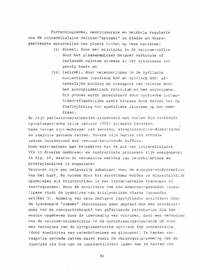
Farmacblogische, neurohumorale en metabole regulatie
van de intracellulaire calcium-"spiegel 11 in gladde en dwars
gestreepte spiercellen kan plaats vinden op twee manieren:
(i) direkt, door een wijziging in de calcium-influx
door het plasrnamernbraan hetgeen verhoogde of
verlaagde calcium niveaus in het cytoplasma tot
gevolg heeft en
(ii) indirekt, door veranderingen in de cyclische
nucleotiden (cyclisch AHP en cyclisch G!vlP) af
hankelijke binding en transport van calcium door
het sarcoplasrnatisch reticulum en het sarcolemma.
Dit proces wordt gereguleerd door cyclische nucleo
tiden-afhankelijke eiwit kinasen door middel van de
fosforylering van specifieke plaatsen op het mem
braan.
Er zijn perfusie-experimenten uitgevoerd met harten die verhoogde
cytoplasmatische vrije vetzuur (VVZ) spiegels bevatten.
Deze harten zijn verkregen uit gevaste, streptozotocine-diabetische
en raapolie gevoede ratten. Tevens zijn harten uit normale
ratten doorstroornd met vetzuur-bevattende buffers.
Onze waarnemingen met betrekking tot de rol van intracellulaire
VVZ in diverse mernbraan- en contractiele processen zijn weergegeven
in fig. 10, waarin de secundaire werking van catecholamines en
prostaglandines is weggelaten
Vetzuren zijn een belangrijk substraat voor de energie-stofwisseling
van het hart. Na opname door het sarcolemma worden ze waarschijnlijk
opgeslagen als triglyceriden in met lipide-gevulde lysosornen of
autofagosornen. Door de activiteit van een rnembraan-gebonden (zure)
lipase vindt de hydrolyse van triglyceriden plaats (appendix
artikel 3). Rernrning van deze endogene lipoly'tische activiteit door
de lysosomale "remmer 11 chloroquine gaat gepaard met een achteruit
gang van de contractiekracht van gelsoleerde ratteharten die kan
worden opgeheven door de toevoeging van vetzuren, door een verhoging
van de calcium-concentratie in de doorstromingsvloeistof of door
een verhoging van de cytoplasmatische cyclisch A}1P concentratie
(door toediening van catecholamines en glucagon) . In harten van
raapolie gevoede ratten waren zowel de chloroquine-remming van de
lipolyse als die van de contractiliteit lager dan in harten van
81
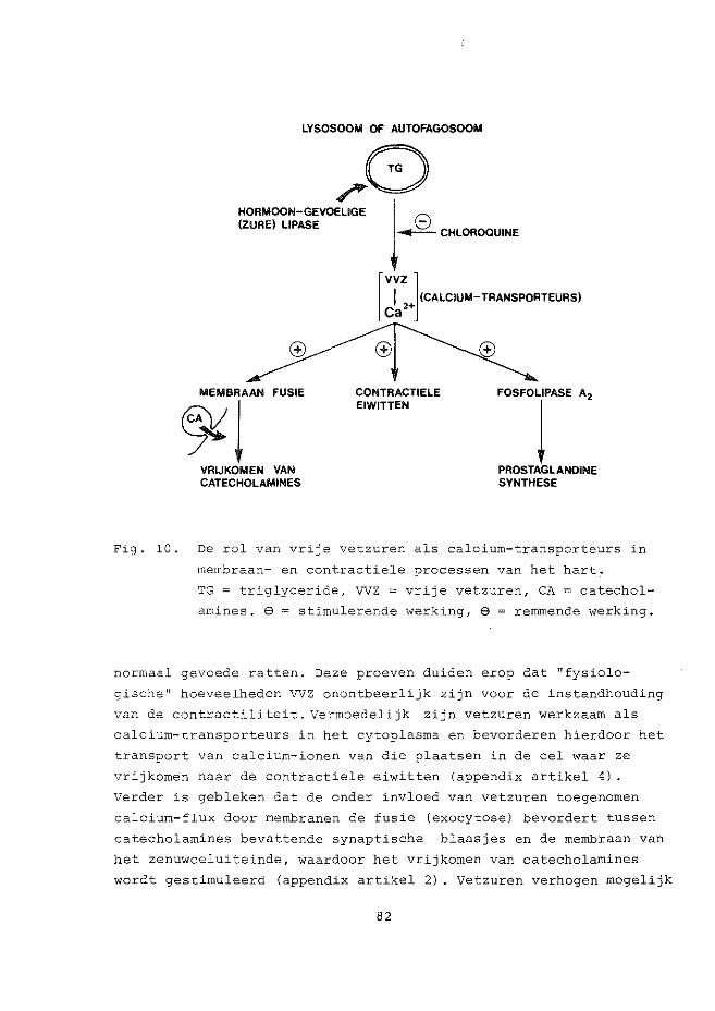
LYSOSOOM OF AUTOFAGOSOOM
/0 (ZURE) LIPASE __:;:__ CHLOROQUINE HORMOON-GEVOELIGE j 8
MEMBRAAN FUSIE
~~ VRIJKOMEN VAN CATECHOLAMINE$
rvvz ] C~
2+ (CALCIUM-TRANSPORTEURS)
CONTRACTIELE EIWITTEN
FOSFOLIPASE A2
j PROSTAGLANOINE SYNTHESE
Fig. 10. De rol van vrije vetzuren als calcium-transporteurs in
membraan- en contractiele processen van het hart.
TG = triglyceride, VVZ = vrije vetzuren, CA = catechol
amines. $ = stimulerende werking, e = rernmende werking.
normaal gevoede ratten. Deze proeven duiden erop dat "fysiolo
gische" hoeveelheden VVZ onontbeerlijk zijn voor de instandhouding
van de contractiliteit. Vermoedelijk zijn vetzuren werkzaarn als
calcium-transporteurs in het cytoplasma en bevorderen hierdoor het
transport van calcium-ionen van die plaatsen in de eel waar ze
vrijkornen naar de contractiele eiwitten (appendix artikel 4) .
Verder is gebleken dat de onder invloed van vetzuren toegenomen
calcium-flux door membranen de fusie (exocytose) bevordert tussen
catecholamines bevattende synaptische blaasjes en de membraan van
het zenuwceluiteinde, waardoor het vrijkomen van catecholarnines
wordt gestirnuleerd (appendix artikel 2) . Vetzuren verhogen mogelijk
82
ook de activiteit van het calcium-afhankelijke fosfolipase A2 hetgeen aanleiding is tot de vorming van prostaglandines uit
membraan-fosfolipiden.
Een verhoogde catecholamine-"aktiviteit" en het vrij-komen van
meer prostaglandines, gepaard gaande met een toenarne in de
coronaire doorstrorning, werd waa,rgenomen in harten van gevaste
en streptozotocine-diabetische ratten (appendix artikel 1).
Endogene catecholarnines die vrijkornen tijdens de 9erfusie bleken
betrokken te zijn bij de instandhouding van de coronaire door
stroming onder normoxische ornstandigheden (a?pendix artikelen l
en 2). Deze vaat-verwijdende werking van (exogene en endogene)
catecholarnines, maar ook die van prostaglandine-achtige ver
bindingen wordt waarschijnlijk veroorzaakt door een toename van
het cyclisch AMP niveau in het cytoplasma van de gladde spier
cellen van de coronair vaten. In tegenstelling echter tot deze
gladde spiercellen, die relaxeren bij verhoogde cytoplasmatisch
cyclisch AMP spiegels 1 neemt de contractiliteit van de dwars
gestreepte hartspiercellen toe na de toediening van norepine
phrine, glucagon en prostaglandine E1 (appendix artikel 4).
Met hun calcium-ionofore eigenschappen zijn vetzuren en sommige
prostaglandines betrokken in de coronaire vaatverwijding omdat
ze de beschikbare hoeveelheid calcium in de gladde spiercellen
verlagen. De toegenornen coronaire doorstromingssnelheid in harten
van raapolie gevoede ratten staat daarom mogelijk in verband met
de waargenornen verhoging van de basale (en horrnoon-gevoelige)
lipolytische activiteit. De te verwachten toename in vetzuur
productie en ui tscheiding door de vervette dvmrsgestreepte
hartspiercellen is vermoedelijk verantwoordelijk voor de relaxatie
van de gladde vaatmusculatuur door dezelfde calcium-verwijderende
werking.
Dit proefschrift beschrijft experimenten die zijn uit
gevoerd om de effecten van vetzuur-perfusies onder normoxische
en ischemische condities te bestuderen. De verlaging van de coro
naire doorstroming door embolisatie van de coronair arterien (met
behulp van zeer kleine polysaccharide Sephadex partikels) werd
gebruikt en gekarakteriseerd als een experimenteel model voor
myocardiale ischernie (appendix artikel 5) . De afnarne in creatine
fosfaat en ATP, en intracellulaire acidose, die optreden tijdens
83
perioden van anoxie, hypoxie en ischemie z~Jn verantwoordelijk
voor de verlaagde activiteit van de energie-afhankelijke calcium-2+ pomp systernen (Ca -ATPases) van het sarcolemma, het sarcoplasma-
tisch reticulum en van de mitochondrien, hetgeen rnogelijk een
verstard ("stenen") hart tot gevolg heeft. De productie en uit
scheiding van ATP-catabolieten (adenosine, inosine en hypoxan
thine) die plaats vindt gedurende zuurstof (en/of substraat)-arrne
ornstandigheden kan mogelijk gebruikt worden als een aanwijzing
voor hart- en vaatziekten bij de mens.
De doorstroming van ratteharten met een vetzuur
bevattende buffer (in een relatief hoge rnolaire vetzuur:albumine
ratio, hetgeen met name bepalend is voor de vetzuur opnarne door
het hart) leidt tot een verlaging van de contractiliteit. De
schadelijke effecten van verhoogde cytoplasrnatische spiegels van
vetzuren en hun CoA en carnitine esters, zowel onder norrnoxische
als ischemische condities, zijn beschreven op basis van hun
chao trope rol als rnembraandetergentia ( zie fig. l 0 en appendix
artikelen 2 en 6). Ook catecholamines, onder invloed van vetzuren
vrijgekomen uit de zenuwuiteinden spelen rnogelijk een rol in de
achteruitgang van de hartfunctie. In dit kader zijn observaties
orntrent de nadelige werking van catecholamines op cellulaire mem
branen en stofwisseling besproken. Gevonden werd dat de afnarne in
contractiliteit onder invloed van vetzuren inderdaad kon worden
"voorkomen" wanneer de endogene catecholarnines werden gedepleteerd.
Ook de toevoeging van glucose, dat door te voorzien in glycerol-3-
fosfaat, de triglyceride synthese stirnuleert en de intracellulaire
VVZ (en CoA en carnitine ester) spiegels verlaagt, voorkwam
(gedeeltelijk)
(i) het vrijkornen van catecholamines,
(ii) de achteruitgang van de rnyocardiale energie
huishouding en
(iii) de afnarne in contractiliteit
die optraden gedurende vetzuur-perfusie onder zuurstof-rijke en
zuurstof-arrne condities.
Onze resultaten duiden op een belangrijke rol van vetzuren
in de functie van het hart onder norrnale en pathologische omstandig
heden, een rol waarin hun ionofore eigenschappen van wezenlijk
belang zijn.
84
CURRICULUM VITAE
7-12-1948 Geboren te Abbekerk (Noord-Ho11and) .
1954 - 1961 Dominicus College, Oranjestad, Aruba (Ned. Antillen). St. Anthonius lagere school, Amstelveen.
1961 - 1966 St. Nicolaas Lyceum, Amsterdam. Eindexamen HBS B.
1966 - 1975 Universiteit van Amsterdam. Kandidaats-exarnen biologie (B4) Doctoraalexarnen met hoofdvak biochernie, bijvak (uitgebreid) neuro-anatomie/biochemie en nevenrichting plantenfysiologie.
1969 - 1975 Part-time leraar biologie en scheikunde aan de Jan Philip Roothaan ~~VO en het Keizer Karel College, beide te Amstelveen,
1973 - 1975 Kandidaats-assistent op de afdeling Neurobiochemie van het Centraal Nederlands Instituut voor Hersenonderzoek.
1975 - 1978 Erasmus Universiteit Rotterdam. Afdelingen Biochemie I en Cardiovasculaire Research (Thoraxcentrum), wetenschappelijk medewerker.
85


APPENDIX


Paper l. Effect of fasting and streptozotocin-diabetes on
the coronary flow in isolated rat hearts: A
possible role of endogenous catecholamines and
prostaglandins
Erratum
H. Starn & W.C. Hillsmann
Basic Res. Cardiol. 72, 1977, 365-375
p. 374 ref. 24. .... guinea-pig isolated heart
preparation.

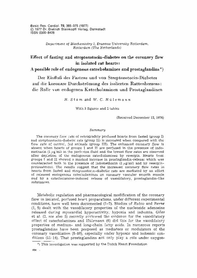
Basic Res. Cardiel. 72, 365-375 {1977) © 1977 Dr. Dietrich Steinkopff Verlag, Darmstadt ISSN 0300-8428
Department of Biochemistry I, Erasmus University Rotterdam, Rotterdam (The Netherlands)
Effect of fasting and streptozotocin -diabetes on the coronary flow in isolated rat hearts:
A possible role of endogenous catecholamines and prostaglandins*)
Der EinfluB des Fastens unci von Streptozotocin-Diabetes
auf die koronare Durchstriimung des isolierten Rattenherzens:
die Rolle von endogenen Katecholaminen unci Prostaglandinen
H. Stamand W. C. HU~smann
With 3 figures and 2 tables
(Received December 12, 1976)
Summary
The coronary flow rate of retrogradely perfused hearts from fasted (group I) and streptozotocin-diabetic rats (group II) is increased when compared with the flow rate of control, fed animals (group III). The enhanced coronary flow is absent when hearts of groups I and II are perfused in the presence of indomethacin (1 ,llg,...ml) in the perfusion. fluid and the lowest flow rates are observed after depletion of the endogenous catecholamines by reserpin. Hearts from groups I and II showed a marked increase in prostaglandin-release which was counteracted both in the presence of indomethacin (1 f1g/ml) and by reserpinpretreatment. The results suggest that the increased coronary flow rates in hearts from fasted and streptozotocin-diabetic rats are mediated by an effect of released endogenous catecholamines on coronary vascular smooth muscle and by a catecholamine-induced release of vasodilatory, prostaglandin-like substances.
Metabolic regulation and pharmacological modification of the coronary flow in isolated, perfused heart preparations, under different experimental conditions, have well been documented (1~7). Studies of Rubio and Berne (1, 5) dealt with the vasodilatory properties of the nucleoside adenosine released during myocardial hyperactivity, hypoxia and ischemia. Giles et al. (2, see also 3) recently reviewed the evidence for the vasodilatory effect of catecholamines and Hiilsmann {6) did this for the vasodilatory properties of medium- and long-chain fatty acids. In numerous reports prostaglandins have been proposed as mediators or modulators of the coronary vasodilation (8~10), especially under hypoxic and ischemic conditions (11~14). That prostaglandins not only play a role under oxygen-
"'} This investigation was supported by the Dutch Heart Foundation
694
366 Basic Research in Cardiology, Vol. 72, No.4 (1977)
limited conditions is pointed out by Schri5r et al. (15-17), who presented evidence that in guinea-pig hearts endogenous prostaglandin-like substances are possibly involved for the maintenance of the coronary vascular resistance since indomethacin, a potent inhibitor of prostaglandin synthesis (18), significantly increased the coronary vascular pressure under optimal perfusion conditions.
Recently Markelonis and Garbus (19) reviewed some mechanisms serving as stimuli evoking prostaglandin biosynthesis and release. Mediators of stress (catecholamines) were proposed to stimulate prostaglandin synthesis since infusion of norepinephrine in the isolated, perfused rabbit heart caused an efflux of prostaglandin E=' (20). Similar observations were done for the dog spleen (21) and rabbit kidney (22), while Broadley (23) mentioned the release of a coronary vasodilator metabolite from the guinea-pig isolated heart stimulated by catecholamines, histamine and electrical pacing, possibly a prostaglandin-like substance.
The important role of endogenous catecholamines as a determinating factor of the coronary flow follows from an observation of Krebs and SchrOr (24) that depletion of endogenous myocardial catecholamine stores after reserpin-pretreatment leads to a significant decrease in coronary vascular resistance in isolated, perfused, paced guinea-pig hearts. The significance of catecholamines in spontaneously beating, perfused rat hearts was pointed out by Schaffer et a!. (25) whose data suggested a clear link between the vasodilatory effect of the calcium ionophore X-537 A and the release of norepinephrine from endogenous stores, since reserpin pretreatment und the ;J-adrenergic blocking agent propranolol abolished the effect of X-537 A.
The study presented here deals with the observation that the coronary flow in isolated, perfused hearts from fasted and streptozotocin-diabetic rats is markedly higher than the flow in control, fed animals. The possible roles of endogenous catecholamines and prostaglandins as responsible factors for this enhanced flow rate have been studied.
Methods and materials
Animals
Three groups of male Wistar rats (200-250 g) were used. The first group (I) of rats was fasted for 48 hours but had free access to water. The second group (II) of rats was made diabetic by an intravenous (tailvein) injection of streptozotocin (50 mg/kg body weight) (26), and the third group (III) consisted of normally fed animals (controls). The rats were anesthetized with an intraperitoneal injection of sodium pentobarbital (70 mg/kg body weight) and subsequently heparinized intravenously with 500 I.U. Depletion of endogenous catecholamines was induced by an intraperitoneal injection of reserpin (10 mg/kg body weight) 6 hours before sacrifice (25).
Perfusion procedure
The hearts were perfused at 37 °C and pH 7.4 with a modified tyrode solution (27) equilibrated with 95 Ofo 0 2 + 5 °/o C02 , according to the Langendorff procedure. All non-ventricular tissue was removed. Isolated hearts were paced at 300 pulses/minute after a total atrio-ventricular block was made by cutting the bundle of His. After a thirty minute control perfusion at a pressure
Stam and Hillsmann, Effect of fasting and streptozotocin-diabetes 367
of 100 em H~O the perfusion pressure was varied from 40 to 80 mm Hg as measured in the coronary inflow tract, using a Statham P 23 Gb pressure transdu.cer. The coronary flow rate was measured by collecting the fluid dripping from the heart during a fixed period of time of by drop counting. After the experiment the hearts were blotted dry and weighed.
Indomethacin was neutralized with equimolar amounts of sodium hydroxide and added to the perfusion medium in a final concentration of 1 _ug/ml.
Extraction of prostaglandins from the effluents
After the control perfusion period, the coronary effluents were collected for thirty minutes under N~ gas at 0 °C, acidified with HCl to pH 3.0 and extracted twice with ethyl acetate. The combined extracts were evaporated to dryness in vacuo at 50-60 °C using a rotary evaporator (Rotavap, Btichi, Germany). The dried extracts were dissolved in saline (500 _ul) and stored at -20 °C. Bioassay of effluent prostaglandins took place within 48 hours after the extraction.
Bioassay of the extracted prostaglandins
Prostaglandins recovered from the extraction procedure were detected and assayed on a rat stomach strip and a rat colon, superfused at 37 °C at pH 7.4 with a Krebs buffer solution (21). The changes in length of the assay tissues were detected by a Harvard isotonic heart/smooth muscle transducer and recorded on a Rikadenki multi-pen recorder. The contractions of the tissues after addition of 100 ,ul of the effluent extract were compared with contractions occurring after the addition of the same volume of a known prostaglandin E~ solution to the superfusion fluid.
Materials
Prostaglandin E~ was a gift of Dr. J. E. Pike from the Upjohn Company (Kalamazoo, U.S.A.). Heparin was purchased from Organon (Oss, The Netherlands), reagents (all of analytical grade) and reserpin from Merck (Darmstadt, Germany) and streptozotocin from Calbiochem (Luzern, Switzerland).
Statistical analysis
Most results are given in mean values ± standard error of the mean. n is the number of observations. Significance was calculated with Student's t-test. P > 0.05 was considered to be not significant.
Results
During the perfusion of hearts, isolated from diabetic rats for other purposes, it was noticed that the coronary flow rate had increased compared with the flow rate in control hearts. Since at least a number of metabolic alterations that occur in diabetes may also be observed in the fasted non-diseased state, also hearts from fasted rats were tested.
The relation between the coronary flow and perfusion pressure in perfused Langendorff hearts from streptozotocin-diabetic, fasted and control hearts is presented in figure 1. The flow rate in hearts from diabetic and fasted rats was significantly higher than the coronary flow in control hearts. From earlier studies it appeared that endogenous catecholamines (25) and prostaglandin-like substances (15) might be involved in the maintenance of coronary flow in isolated perfused hearts. Therefore we studied the effects of indomethacin (a potent inhibitor of prostaglandin
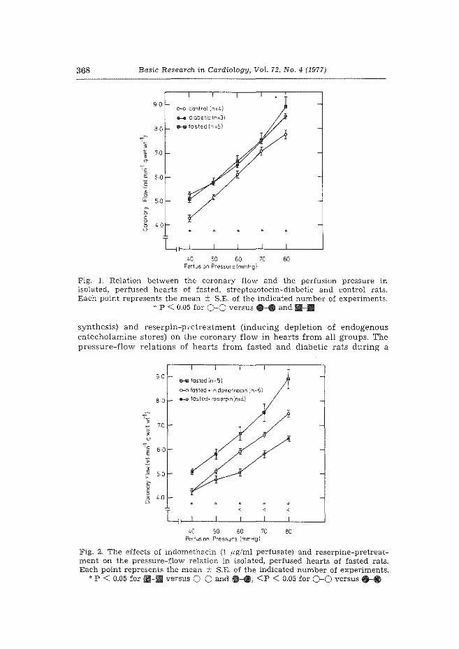
368 Basic Research in Cardiology, Vat. 72, No.4 (1977)
9 0 o-o control In~ I.)
e-e diabetic ln=3l
8 0 11--111 tasted {n=Sl
'c E 6-0 E
~ LL 5-0
40 50 60 PerfusiOn Pressure{mmHgl
70 80
Fig. 1. Relation between the coronary flow and the perfusion pressure in isolated, perfused hearts of fasted, streptozotocin-diabetic and control rats. Each point represents the mean ± S.E. of the indicated number of experiments.
* P < 0.05 for 0-0 versus 8-· and !11-.11
synthesis) and reserpin-pretreatment (inducing depletion of endogenous catecholamine stores) on the coronary flow in hearts from all groups. The pressure-flow relations of hearts from fasted and diabetic rats during a
9 0 m-m fasted ln=Sl
o-o fasted • 1n doml?tho.cm { n=61
8 .Q 0-4 las!E.>d• mserp1nln=L.l
,_ ' 0 70
' ~ 'c 50 E :!'_
~ 50 ~
c 0 c
~ 40 u •
L 40 50 60 70 80
Perfus1on Pr12ssure lmmHg)
Fig. 2. The effects of indomethacin (1 ,ug/ml perfusate) and reserpine-pretreatment on the pressure-flow relation in isolated, perfused hearts of fasted rats. Each point represents the mean ± S.E. of the indicated number of experiments.
' P < 0.05 for !11-1111!1 versus 0-0 and e-e, <P < 0.05 for 0-0 versus ....
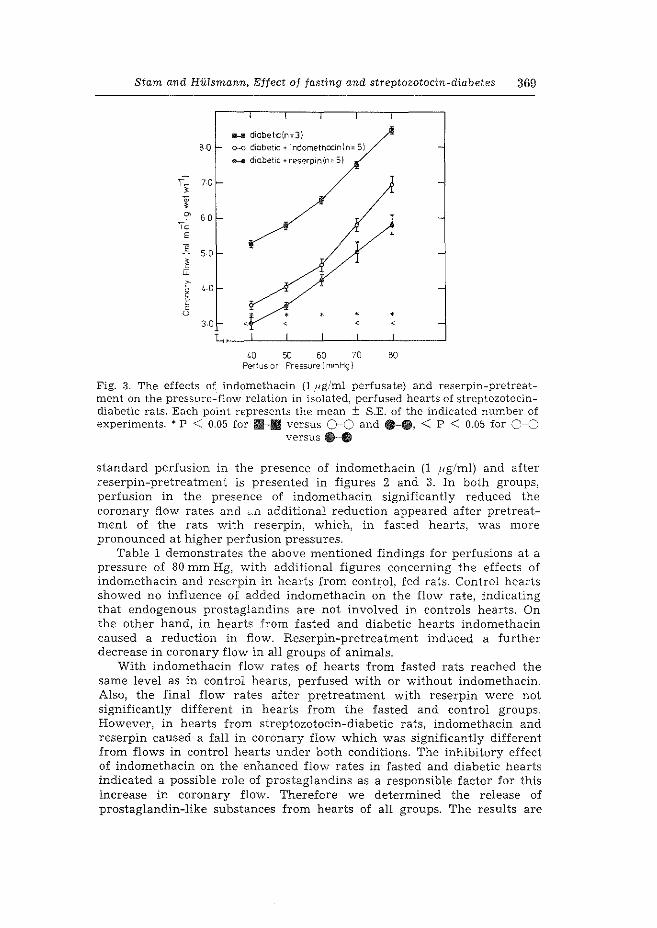
Stam and Hillsmann, Effect of fasting and streptozotocin-diabetes 369
D-Ill diabellc{n=3)
B 0 o-o diabetic+ indomethacin In= 5)
.._. d1obet1c + reserp1n(n= 5)
~ 60 'Cc
E
E 50
~ ~
'0
3 0
L~J_--~--~--L---L---~
50 60 70 BO Perfus1on Pressure I mmHg l
Fig. 3. The effects of indomethacin (1 pg/ml perfusate) and reserpin-pretreatment on the pressure-flow relation in isolated, perfused hearts of streptozotocindiabetic rats. Each point represents the mean ± S.E. of the indicated number of experiments. * p < 0.05 for 111-11 versus 0-0 and --·· < p < 0.05 for c-o
versus e-e
standard perfusion in the presence of indomethacin (1 pg/ml) and after reserpin-pretreatment is presented in figures 2 and 3. In both groups, perfusion in the presence of indomethacin significantly reduced the coronary flow rates and <:.-n additional reduction appeared after pretreatment of the rats with reserpin, which, in fasted hearts, was more pronounced at higher perfusion pressures.
Table 1 demonstrates the above mentioned findings for perfusions at a pressure of 80 mm Hg, with additional figures concerning the effects of indomethacin and reserpin in hearts from control, fed rats. Control hearts showed no influence of added indomethacin on the flow rate, indicating that endogenous prostaglandins are not involved in controls hearts. On the other hand, in hearts from fasted and diabetic hearts indomethacin caused a reduction in flow. Reserpin-pretreatment induced a further decrease in coronary flow in all groups of animals.
With indomethacin flow rates of hearts from fasted rats reached the same level as in control hearts, perfused with or without indomethacin. Also, the final flow rates after pretreatment with reserpin were not significantly different in hearts from the fasted and control groups. However, in hearts from streptozotocin-diabetic rats, indomethacin and reserpin caused a fall in coronary flow which was significantly different from flows in control hearts under both conditions. The inhibitory effect of indomethacin on the enhanced flow rates in fasted and diabetic hearts indicated a possible role of prostaglandins as a responsible factor for this increase in coronary flow. Therefore we determined the release of prostaglandin-like substances from hearts of all groups. The results are
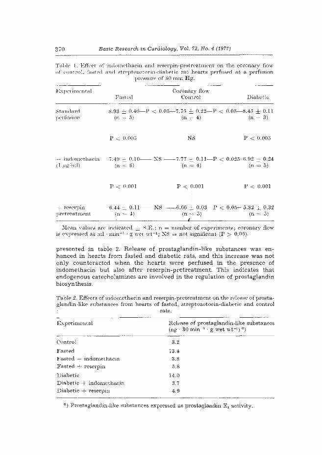
370 Basic Research in Cardiology, Vol. 72, No.4 (1977)
Tnlllt' l. Effect nf iwhmwthcwin and rC'serpin-prEOtreatnwnt on the coronary flow ()f controL fac:;tt•d nnd ~tt'eptnzntocin-dia.botic rat hearts perfused at a. perfusion
pressurE' of 80 mm Hg.
E:s:Jlt'l'itm•nt<:\1
:-\t<HHbnl pel'ftJ:-;ini1
-- indnnwtlwein ( l ,ng 'ml)
- rec:.erpin prt'trC'atnwnt
Coronar;-,r flo-w Control Diabetic
8.0:1 =:::_ 0.40-P < O.Oi'i-7.7:3.::: 0.:22--P < 0.05-8.45 ± 0.11 (n ~ ''I (n ~ 41 (n ~ 31
p -...:.: 0.00;) NS
7.40 = 0.10-- i\S ---7.77 = 0.11-P (n "'-" (i) (n = 4)
p < 0.001 p < 0.001
p < 0.00;'5
< 0.02;)-(i.0:2 ± 0.24 (n ~,;I
p < 0.001
ii.44 = 0.11-- KS ---6.66 :i: 0.03-P < 0.0;'5-5.8:2 ::::: 0.32 (n = 4) (n = 5) (n = 5)
::\h•an \·ahws arc indica.t('(l :::i:: ~-E.: n = nmnber of expC'rimcnts; coronary flow is expressed as ml · min- 1 · g \\'d \Yt-1 ; KS = not significant (P > 0.05).
presented in table 2. Release of prostaglandin-like substances was enhanced in hearts from fasted and diabetic rats, and this increase was not only counteracted when the hearts were perfused in the presence of indomethacin but also after reserpin-pretreatment. This indicates that endogenous catecholamines are involved in the regulation of prostaglandin biosynthesis.
Table :2. Effects of indomethacin and reserpin-pret-rea.tment- on thf' n'ka~e of prostaglandin-like substances from hearts of fasted, strept-ozotocin-dialwtic and control
rats.
Experirnental
Control
Fasted
Fasted - indomethacin
Fasted -;- reserpin
Diabetic
Diabetic + indomethacin
Diabetic + reserpin
Release of prostaglandin-like substances (ng · 30 min-1 · g \vet wt-J.) *)
3.2
13.4
3.8 5.8
14.0
3.7
4.9
*)Prostaglandin-like substances expressed as prostaglandin E 2 activity.
Stam and Hillsmann, Effect of fasting and streptozotocin-diabetes 3i1
Discussion
In recent years investigations on the mode of action of prostaglandins and catecholamines on the coronary vascular system have been subjects of many studies [see for references Dempsey and Cooper (3) and Nakano (28)]. Most of the studies presented, however, dealt with interactions occurring during hypoxia, ischemia or other pathological conditions. About the role of prostaglandins and that of circulating and endogenous catecholamines in the regulation of coronary flow, under normoxic conditions, much uncertainty exists.
In the isolated, perfused rabbit heart Block et al. (11) did not observe indomethacin-induced alterations in the coronary flow while SchTOr et al. (16) noticed an increase in coronary vascular pressure caused by indomethacin in isolated guinea-pig hearts under conditions of constant perfusion rate. During normoxic perfusions we only observed a reduction in coronary flow by indomethacin in hearts from fasted and streptozotocin-diabetic rats while in control hearts there was no diminution in flow rate. This indicates that in hearts from control, fed, rats prostaglandin-like substances are not involved in the maintenance of the coronary flow, while, on the other hand, they are in hearts from fasted and diabetic rats.
The involvement of endogenous catecholamines in the regulation of the coronary flow under control conditions in spontaneously beating or electrically stimulated hearts is far from clear.
Significant norepinephrine release was found to occur from isolated guinea-pig hearts under conditions of electrical stimulation (16). For rats, less is known about the release of catecholamines from endogenous nerveending storage granules, ';ut that the stored amounts are very well capable to induce a net coronary vasodilation has been observed by Schaffer et al. (25). The ionophore X-537 A induced a markable increase in coronary flow in spontaneously beating, perfused working heart preparations which was absent after depletion of endogenous catecholamines with reserpin. As Schaffer et aL (25) suggested, the enhanced flow is probably caused by the facilitated release of catecholamines from endogenous stores induced by the ionophore.
Our finding that reserpin-pretreatment led to a significant drop in coronary flow rate in electrically driven, perfused, hearts from control, fed, as well as fasted and diabetic rats, indicates that catecholamines, likely to be released from the endogenous granule stores, serve as a supplementary factor in the regulation of the coronary flow under normoxic conditions.
The possibility that free fatty acids, which possess coronary vasodilatory properties (6), were involved in the increased flow of fasted and diabetic hearts was also investigated (not presented).
The release of free fatty acids under normoxic conditions from hearts of all three groups was found to be very low and since only minor differences were found, it was concluded that fatty acids do not contribute significantly to the increased coronary flow rate in hearts from fasted and diabetic rats.

37~ Basic Research in Cardiology, Vol. 72, No.4 (1977)
. Our results concerning the effects of indomethacin and reserpin-pretreatment during normoxic conditions· in hearts from control, fed, rats are in clear contrast with the results of SchrOr et al. (16) and those of Krebs and SchrOr (24) in isolated guinea-pig hearts, perfused at a constant flow rate.
The indomethacin effect resembles that observed in isolated rabbit hearts (11, 13). As the experiments with indomethacin indicated, prostaglandin-like substances probably were involved in the regulation of \Tascular smooth muscle tone in hearts from fasted and diabetic rats while no influence was present in control hearts. The role of endogenous catecholamines was evident in all groups of hearts. Therefore we tested the effect of indomethacin as inhibitor of the prostaglandin biosynthesis, and depletion of endogenous catecholamines upon the release of prostaglandin-like substances from hearts of all groups. From the literature only little is known about the release of prostaglandins from isolated, perfused rat hearts under normoxic conditions. We observed that prostaglandin release from one heart during control conditions is rather low and in the lowest range of the bioassay-sensitivity. When effluents of four hearts were pooled clearly measurable amounts could be extracted and assayed (see table 2).
The release of prostaglandin-like substances from hearts from fasted and diabetic rats is about four times higher than the release from control hearts. This confirms the effect of indomethacin on coronary flow rates in the perfusion experiments. Both indomethacin and pretreatment with reserpin abolished this enhanced release of prostaglandin-like substances, indicating the involvement of endogenous catecholamines in the biosynthesis and release of prostaglandins in hearts from fasted and diabetic rats. The regulatory role of catecholamines in the prostaglandin synthesis has already been described by Junstadt and Wennmalm (20) who observed an increased prostaglandin release from the rabbit heart evoked by an infusion with norepinephrine.
Coronary flow rates and cardiac or arterial smooth muscle levels of cyclic AMP are very well related. HUlsmann (6) detected a significant rise in adenylcyclase activity of cultured rabbit aorta smooth muscle cells incubated with prostaglandin E 1 in a concentration that fairly increased the coronary flow rate in retrogradely perfused rat hearts. These observations are confirmed by Sen et al. (29) who clearly demonstrated a good correlation between myocardial cyclic AMP levels and the coronary flow rate in the presence of prostaglandin E:: and/or exogenous norepinephrine. The influence of prostaglandins and endogenous catecholamines on thr coronary flow might have a common denominator in cyclic AMP as a dilatory substance for vascular smooth muscle cells. About the level and release of catecholamine's from endogenous stores in hearts from fasted and diabetic rats little is known.
Under conditions of stress, however, the rate of synthesis of norepinephrine is increased (30) and the availability of catecholamines at the receptor site is enhanced {31}. On the basis of the experiments presented we propose that under fasting and "acute", streptozotocin-induced, diabetic conditions also an increase in catecholamine release from endogenous
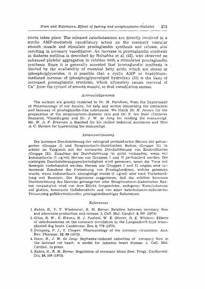
Stam and Hiasmann, Effect of fasting and streptozotocin-diabetes 373
stores takes place. The released catecholamines are directly involved in a (cyclic AMP-mediated) vasodilatory action on the coronary vascular smooth muscle and stimulate prostaglandin synthesis and release, also resulting in coronary vasodilation. An increase in prostaglandin synthesis in diabetes mellitus is described Qy Halushka et aL (32), who observed an enhanced platelet aggregation in relation with a stimulated prostaglandin synthesis. Since it is generally accepted that prostaglandin synthesis is limited by the availability of essential fatty acids, which are stored in (phospho)glycerides, it is possible that a cyclic AMP or bradykininmediated increase of (phospho)glycerolipid hydrolysis (33) is the basis of increased prostaglandin synthesis, which ultimately causes removal of Ca++ from the cytosol of smooth muscle, so that va~odilation ensues.
Acknowledgements
The authors are greatly indebted to Dr. M. Farnham, from the Department of Pharmacology of our faculty, for help and advice concerning the extraction and bioassay of prostaglandin-like substances. We thank Dr. H. Jansen for the preparation of the streptozotocin-diabetic rats and Dr. F. ten Hoar (Unilever Research, Vlaardingen) and Dr. J. W. de Jong for reading the manuscript. Mr. W. A. P. Breeman is thanked for his skilled technical assistance and Miss A. C. Hanson for typewriting the manuscript.
Zusammenfassung
Die koronare DurchstrOmung der retrograd perfundierten Herzen der gehungerten (Gruppe I) und Streptozotocin-diabetischen Ratten (Gruppe II) ist erh6ht im Vergleich mit der koronaren Durchstr6mung von Kontrolltieren (Gruppe III). Zunahme der Durchstr6mung ist nicht vorhanden, wenn mit Indomethacin (1 f~g/ml) Herzen aus Gruppen I und II perfundiert werden. Die niedrigste Durchstr6mungsgeschwindigkeit wird gemessen, wenn die Tiere mit Reserpin vorbehandelt werden. Herzen aus Gruppen I und II zeigten eine bedeutende Zunahme der Freisetzung von Prostaglandinen, welche gehemmt wurde, wenn Indomethacin hinzugefiigt wurde (1 ,ug/ml) oder nach Vorbehandlung mit Reserpin. Die Ergebnisse suggerieren, daB die erh6hte koronare Durchstr6mung des Herzens gehungerter oder Streptozotocin-diabetischer Ratten veranstaltet wird von dem Effekt freigesetzter, endogener Katecholamine auf glatten, konoraren Gefi:i.Bmuskeln und von einer katecholamin-induzierten Freisetzung gefi:i.Berweiternder, prostaglandinartiger Substanzen.
References
1. Rubio, R., V. T. Wiedmeier, R. M. Berne: Relation between coronary flow and adenosine production and release. J. CelL Mol. CardieL 6, 561 (1974).
2 .. Giles, R. W., E. Eikens, H. J. Paoloni, W. E. Glover, D. E. WHcken: Effects of catecholamines on the coronary circulation in the Langendorff-type transplanted dog heart. Cardiovasc. Res. 9, 779 (1975).
3. Dempsey, P. J., T. Cooper: Pharmacology of the coronary circulation. Ann. Rev. Pharmac. 12, 99 (1972).
4. Stam, H., J. W. de Jong: Sephadex-induced reduction of coronary flow in the isolated rat heart: A model for ischemic heart disease. J. Cell. MoL Cardiel., in press.
5. Rubio, R., R. M. Berne: Regulation of coronary blood flow. Progr. Cardiovasc. Dis. 18, 105 (1975).
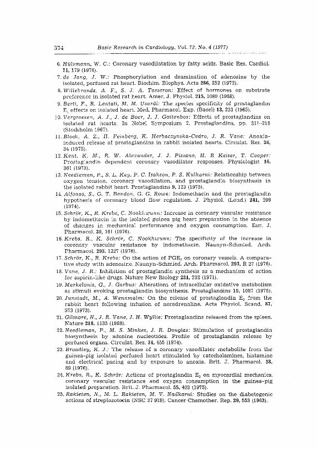
374 Basic Research in Cardiology, VoL 72, No.4 (1977)
6. HULsmann, W. C.: Coronary vasodilatation by fatty acids. Basic Res. Cardiol. 71, 179 (1976).
7. de Jong, J. W.: Phosphorylation and deamination of adenosine by the isolated, perfused rat heart. Biochim. Biophys. Acta 286, 252 (1972).
8. Willebrands, A. F., S. J. A. Tasseron: Effect of hormones on substrate preference in isolated rat heart. Amer. J. Physiol. 215, 1089 (1968).
9. Berti, F., R. Lentati, M. M. Usardi: The species specificity of prostaglandin E 1 effects on isolated heart. Med. Pharmacal. Exp. (Basel) 13, 233 (1965).
10. Vergroesen, A. J., J. de Boer, J. J. Gottenbos: Effects of prostaglandins on isolated rat hearts. In Nobel Symposium 2. Prostaglandins, pp. 21J-218 (Stockholm 1967).
11. Block, A. Z., H. Feinberg, K. Herbaczynska-Cedro, J. R. Vane: Anoxiainduced release of prostaglandins in rabbit isolated hearts. Circulat. Res. 36, 34 (1975).
12. Kent, K. M., R. W. Alexander, J. J. Pissano, H. R Keiser, T. Cooper: Prostaglandin dependent coronary vasodilator responses. Physiologist 16, 361 (1973).
13. Needleman, P., S. L. Key, P. C. Isakson, P. S. Kulkarni: Relationship between oxygen tension, coronary vasodilation, and prostaglandin biosynthesis in the isolated rabbit heart. Prostaglandins 9, 123 (1975).
14. Alfonso, S., G. T. Bandon, G. G. Rowe: Indomethacin and the prostaglandin hypothesis of coronary blood flow regulation. J. Physiol. (Lond.) 241, 299 (1974).
15. SchrOr, K., R. Krebs, C. Nookhwunn: Increase in coronary vascular resistance by indomethacin in the isolated guinea pig heart preparation in the absence of changes in mechanical performance and oxygen consumption. Eur. J. Pharmacal. 39, 161 (1976).
16. Krebs, R., K. SchrOr, C. Nookhwunn: The specificity of the increase in coronary vascular resistance by indomethacin. Naunyn-Schmied. Arch. Pharmacal. 293, 1227 (1976).
17. SchrOr, K., R. Krebs: On the action of PGE2 on coronary vessels. A comparative study with adenosine. Naunyn-Schmied. Arch. Pharmacol. 293, R 27 {1976).
18. Vane, J. R.: Inhibition of prostaglandin synthesis as a mechanism of action for aspirin-like drugs. Nature New Biology 231, 232 (1971).
19. Markelonis, G., J. Garbus: Alterations of intracellular oxidative metabolism as stimuli evoking prostaglandin biosynthesis. Prostaglandins 10, 1087 (1975).
20. Junstadt, M., A. Wennmaim: On the release of prostaglandin E~ from the rabbit heart following infusion of noradrenaline. Acta Physiol. Scand. 87, 573 (1973).
21. Gilmore, N., J. R. Vane, J .. H. WyUie: Prostaglandins released from the spleen. Nature 218, 1135 (1968).
22. NeedLeman, P., M. S. Minkes, J. R. DougLas: Stimulation of prostaglandin biosynthesis by adenine nucleotides. Profile of prostaglandin release by perfused organs. Circulat. Res. 34, 455 (1974).
23. Broadley, K. J.: The release of a coronary vasodilator metabolite from the guinea-pig isolated perfused heart stimulated by catecholamines, histamine and electrical pacing and by exposure to anoxia. Brit. J. Pharmacol. 58, 89 (1976).
24. Krebs, R., K. SchrOr: Actions of prostaglandin E 2 on myocardial mechanics, coronary vascular resistance and oxygen consumption in the guinea-pig isolated preparation. Brit. J. Pharmacol. 55, 403 (1975).
25. Rakieten, N., M. L. Rakieten, M. V. Nadkarni: Studies on the diabetogenic actions of streptozotocin {NSC 37 919). Cancer Chemother. Rep. 29, 553 (1963).
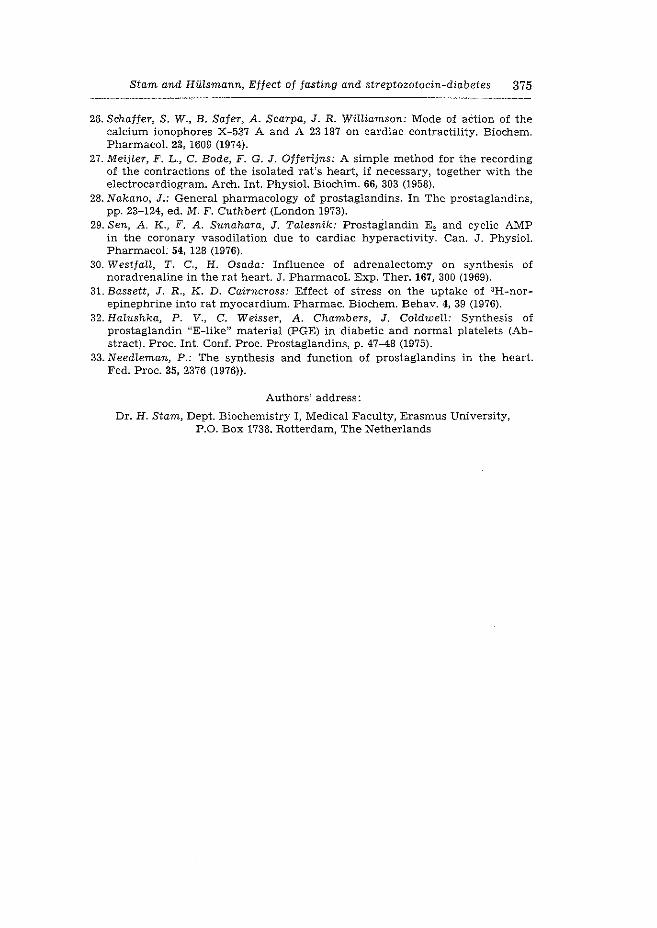
Stam and Hillsmann, Effect of fasting and streptozotocin-diabetes 375
26. Schaffer, S. W., B. Safer, A. Scarpa, J. R. Williamson: Mode of aCtion of the calcium ionophores X-537 A and A 23 187 on cardiac contractility. Biochem. Pharmacal. 23, 1609 (1974).
27. Meijler, F. L., C. Bode, F. G. J. Offerijns: A simple method for the recording of the contractions of the isolated rat's heart, if necessary, together with the electrocardiogram. Arch. Int. Physiol. Biochim. 66, 303 (1958).
28. Nakano, J.: General pharmacology of prostaglandins. In The prostaglandins, pp. 23-124, ed. M. F. Cuthbert (London 1973).
29. Sen, A. K., F. A. Sunahara, J. Talesnik: Prostaglandin E2 and cyclic AMP in the coronary vasodilation due to cardiac hyperactivity. Can. J. Physiol. Pharmacal. 54, 128 (1976).
30. Westfall, T. C., H. Osada: Influence of adrenalectomy on synthesis of noradrenaline in the rat heart. J. Pharmacal. Exp. Ther. 167, 300 (1969).
31. Bassett, J. R., K. D. Cairncross: Effect of stress on the uptake of 3H-norepinephrine into rat myocardium. Pharmac. Biochem. Behav. 4, 39 (1976).
32. Halushka, P. V., C. Weisser, A. Chambers, J. ColdweU: Synthesis of prostaglandin "E-like" material (PGE) in diabetic and normal platelets (Abstract). Proc. Int. Conf. Proc. Prostaglandins, p. 47-48 (1975).
33. Needleman, P.: The synthesis and function of prostaglandins in the heart. Fed. Proc. 35, 2376 (1976)).
Authors' address:
Dr. H. Stam, Dept. Biochemistry I, Medical Faculty, Erasmus University, P.O. Box 1738. Rotterdam, The Netherlands

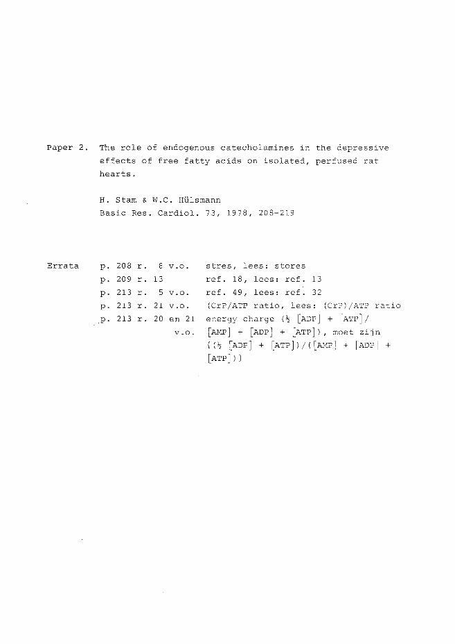
Paper 2. The role of endogenous catecholamines in the depressive
effects of free fatty acids on isolated, perfused rat
hearts.
H. Starn & W.C. Hlilsmann
Basic Res. Cardiol. 73, 1978, 208-219
Errata p. 208 r. 6 v.o. stres, lees: stores
p. 209 r. 13 ref. 18' lees: ref. 13
p. 213 r. 5 v.o. ref. 49' lees: ref. 32
p. 213 r. 21 v.o. (CrP/ATP ratio, lees: (CrPI/ATP ratio
p. 213 r. 20 en 21 energy charge I 'I [ADP j + IATP]/
v.o. [AHPj + [ADP] + [ATP] I, moet zij n
{ (!;; [ADP] + [ATPj I/ I [AHP] + [ADP I +
[ATP]Il

Basic Res. Cardiel. 73, 208-219 (1978) © 1978 Dr. Dietrich Steinkopff verlag, Darmstadt ISSN 0300-8428
Departments of Cardiovascular Research (Thoraxcenter) and Biochemistry I, Medical Faculty,
Erasmus University Rotterdc.m, Rotterdam (The Netherlands)
The role of endogenous catecholamines in the depressive effects of free fatty acids on isolated, perfused rat hearts*)
Die Rolle von endogenen Katecholaminen
in der depressiven Wirkung von freien Fettsauren
auf isolierte, perfundierte Rattenherzen
H. Starn and W. C. Hidsmann
With 4 figures and 1 table
(Received July 5. 1917)
Summary
Langendorff perfusion of hearts with 0.5 mM palmitate complexed with albumin in a molar ratio of six showed a depression of contractility to about 50°/o of control vs.J.ues. The coronary flow rate was enhanced and no arrhythmias cccured. This decre<Jse in contractility during fatty acid perfusion •,vas irreversible smce reperfusion "1.'-Iith control medium containing ll mM glucose as substrate did not lead to a recovery in contractile behaviour, while coronary flow rates reached va~ues equal to, or below control levels. The deleterious effects of pcdr:1ita'ce v..;er-e partly overcome by adding 11 mM glucose to the fatty aciC. conta~ning nedium. No significant change in energy charge of ventrin1lar tissue was observed alter a i.hirty minute recirculating perfusion v:,rith palmitate \vhen compared wi cl!_ th-e energy charge of hearts perfused with pahnit2rte p~c.cs 11 lT'ti\T glucose.
In hearts_ C."-'r:;leteci fr0m lheir endogenous c-atec-holamines by preperfusi:::rn with ty:-anine, thE fa'c'c:;r aciC, ind'--lced dec2'ease in contractility was signif,::-ar/lly less compared v-Iitl:l coc-:trol ·hearts during perfusion witD. palmitate. Reperfusion of tyramine-pretreated he8.rts with control medium vvas followed by a slow recovery of contrc.ctility. Hearts, _9I'eloaded with C'H] - norepinephrine showed a sudden release of radioactivity in the perfusate upon introduction of 0.5 mM palmitate, while no release occurred upon introduction of 05 mM palmitate ir; the presence of U :'Y'.J'v1 glucose. These findings suggest that cate~'holamines
released from endcger'-nt:s stres _may play a~ important role in the depressive actions of iatty acids. T~w rc!e of the intracel:ular levels of fatty acids and their possible ionophoTic r<)le ~n 'she release of catecholamines is discussed in ::-elation to the detaiOTaEor: oi' .l:."lyocardia.l function. Prostaglandin E 1, of which ionophoric p:rope:ties t.ave also been sho-~Nn, added instead of ·fatty acids, was found to have a simEar effect.
*) Support was obtained from th,e Dutch Heart Foundation.
734
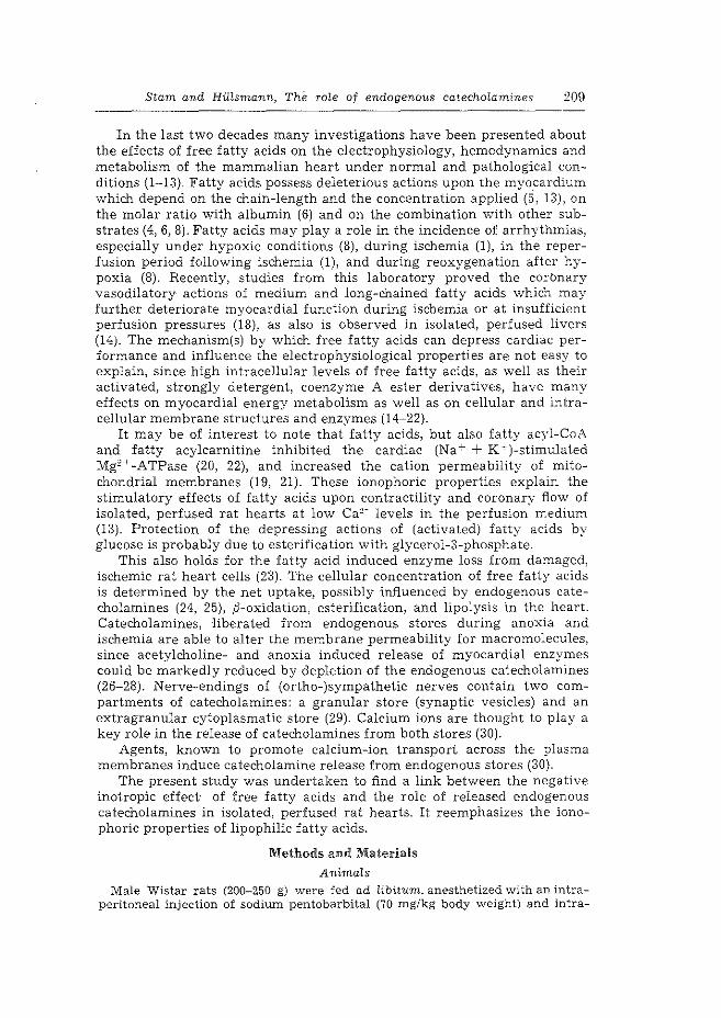
Stam and Hiasmann, The role of endogenous catecholamines 209
In the last two decades many investigations have been presented about the effects of free fatty acids on the electrophysiology, hemodynamics and metabolism of the mammalian heart under normal and pathological conditions (1-13). Fatty acids possess deleterious actions upon the myocardium which depend on the chain-length and the concentration applied (5, 13), on the molar ratio with albumin (6) and on the combination with other substrates (4, 6, 8). Fatty acids may play a role in the incidence of arrhythmias, especially under hypoxic conditions (8), during ischemia (1), in the reperfusion period following ischemia (1), and during reoxygenation after hypoxia (8). Recently, studies from this laboratory proved the corbnary vasodilatory actions of medium and long-chained fatty acids which may further deteriorate myocardial function during ischemia or at insufficient perfusion pressures (18), as also is observed in isolated, perfused livers (14). The mechanism(s) by which free fatty acids can depress cardiac performance and influence the electrophysiological properties are not easy to explain, since high intracellular levels of free fatty acids, as well as their activated, strongly detergent, coenzyme A ester derivatives, have many effects on myocardial energy metabolism as well as on cellular and intracellular membrane structures and enzymes (14-22).
It may be of interest to note that fatty acids, but also fatty acyl-CoA and fatty acy1carnitine inhibited the cardiac (Na+ + K-)-stimulated Mg2 '~-ATPase (20, 22), and increased the cation permeability of mitochondrial membranes (19, 21). These ionophoric properties explain the stimulatory effects of fatty acids upon contractility and coronary flow of isolated, perfused rat hearts at low Ca2 ~ levels in the perfusion medium (13). Protection of the depressing actions of (activated) fatty acids by glucose is probably due to esterification with glycerol-3-phosphate.
This also holds for the fatty acid induced enzyme loss from damaged, ischemic rat heart cells (23). The cellular concentration of free fatty acids is determined by the net uptake, possibly influenced by endogenous catecholamines (24, 25), f)-oxidation, esterification, and lipolysis in the heart. Catecholamines, -liberated from endogenous stores during anoxia and ischemia are able to alter the membrane permeability for macromolecules, since acetylcholine- and anoxia induced release of myocardial enzymes could be markedly reduced by depletion of the endogenous catecholamines (26-28). Nerve-endings of (ortho-)sympathetic nerves contain two compartments of catecholamines: a granular store (synaptic vesicles) and an extragranular cytoplasmatic store (29). Calcium ions are thought to play a key role in the release of catecholamines from both stores (30).
Agents, known to promote calcium-ion transport across the plasma membranes induce catecholamine release from endogenous stores (30).
The present study was undertaken to find a link between the negative inotropic effect· of free fatty acids and the role of released endogenous catecholamines in isolated, perfused rat hearts. It reemphasizes the ionophoric properties of lipophilic fatty acids.
Methods and Materials
Animals
Male Wistar rats (200-250 g) were fed ad libitum, anesthetized with an intraperitoneal injection of sodium pentobarbital (70 mg/kg body weight) and intra-
210 Basic Research in Cardiology, VoL 73, No. 2 ( 1978)
venously heparinized with 500 I.U. If depletion of endogenous catecholamines took place with reserpin, the drug was injected intraperitoneally (10 mg/kg body Weight) six hours before sacrifice.
Perfusion procedure
The hearts were quickly excised, cannulated and connected to the perfusion apparatus for retrograde perfusion (Langendorjj) as described earlier (31, 32). Contractility was measured as apex displacement and registered as described previously (31).
Apex displacement after 30 min perfusion was taken as control. Coronary flo\v rates were measured by timed collection. After the experiments the hearts \Vere cut open, blotted dry thoroughly and weighed. The standard perfusion medium (SPM) consisted of a modified Tyrode solution (13, 31), pH 7.4, containing 11 mM glucose and/or 0.5 mM potassium palmitate complexed with fatty acid free bovine serum albumin (BSA) (in a molar ration of 6:1). The perfusion fluids were equilibrated with 95 °/o 0 2 + 5 Ofo C02, and foaming of the albumin containing solutions was prevented by a silicone defoamer (Antifoam A).
The palmitate-albumin complex was prepared by adding recristallized potassium palmitate in water of 80° C to an albumin solution under vigorous stirring. Before use, the solution \vas passed through a washed, 1.2 ,u Millipore membrane filter (Sartorius, Gtittingen, Germany).
Perfusion of the hearts occurred in three periods. All experiments were started with an initial recovery period of 30 min (period I) in which perfusion took place \vith SPIVI containing 11 mM glucose (31, 32). It was followed by a 30 min recirculating perfusion (period ID with 75 ml of SPM containing 0.5 mlVI palmitate (complexed to 0.08 mM BSA) ± 11 mM glucose or, in control experiments, SPM containing 11 mM glucose ± 0.08 mM BSA. Subsequently perfusion took place, again for 30 min (period III), with SPIVI containing 11 mM glucose.
Depletion of endogenous catecholamines was induced by an infusion of tyramine (6.5 .u/min) in the aortic cannula during the first 15 min of period I (34). Control experiments proved that tyramine washout during perfusion was complete at the end of the period I.
Radioactive experiments
After a 15 min equilibration perfusion, the transmitter stores of the hec.rt \\rere labelled in a 15 min perfusion with glucose-medium, containing 5.10-~ M DL-[7~:3HJ norepinephrine ([3H]NE).
The specific Etctivity of the PHJNE was 11 Ci/mmol. Efflux of [:lH]-labelled compounds \vas follovled by collection of effluents during perfusion with control medium for 10 min. Subsequently the rlH]NE-labelled hearts were exposed to SPM either containing a) 11 mM glucose + 0.08 mM BSA, or b) 0.5 mM palmitate ..L 0.08 mM BSA, or c) 11 mM glucose, 0.5 palmitate + 0.08 mM BSA, or d) 11 mM glucose + 10-G M prostaglandin Er (PGE1). During these nonrecirculating perfusions effluent samples were collected for 20 min and 2 ml of all samples wc.s added to 10 ml Instagel. Radioactivity was determined by liquid scintillation cmJ.nting.
AnaLyses
At the end of pe:·~·usion ~.,eriod I beating hearts were freeze-clamped between aluminium blccks at ~h2 temperature of liquid nitrogen (35) and extracted as previously described {33).
The tissue extracts were determined for creatine phosphate, ATP, ADP and AI'viP spectrophotometrically {36).
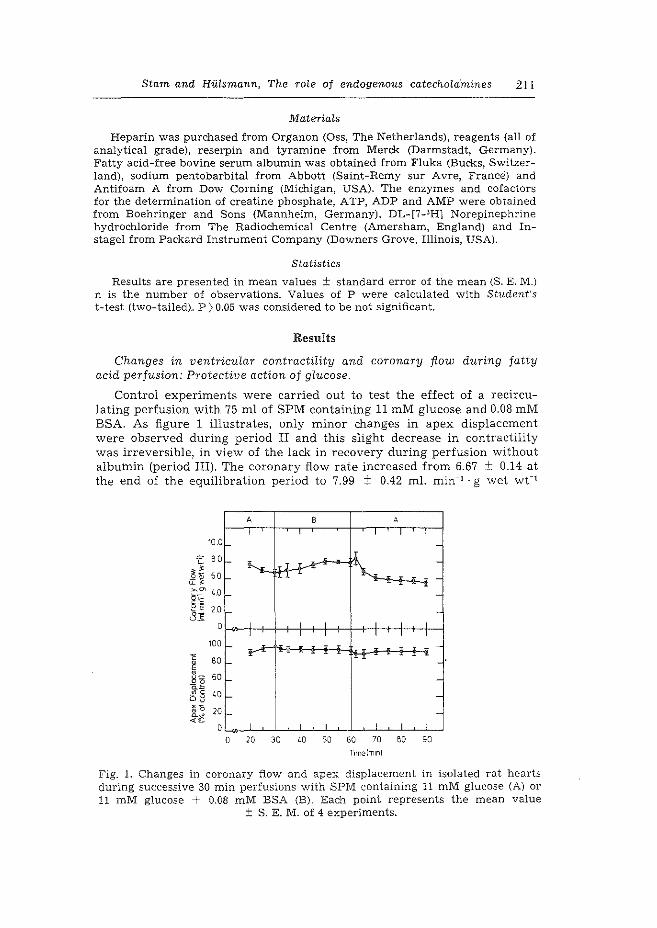
Stam and HiHsmann, The ro~e of endogenous catecholafnines 211
Materials
Heparin was purchased from Organon (Oss, The Netherlands), reagents (all of analytical grade), reserpin and tyramine from Merck (Darmstadt, Germany). Fatty acid-free bovine serum albumin was obtained from Fluka (Bucks, Switzerland), sodium pentobarbital from Abbott (Saint-Remy sur Avre, France) and Antifoam A from Dow Corning (Michigan, USA). The enzymes and cofactors for the determination of creatine phosphate, ATP, ADP and AMP were obtained from Boehringer and Sons (Mannheim, Germany), DL-[7-3H) Norepinephrine hydrochloride from The Radiochemical Centre (Amersham, England) and Instage! from Packard Instrument Company (Downers Grove, Illinois, USA).
Statistics
Results are presented in mean values ± standard error of the mean (S. E. M.) n is the number of observations. Values of P were calculated with Student's t-test (two-tailed). P > 0.05 was considered to be not significant.
Results
Changes in ventricular contractility and coronary flow during fatty acid perfusion: Protective action of glucose.
Control experiments were carried out to test the effect of a recirculating perfusion with 75 ml of SPM containing 11 mM glucose and 0.08 mM BSA. As figure 1 illustrates, only minor changes in apex displacement were observed during period II and this slight decrease in contractility was irreversible, in view of the lack in recovery during perfusion without albumin (period III). The coronary flow rate increased from 6.67 ± 0.14 at the end of the equilibration period to 7.99 ± 0.42 ml. min_, · g wet wt-1
10
A B A
I I I I I I 0
0 ~1 ;1-.V"' [l
~ . -
0 I
,..J._-I I
0 .-I
cy
0
I
o_ o_ 0 I . I I I
o 20 Jo w ~ w ro w w T•m8lm1nl
-
-· -
-
-
Fig. 1. Changes in coronary flow and apex displacement in isolated rat hearts during successive 30 min perfusions with SPM containing 11 mM glucose (A) or 11 mM glucose + 0.08 mM BSA (B). Eac.'IJ. point represents the mean value
± S. E. M. of 4 experiments.
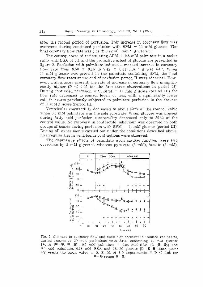
Basic Research in Cardiology, VoL 73, No. 2 (1978)
after the second period of perfusion. This increase in coronary flow was overcome during continued perfusion with SPM + 11 mM glucose. The final coronary flow rate was 5.54 ± 0.22 ml· min- 1 · g wet wt-1.
The consequences of recirculating SPM + 0 .. 5 mM palmitate in a molar ratio with BSA of 6:1 and the protective effect of glucose are presented in figure 2. Perfusion with palmitate induced a marked increase in coronary flow rate from 6.58 ± 0.16 to 9.42 ± 0.81 · min- 1 · g wet wt-1 • When 11 mM glucose was present in the palmitate containing SPM, the final coronary flow rates at the end of perfusion period II were identicaL However, with glucose present, the rate of increase in coronary flow is significantly higher (P < 0.05 for the first three observations in period II). During continued perfusion with SPM + 11 mM glucose (period III) the flow rate decreased to control levels or less, with a significantly lower rate in hearts previously subjected to palmitate perfusion in the absence of 11 mM glucose (period II).
Ventricular contractility decreased to about 50 °/o of the control value vvhen 0.5 mM palmitate was the sole substrate. When glucose was present during fatty acid perfusion contractility decreased only to 80 °/o of the control value. No recovery in contractile behaviour was observed in both groups of hearts during perfusion with SPM + 11 mM glucose (period III). During all experiments carried out under the conditions described above, no irregularities in ventricular contractions were observed.
The depressive effects of palmitate upon cardiac function were also overcome by 5 mM glycerol, whereas pyruvate (5 mM), lactate (5 mM),
T: ~
~· -' ~~
~c o·s §E OE U-
c 0 E o_ u-£2 ~-':!"15 Ou 'a ~~
10 0
80
60
'0
20
0
100
80
60
'0
20
0
0
A
0
"' 20
.. ' * "' * * *
**** *******
30 40 50 60 70 00 90
Timcelmml
Fig. 2. Changes in coronary flow and apex displacement in isolated rat hearts, during successive 30 min perfusions with SPM containing 11 mM glucose [A, A (e -Ill, !II- Ill)], 0.5 mM palmitate + 0.08 mM BSA [C (111-111)] and 0.5 mM palmitate, 0.08 ml\'1 BSA and llmM glucose [D (1!6 -!li)J.Each point represents the mean value ± S. E. M. of 6-9 experiments. * P < 0.05 for
111- #Jj versus !ml- ili!L
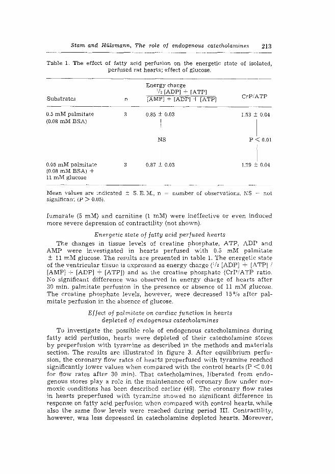
Stam and HiUsmann, The role of endogenous catecho[amines 213
Table 1. The effect of fatty acid perfusion on the energetic state of isolated, perfused rat hearts; effect of glucose.
Substrates
0.5 mM palmitate (0.08 mM BSA)
0.05 mM palmitate (0.08 mM BSA) + 11 mM glucose
n
3
3
Energy charge '/, [ADP] + [ATP]
[AMP]+ [ADP] + [ATP] CrP!ATP
0.85 ± 0.03 1.53 ± 0.04
NS p 0.01
0.87 0.03 1.79 0.04
----------------------------Mean values are indicated ± S. E. M., n = number of observations. NS not significant (P > 0.05).
fumarate (5 mM) and carnitine (1 mM) were ineffective or even induced more severe depression of contractility (not shown).
EneTgetic state of fatty acid perfused hearts The changes in tissue levels of creatine phosphate, ATP, ADP and
AMP were investigated in hearts perfused with 0.5 mM palmitate ± 11 mM glucose. The results are presented in table l. The energetic state of the ventricular tissue is expressed as energy charge Ch [ADPJ + [ATP] / [AMP]+ [ADP] + [ATP]) and as the creatine phosphate (CrP/ATP ratio. No significant difference was observed in energy charge of hearts after 30 min. palmitate perfusion in the presence or absence of 11 mM glucose. The creatine phosphate levels, however, were decreased 15 °/o after palmitate perfusion in the absence of glucose.
Effect of palmitate on cardiac function in hearts depleted of endogenous catecholamines
To investigate the possible role of endogenous catecholamines during fatty acid perfusion, hearts were depleted of their catecholamine stores by preperfusion with tyramine as described in the methods and materials section. The results are illustrated in figure 3. After equilibrium perfusion, the coronary flow rates of hearts preperfused with tyramine reached significantly lower values when compared with the control hearts (P < 0.01 for flow rates after 30 min). That catecholamines, liberated from endogenous stores play a role in the maintenance of coronary flow under normoxie conditions has been described earlier (49). The coronary flow rates in hearts preperfused with tyramine showed no significant difference in response on fatty acid perfusion when compared with control hearts. while also the same flow levels were reached during period HI. Contractility, however, was less depressed in catecholamine depleted hearts. Moreover,
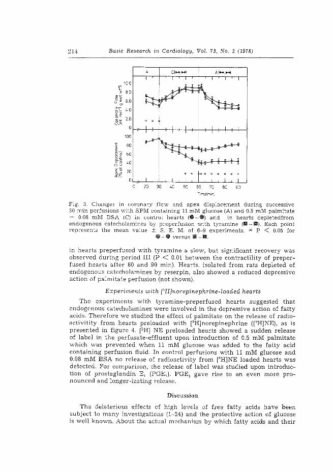
~14 Basic Research in Cardiology, VoL 73, No. 2 (1978)
A C!-.-1 Al,..,-i
10 0 f ' 80
]~ 6 0 ~~ ~
'0 O's §E
20 OE • • • U-0
~
100
c 80 0 E 8'3 60 _gz
~ ~c
'0 ~0 ou ~0 ~~
2D ' ' .. • • '
0 0 20 30 '0 50 60 70 80 90
TimG(mm)
Fig. 3. Changes in coronary flow and apex displacement during successive 30 min perfusions \Vith SPM containing 11 mM glucose (A) and 0.5 mM palmitate -t- 0.08 mM BSA (C) in control hearts (e- e) and in hearts depletedfrom endogenous catecholamines by preperfusion with tyramine (111-111). Each point represents the mean value ± S. E. M. of 6-9 experiments. * P < 0.05 for
~-CD versus Ill - 1111.
in hearts preperfused with tyramine a slow, but significant recovery was observed during period III (P < 0.01 between the contractility of preperfused hearts after 60 and 90 min). Hearts, isolated from rats depleted of endogenous catecholamines by reserpin, also showed a reduced depressive action of palmitate perfusion (not shown).
Experiments with [lH]norepinephrine-loaded hearts
The experiments with tyramine-preperfused hearts suggested that endogenous catecholamines were involved in the depressive action of fatty acids. Therefore we studied the effect of palmitate on the release of radioactivitity from hearts preloaded with rlH]norepinephrine (CH]NE), as is presented in figure 4. [3HJ NE preloaded hearts showed a sudden release of label in the perfusate-effluent upon introduction of 0.5 mM palmitate which was prevented when 11 m.M glucose was added to the fatty acid containing perfusion fluid. In control perfusions with 11 mM glucose and 0.08 mM BSA no release of radioactivity from [3H]NE loaded hearts was detected. For comparison, the release of label was studied upon introduction of prostaglandin E 1 (PGE1). PGE1 gave rise to an even more pronounced and longer-lasting release.
Discussion
The deleterious effects of high levels of free fatty acids have been subject to many investigations (1-24) and the protective action of glucose is well known. About the actual mechanism by which fatty acids and their
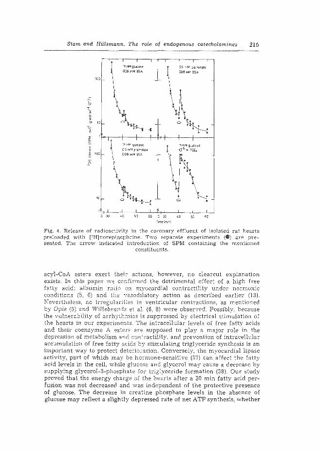
Stam and Hiilsmann, The rofe of endogenous catechoLamines 215
WO
0
~ 100
i
'I-
11 m'-1 g:ucose 008 mM BSA
' ' OS mM palm11a1e
008 rr/'-1 BSA
Fig. 4. Release of radioactivity in the coronary effluent of isolated rat hearts preloaded with r1H]norepinephrine. T"\VO separate experiments (~) are presented. The arrow indicated introduction of SPM containing the mentioned
constituents.
acyl-CoA esters exert their actions, however, no clearcut explanation exists. In this paper we confirmed the detrimental effect of a high free fatty acid: albumin ratio on myocardial contractility under normoxic conditions (5, 6) and the vasodilatory action as described earlier {13). Nevertheless, no irregularities in ventricular contractions, as mentioned by Opie (5) and WiHebrands et al. (6, 8) were observed. Possibly, because the vulnerability of arrhythmias is suppressed by electrical stimulation of the hearts in our experimElts. The intracellular levels of free fatty acids and their coenzyme A esters are supposed to play a major role in the depression of metabolism and con'~ractility, and prevention of intracellular accumulation of free fatty acids by stimulating triglyceride synthesis is an important way to protect dete:-iors.tion. Conversely, the myocardial lipase activity, part of which may be hormone-sensitive (37} can affect the fatty acid levels in the cell, while glucose and glycerol may cause a decrease by supplying glycerol-3-phosphate for triglyceride formation (38). Our study proved that the energy charge of the hearts after a 30 min fatty acid perfusion was not decreased and was independent of the protective presence of glucose. The decrease in creatine phosphate levels in the absence of glucose may reflect a slightly depressed rate of net ATP synthesis, whether
Basic Research in Cardiology, VoL 73, No. 2 (1978)
due to depressed glycolysis or oxidative phosphorylation leading to decreased creatine phosphate synthesis (40, 41).
Our observation that the deleterious effects of palmitate upon the contractility were irreverible illustrates the membrane damaging properties of high levels of lipophilic fatty acids.
Studies of Mathur and Mokler (25) indicated that endogenous catecholamines play an important role in the fatty acid metabolism and for that reason we studied the effects of fatty acid perfusion in rat hearts depleted of their catecholamine stores by preperfusion with tyramine. We observed a marked reduction of the negative inotropic action of palmitate, while continued perfusion with fatty acid free medium resulted in a slow recovery of contractile behaviour. The vasodilatory response to palmitate \Vas identical in control and catecholamine-depleted hearts. This confirms earlier observations that catecholamines were not involved in the vasoactive properties of fatty acids (13).
Catecholamines can interfere with the fatty acid metabolism by mediating their transport through the cellular membrane, whether directly or through increased cytosolic metabolism, or by stimulating the hormonesensitive lipase. Furthermore it is known that catecholamines released from endogenous stores alter the membrane permeability for macromolecules (27, 28).
The secretion of catecholamines from nerve-endings of sympathetic neurons occurring during myocardial ischemia and hypoxia (26), is suggested to be mediated by Ca~+ ions (29, 30), and can be enhanced by ionophores (42).
That lipophilic fatty acids, but also prostaglandin E 1, may possess ionophoric properties has been demonstrated before (13, 19, 21, 4~). In hearts preloaded with r~HlNE, introduction of a high intracellular level of free fatty acids (and of acyl-CoA esters, since fatty acid activation is not limiting for metabolism) resulted in a sudden release of radioactivity from the heart, representing the release of norepinephrine or its metabolites. The release of the neurotransmitter or its metabolites induced by fatty acids may be explained by their Ca2* ionophoric properties. It is likely that, in states of impaired energy metabolism (ischemia, anoxia) when increased neurotransmitter release is observed (26), intracellular fatty acid levels are increased, since fatty acid oxidation will be hampered and lipolysis is increased because of stress.
Upon introduction of prostaglandin Et, from which the ionophoric properties have been described (43), liberation of radioactivity also occurred, which strengthens the idea that the lipophilic fatty acids (or their acylCoA esters), have ionophoric properties. That the released, catecholamines did not induce a positive inotropic effect may be due to the action of long chain fatty acids on membranous processes.
Acknowledgements
The authors wish to thank Mr. A. Roodnat for his contribution to the perfusion experiments, Ms. M. Stewart and Mr. W. A. P. Breeman for skilled technical assistance and Ms. C. Vioo[ and Ms. C. v. d. Kleijn for typewriting the manuscript.
Stam and Hiilsmann, The role of endogenous catecholamines 217
Zusammenfassung
Die Kontraktilitat des retrograd perfundierten Rattenherzens wird urn 50°/o herabgesetzt, wenn 0,5 mM Palmitinsaure, komplexiert mit 0,08 mM Albumin, im Perfusionsmedium vorhanden ist. Die Fettsaureperfusion erhOht die Koronardurchstr6mungsgeschwindigkeit, w1ihrend keine RhythmusstOrungen beobachtet wurden. Die fettsliureinduzierte Kontraktilitatssenkung war nicht umkehrbar, weil nachtragliche Durchstr6mung mit 11 mM Glukose anstatt Fetts3.ure die Kontraktilitat nicht verbesserte. Wenn Glukose wahrend der Fetts3.uredurchstr6mung zu gleicher Zeit hinzugefiigt wurde, war die sch8.dliche Wirkung der Fettsaure beschrfi.nkt. Nach halbsti.indiger DurchstrOmung mit Palmitinsaure als einzigem Brennstoff war der Gehalt energiereicher Adeninnukleotide unver3.ndert, obwohl eine geringe Herabsetzung des Kreatinphosphatgehaltes beobachtet wurde. Wenn Herzen wahrend 15 Minuten mit Tyramine praperfundiert \VUrden. urn die endogenen Katecholamine zu entfernen, hatte PalmitatdurchstrOmung nur einen kleinen Effekt auf die Kontraktilitat, I.V1ihrend die geringe Herabsetzung reversibel war, wenn eine nachtr8.gliche DurchstrOmung mit Glukose erfolgte. Diese Beobachtung machte uns auf die MOglichkeit aufmerksam, daB Fettsaure die endogenen Katecholamine mobilisieren. Wenn Herzen mit PH]-Norepinephrin in de:r frUhen Perfusionszeit aufgeladen wurden, stellte sich heraus, daB nachher die Radioaktivitat auf asymptotische Weise aus dem Herzen freigemacht wurde. Wenn Palmitinsaure (mit Albumin im 6:1-Molar-Verh8.ltnis) angeboten wurde, wurde piOtzlich viel mehr Radioaktivitat freigemacht, was nicht geschieht, wenn zu gleicher Zeit Glukose angeboten wurde. Diese Befunde deuten darauf, daB freie Fettsauren endogene Katecholamine mobilisieren kOnnen. Es ist mtiglich, daB die ionophoren Eigenschaften von Fettsaure dabei eine wesentliche Rolle spielen, weil wir auch fanden, daB Prostaglandin Et (dessen Rolle als ionophore Verbindung be\viesen \\.'urde) den gleichen Effekt zeigte wie Fettsaure, 1.v1ihrend bakterielle Ionophore, wie X-537 A, in der Literatur auch Katecholamine mobilisierende Eigenschaften zeigten.
References
l. Kurien, V. A., P. A .. Yates, M. F. Q[iver: The role of free fatty acids in the production of ventricular c:trrhythmias after acute coronary artery occlusion. Eur. J. Clin. Invest. 1. 225-241 (1971).
2. Opi.e, L. H., R. M. j\fonis, hl. Thomas, A. J. Holland, P. Owen, S. van Noorden: Failure of high ccncen-crations of circulating free fatty acids to prO\'Oke arrhythmias in exper.irTJental n1yocardial infarction. Lancet 1971 .:JL 818--8:?.2.
3. ClwUoner, D. R., D. SLoin.Oerg: Oxidative metabolism of myocardium as influenced by fatty acids zmd. epinephrine. Am. J. Physiol. 211, 897-·902 (1966)
4. Gmsine-r, R. G., C. ..4.ps~eii,,, N. Brachfeld: Effect of palmitate on hypoxic cardiac performance. J. 1/Icl. Cell. Cardiol, 7, 227--235 (1975).
5. Opie, L. H.: Effect of fa. tty acids on contractility and rhythm of the heart. Nature 227, 1055-1056 (1970).
6. WiHebrands, A. F., H. F. Te-r Vi,rene, S. J. A. Tasseron: The effect of a high molar FFA/albumin ratio ir:. the perfusion medium on rhythm and contractility of the isolated rat heart.:. :;'l;=ol. Cell. Cardiol. 5, 259-273 (1973).
7. Wasilewska-Dziubiiiska, E., M. Czarnecka, A. Beresewicz, B. Lewartowski: Influence of sodium palmitate on the cellular action potentials of left ventricle of isolated, perfused guinea-pig j_eart. Acta Fhysiol. Fol. 26, l-11 (1975).
o. WiHebrands, A. F., S. J. A. Tasseron, H. F. Ter WeLte, R. Th. Van Dam: Effects of oleic acid and oxygen restriction followed by reoxygenation on rhythm and contractile activity of the isolated rat heart; protective action of glucose. J. Mol. Cell. Cardiol. 8, 375-388 (1976).
218 Basic Research in Cardio[ogy, Vol. 73, No. 2 (1978)
9. Cowan, J. C., E. M. Vaughan WiLLiams: The effects of palmitate on intracellular potentials recorded from Langendorfj-perfused guinea-pig hearts in normoxia and hypoxia, and during perfusion at reduced flow rate. J. Mol. Cell. Cardiel. 9, 327-342 (1977).
10. Henderson, A. H., E. H. SonnenbHck: Influence of free fatty acids on myocardial mechanics and oxygen consumption .in rat papillary muscles and perfused hearts, under oxygenated and hypoxic conditions. In myocardiology: Recent Advances in studies on Cardiac Structure and Metabolism, Volume 1, Bajusz, E., G. Rona, Eds., pp. 147~153. (Baltimore, London, Tokyo 1972).
J L Bing, J. R.: Cardiac metabolism. Physiol. Rev. 45, 171-213 (1965). 12. Opie, L. H.: Metabolism of the heart in health and disease I. Am. Heart
J. 76, 685-698 (1968). 13. Hillsmann, W. C.: Coronary vasodilation by fatty acids. Basic Res. Cardiel.
71, 179-191 (1976). 14. Hiltsmann, W. C., R. Kuypershoek-Davidov: Fatty acids and metabolism in
the in vitro hemoglobin-free perfused rat liver. Biochim. Biophys. Acta 354, 39-48 (1974).
15. Pande, S. V., J. F. Mead: Inhibition of enzyme activities by free fatty acids. J. Bioi. Chern. 243, 6180-6185 (1968).
16. Pressman, B. C., H. A. Lardy: Effect of surface agents on latent ATPase of mitochondria. Biochim. Biophys. Acta 21, 458-466 (1956).
17. Hiilsmann, W. C.: Over het mechanism van de ademhalingsketenphosphorylering (Academic Thesis) (Amsterdam 1958).
18. Hillsmann, W. C., W. B. Elliot, E. C. Slater: The nature and mechanism of action of uncoupling agents present in mitochrome preparations. Biochim. Biophys. Acta 39, 267-276 (1960).
19. Wojtczak, L.: Effect of long-chain fatty acids and acyl-CoA on mitochondrial permeability, transport, and energy-coupling processes. J. Bioenerg. Biomembr. 8, 293-311 (1976).
2.0. Lamers, J. M. J., W. C. Hillsmann: Inhibition of (Na+ + K+)-stimulated ATPase of heart by fatty acids. J. MoL CelL Cardiel. 9, 343-346 (1977).
21. Asimakis, G. K., L. A. Sordahl: Effects of atractyloside and palmitoyl-Coenzyme a on calcium tr8.nsport in cardiac mitochondria. Arch. Biochem. Biophys. 179, 200-210 (1977).
22. McMillin Wood, J., B. Bush, B. J. R. Pitts, A. Schwartz: Inhibition of bovine heart Na+, K+-ATPase by palmitylcarnitine and palmityl-CoA. Biochem. Biophys. Res. Commun. 74, 677-684 (1977).
23. De Leiris, J., L. H. Opie, W. F. Lubbe: Effects of free fatty acids and glucose on enzyme release in experimental myocardial infarction. Nature 253, 746-747 (1975).
24. Evans, J. R., L. H. Opie, J. C. Shipp: Metabolism of palmitic acid in perfused rat heart. Am. J. Physiol. 205, 766-770 (1963).
25. Mathur, P. P., C. M. Mokler: Subcellular distribution and incorporation of palmitate-U-1·iC into myocardial lipids: Role of endogenous and exogenous catecholamines. J. Mol. Cell. Cardiel. 7, 17-26 (1975).
~6. Shabab, L., A. Wollenberger: Freisetzung von Noradrenalin aus dem isolierten durchstr6mten Herzen bei akuter Anoxie und nach Gabe von Stoffwechselgiften. Acta Bioi. & Med. Germ. 19, 939-959 (1967).
27. Sakai, K., P. G. Spieckerman: Effects of reserpine and propanolol on anoxiainduced enzyme release from the isolated, perfused guinea-pig heart. Naunyn-Schmied. Arch. Pharmacal. 291, 123-130 (1975).
28. De Leiris, J., D. Feuvray, C. Come: Acetylcholine-induced release of lactate dehydrogenase from isolated perfused rat hearts. J. Mol. Cell. CardieL 4. 357-365 (1972).
Stam and Hiilsmann, The ro~e of endogenous catecholamines 219
29. Phillips, J. H.: The mechanism of catecholamine secretion: a Hypothesis for neurotransmitter release. Biochem. Soc. Transact. 4, 1003~1006 (1976).
30. Krueger, B., J. Forn, P. Greengard: Depolarization-induced phosphorylation of specific proteins, mediated by calcium ion influx, in rat brain synaptosomes. J. Biol. Chern. 252, 2764-2773 (1977).
31. Stam, H., J. W. DeJong: Sephadex-induced reduction of coronary flow in the isolated rat heart: A model for ischemic heart disease. J. Mol. Cell. Cardiol. 9, 633-650 (1977).
32. Stam, H., W. C. HiHsmann: Effect of fasting and streptozotocin-diabetes on the coronary flow in isolated rat hearts: A possible role of endogenous catecholamines and prostaglandins. Basic. Res. Cardiol. 72, 365~375 (1977).
33. De Jong, J. W.: Phosphorylation and deamination of adenosine by the isolated, perfused rat heart. Biochim. Biophys. Acta 286, 252-259 (1972).
34. SchUmann, H. J., A. PhiHipu: The mechanism of catecholamine release by tyramine. Int. J. Neuropharmacol. 1,179-182 (1962).
35. Wollenberger, A., 0. Ristau, G. Schofja: Eine einfache Technik der extrem schnellen Abki.ihlung gr6Derer Gewebestlicke. Pflligers Archiv. 270, 399-412 (1960).
36. Lamprecht, W., P. Stein, F. Heinz, H. Weisser: In: Methoden derenzymatischen Analyse (2nd ed.). Bergmeyer, H. U., Ed., pp 1729-1733, 2024-2033, 2051-2056. (Weinheim/BergstraDe, Germany 1970).
37. Crass III, M. F., J. C. Shipp, G. M. Pieper: Effects of catecholamines on myocardial endogenous substrates and contractility. Am. J. Physiol. 228, 618-627 (1975).
38. Lochner, A., J. L. N., Kotze, W. Gevers: Mitochondrial oxidative phosphorylation in myocardial ischemia: Effects of glycerol, glucose and insulin on anoxic hearts perfused at low pressure. J. Mol. Cell. Cardiol. 8, 575-584 (1976).
40. Scholte, H. R.: On the triple localization of creatine kinase in heart and skeletal muscle cells of the rat: Evidence for the existence of myofibrillar and mitochondrial isoenzymes. Biochim. Biophys. Acta 305, 413-427 (1973).
41. Saks, V. A., N. V. Lipina, N. V. Lyulina, G. B. Chernousova, R. Fetter, V. N. Smirnov, E. I. Chazov: Functional characterization of creatine phosphokinase reactions in heart mitochondria and myofibrils. Biochemistry 41, 1191-1199 (1976).
42. Schaffer, S. W., B. Safer, A. Scarpa, J. R. Winiamson: Mode of action of calcium ionophores X-537 A and A-23187 on cardiac contractility. Biochem. Pharmacal. 23, 1609-1617 (1974).
43. Kirtland, S. J., H. Baum: Prostaglandin Et may act as a "calcium ionophore". Nature (New Biology) 236, 47-49 (1972).
Authors' address:
Dr. H. Stam, Cardiochemical Laboratory, Department of Cardiovascular Research,
Medical Faculty, Erasmus University Rotterdam, P. 0. Box 1738, Rotterdam (The Netherlands)

Paper 3.
Erratum
Intracellular origin of hormone-sensitive lipolysis
in the rat
H. C. HU l srnann & H . S tam
Biochern. Biophys. Res. Corrmun. 82, 1978, 53-59
p. 57 Table I, sub. a, The adipocytes were incubated
at 37°C in Krebs-Ringer bicarbonate buffer cont~~~~ng 3% bovine serum albumin (BSA) ,

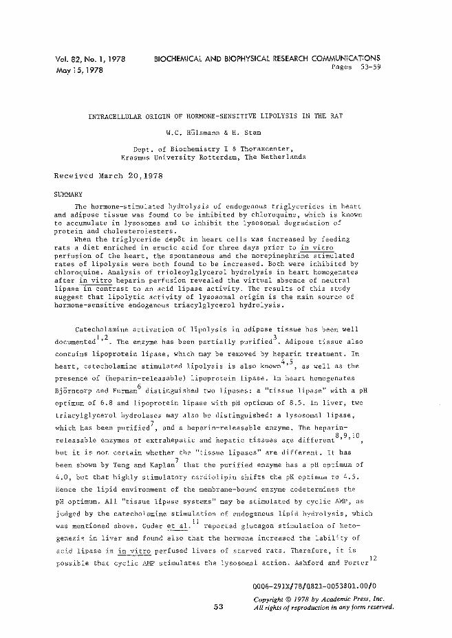
Vol. 82, No. 1, 1978 May15, 1978
BIOCHEMICAl AND BIOPHYSICAl RESEARCH COMMUNICATIONS Pages 53-59
INTRACELLULAR ORIGIN OF HORMONE-SENSITIVE LIPOLYSIS IN THE RAT
W.C. Hlllsmann & H. Starn
Dept. of Biochemistry I & Thoraxcenter, Erasmus University Rotterdam, The Netherlands
Received March 20,1978
SUMMARY
The hormone-stimulated hydrolysis of endogenous triglycerides in heart and adipose tissue was found to be inhibited by chloroquine, which is known to accumulate in lysosomes and to inhibit the lysosomal degradation of protein and cholesterolesters.
When the triglyceride depOt in heart cells was increased by feeding rats a diet enriched in erucic acid for three days prior to in vitro perfusion of the heart, the spontaneous and the norepinephrine stimulated rates of lipolysis were both found to be increased. Both were inhibited by chloroquine. Analysis of trioleoylglycerol hydrolysis in heart homogenates after in vitro heparin perfusion revealed the virtual absence of neutral lipase in contrast to an acid lipase activity. The results of this study suggest that lipolytic activity of lysosomal origin is the main source of hormone-sensitive endogenous triacylglycerol hydrolysis.
Catecholamine activation of lipolysis in adipose tissue has been well
documented 1•2 . The enzyme has been partially purified 3 . Adipose tissue also
contains lipoprotein lipase, which may be removed
heart, catecholamine stimulated lipolysis is also
by heparin treatment. In 4 5 known ' , as well as the
presence of (heparin-releasable) lipoprotein lipase. In heart homogenates
BjOrntorp and Furman6 distinguished two lipases: a "tissue lipase" with a pH
optimum of 6.8 and lipoprotein lipase with pH optimum of 8.5. In liver, two
triacylglycerol hydrolases may also be distinguished: a lysosomal lipase,
which has been purified 7 , and a heparin-releasable enzyme. The heparin-. 8 9 10
releasable enzymes of extrahepatic and hepatic tissues a~e d~fferent ' ' ,
but it is not certain
been shown by Teng and
whether the "tissue lipases" are different. It has 7 Kaplan that the purified enzyme has a pH optimum of
4.0, but that highly stimulatory c~rdi~lipin shifts the pH optimum to 4.5.
Hence the lipid environment of the membrane-bound enzyme codetermines the
pH optimum. All "tissue lipase systems" may be stimulated by cyclic AMP, as
judged by the catecholamine stimulation of endogenous lipid hydrolysis, which 11
was mentioned above. Guder et al. reported glucagon stimulation of keto-
genesis in liver and found also that the hormone increased the lability of
acid lipase in in vitro perfused livers of starved rats. Therefore, it is . . . 1 . 12 possLble that cycllc AMP stlmulates the lysosoma actlon. Ashford and Porter
QG06-291X/78/0821-0053$01.00/0
Copyright© 1978 by Academic Press, Inc. 53 All rights of reproduction in any form reserved.
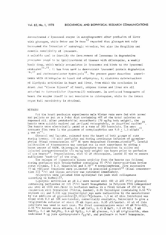
Vol. 82, No. 1, 1978 BIOCHEMICAl AND BIOPHYSICAl RESEARCH COMMUNICATIONS
demonstrated a· lysosomal enzyme in autophagosomes after perfusion of liver 13
with glucagon, Hhile Deter and De Duve reported that glucagon not only
increased the formation of autophagic vacuoles, but also the fragility and
osmotic sensitivity of lysosomes.
A suitable tool to identify the involvement of lysosomes in degradative
processes seems to be (pre)treatment of tissues with chloroquine, a weakly
basic drug, which mainly accumulates in lysosomes and binds to the lysosomal 14 15
membranes ' . It has been used to demonstrate lysosomal protein degradation 16,17 d l . IS . . an cholesterolester hydro ys~s . The present paper descn.bes experl-
ments with chloroquine on heart and adipocytes, it discusses determinations
of lipolytic activities in hea~t and liver, from which the conclusion is
drawu that "tissue lipases" of heart, adipose tissue and liver are all
atL1ched to intracellular (lysosomal?) membranes. In sonicated homogenates of
heart the enz;"TTle itself is not sensitive to chloroquine, while in the intact
organ full sensitivity is obtained.
HETHODS
For the heart perfusion experiments male Wistar rats were fed with normal rat pellets or put on a 3-day diet containing 40% of the total calories as rapeseed oil. After pentobarbital anaesthesia (70 mg/kg body weight), the 19 hearts were quickly excised and perfused retrogradely, as described earlier The hearts "'ere electrically paced at a rate of 300 beats/min. The mean _ 1 coronary flot·l rate in the presence of norepinephrine was 9.0 + 1.3 ml/min g wet v:t- 1 . -
Glycerol and lactate, released from the heart of both groups of rats during control (IS min) perfusion and during continuous infusion of norepinephrine (final concentration 10-7M) were determined fluoromecrically20. Careful calibration of fluorescence was carried out in each experiment by adding a knm,'TI amount of NADH, Chloroquine diphosphate was dissolved in saline and injected intraperitoneally (75 mg/kg body weight) two hours prior to perfusion of the hearts 21 . Preperfusions, with 50 uM chloroquine, lasted 30 min to ensure sufficient "wash-in" of the drug.
The release of lipoprotein lipase activity from the hearts v.•as followed during perfusion with Tyrode-buffer containing 1% (Wfv) fatty-acid-free bovine serum albumin, 5 I.U. heparin/ml and 5.10-7 M norepinephrine. Five successive 20 ml effluent fractions were collected in precooled glycerol22 (final concentration 20% Vfv) and lipase activity was estimated immediately.
Adipocytes were isolated from epididymal fat pads with collagenase according to Rodbell23.
Lipolytic activities at pH 8.2 were tested with 100 ~1 20% (W/v) glycerol containing perfusates or 10% (W/v) heart homogenates (a Polytron PTIO homogenizer was used at 4000 rev./min) in perfusion medium in a final volume of 250 ~1 by incubation with Intralipid (Vitrum, Sweden), 0.5% Intralipid (containing 0.5% Wfv soybean oil and 0.03% egg phospholipid) was made radioactive by the manufacturer by the addition of glyceroltri[9,JO(n)-3H]oleate. 1.9 ml of this Intralipid was mixed with 0.5 ml 10% non-labeled, commercially available, Intralipid to give a triglyceride solution of about 28 mM (spec.act. 0.35 wCi/wmole). 40 wl of this substrate was used in each test. The final concentrations were: 40 mM Tris-HCl, 2.5% Wfv bovine serum albumin, 0.8 mM CaCl 2 , 0.15 M NaCl, I .9 mM KCl, 8 mM NaHC03, 0.1 mM NaH2P04, 0.4 m}f MgCl2, 4.4 mM glucose, 4.5 mM triglyceride, when indciated 8 wg pure apolipoprotein c11 /ml, and perfusate or heart homogenate.
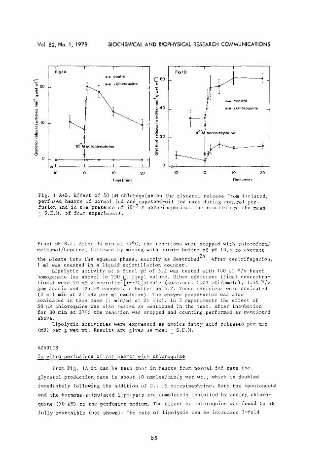
Vol. 82, l<!o. 1, 1978 BIOCHEMICAl AND BIOPHYSICAl RESEARCH COMMUNICATIONS
Fig.1A
. • 0
I
.._.. control
+chloroquine
~~ 60 ';
~
Fig.1B
- control
+chloroquine
E 510
~ ~I ., t
]
• ] ~ a
0
0
,t 10 M norepinephrine
t .: "p:: --:. -10 0 10
Time(min)
20 -10 0 20
Time(min)
Fig. I A+B, Effect of 50 ~M chloroquine on the glycerol release from isolated, perfused hearts of normal fed and rapeseed-oil fed rats during control perfusion and in the presence of JQ-7 M norepinephrine. The results are the mean + S.E.M. of four experiments.
Final pH 8.2. After 30 min at 37°C, the reactions were stopped with chloroform/ methanol/heptane, followed by mixing with borate buffer of pH 10.5 to extract
. h 1 . b 24 f . f . the oleate ~nto the aqueous p ase, exact y as descr~ ed A ter centr~ ugac~on, I ml was counted in a liquid scintillation counter.
Lipolytic activity at a final pH of 5.2 was tested with 100 1-'1 Wfv heart homogenate (as above) in 250 ].:1 final volume. Other additions (final concentrations) were 50 Illi'1 glyceroltri[J-l4c]oleate (spec.act. 0.03 wCi/]Jmole), 1.5% w;v gum acacia and 125 mM cacodylate buffer pH 5.2. These additions were sonicated (3 x 1 min at 21kHz per ml emulsion). The enzyme preparation was also sonicated in this case (I min/ml at 21 kHz). In 3 experiments the effect of 50 )JM chloroquine was a1so tested as menLioned in the text. After incubation for 30 min at 37°C the reaction was stopped and counting performed as mentioned above.
Lipolytic activities were expressed as nmoles fatty~acid released per min (mU) per g wet wt. Results are given as mean+ S.E.M.
RESULTS
In vitro perfusions of rat hearts with chloroquine
From Fig. JA it can be seen that in hearts from normal fed rats the
glycerol production rate is about 10 nmoles/min/g "Tet wt., which is doubled
immediately following the addition of 0.1 \-1M norepinephrine. Both the spontaneous
and the hormone~stimulated lipolysis are completely inhibited by adding chloro
quine (50 JJM) to the perfusion medium. The effect of chloroquine was found to be
fully reversible (not shown). The rate of lipolysis can be increased 3-fold
55
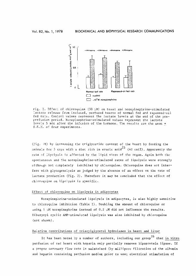
Vol. 82, No. 1, 1978 BIOCHEMICAl AND BIOPHYSICAl RESEARCH COMMUNICATIONS
Normal led rats
D control
Rapeseed-oil fed rats
CJ +10-7M norepinephrine
Fig. 2. Effect of chloroquine (50 wM) on basal and norepinephrine-stimulated lactate release from isolated, perfused hearts of normal fed and rapeseed-oil fed rats. Control values represent the lactate levels at the end of the preperfusion period. Norepinephrine-stimulated values represent the lactate levels 5 min after the infusion of the hormone. The results are the mean + S.E.H. of four experiments.
(Fig. lB) by increasing the triglyceride content of the heart by feeding the
~nimals for 3 days with a diet rich in erucic acid25
(40 cal%). Apparently the
rate of lipolysis is affected by the lipid store of the organ. Again both the
spontaneous and the norepinephrine-stimulated rates of lipolysis were strongly
although not completely inhibited by chloroquine. Chloroquine does not inter
fere with glycogenolysis as judged by the absence of an effect on the rate of
lactate production (Fig. 2). Therefore it may be concluded that the effect of
chloroquine on lipolysis is specific.
Effect of chloroquine on lipolysis in adipocytes
Norepinephrine-stimulated lipolysis in adipocytes, is also highly sensitive
to chloroquine inhibition (Table I). Doubling the amount of chloroquine or
using 1 ~M norepinephrine instead of 0.2 ~M did not influence the results.
Dibutyryl cyclic AMP-stimulated lipolysis was also inhibited by chloroquine
(not shown).
Relative contributions of triacylglycerol hydrolases in heart and liver
26 It has been noted by a number of authors, including our group that in vitro
perfusion of rat heart with heparin only partially removes lipoprotein lipase. If
a proper coronary flow rate is maintained (by millipore filtration of the albumin
and heparin containing perfusion medium prior to use; electrical stimulation of
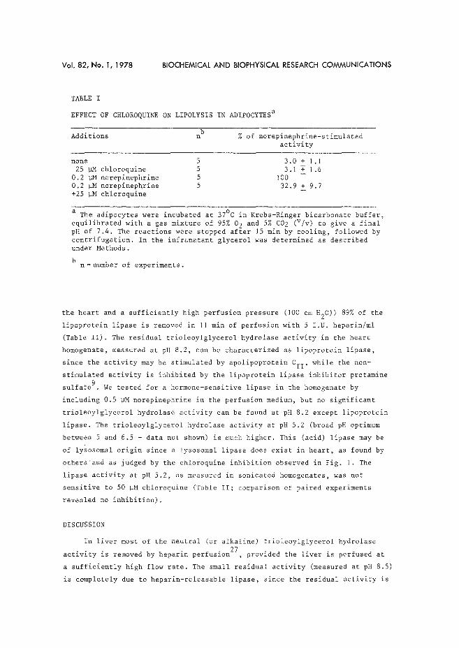
Vol. 82, No. 1, 1978 BIOCHEMICAL AND BIOPHYSICAL RESEARCH COMMUNICATIONS
TABLE I
EFFECT OF CHLOROQUINE ON LIPOLYSIS IN ADIPOCYTESa
Additions
none 25 ~ chloroquine
0.2 ~norepinephrine 0.2 ~M norepinepqrine +25 ~M chloroquine
b n
5 5 5 5
% of norepinephrine-stimulated activity
3.0+1.1 3.1 + 1.6
100 32.9 + 9.7
a The adipocytes were incubated at 37°C in Krebs-Ringer bicarbonate buffer, equilibrated with a gas mixture of 95% 02 and 5% COz (v/v) to give a final pH of 7.4. The reactions were stopped after IS min by cooling, followed by centrifugation. In the infranatant glycerol was determined as described under Methods.
b . n"' number of expen_ments.
the heart and a sufficiently high perfusion pressure (100 em H20)) 89% of the
lipoprotein lipase is removed in II min of perfusion with 5 I.U. heparin/ml
(Table II). The residual trioleoylglycerol hydrolase activity in the heart
homogenate, measured at pH 8.2, can be characterized as lipoprotein lipase,
since the activity may be stimulated by apolipoprotein CII' while the non
stimulated activity is inhibited by the lipoprotein lipase inhibitor protamine
sulfate 9 . We tested for a hormone-sensitive lipase in the homogenate by
including 0.5 ~M norepinephrine in the perfusion medium, but no significant
trioleoylglycerol hydrolase activity can be found at pH 8.2 except lipoprotein
lipase. The trioleoylglycerol hydrolase activity at pH 5.2 (broad pH optimum
between 5 and 6.5 - data not shown) is much higher. This (acid) lipase may be
of lysosomal origin since a lysosomal lipase does exist in heart, as found by
others'and as judged by the chloroquine inhibition observed in Fig. l. The
lipase activity at pH 5,2, as measured in sonicated homogenates, was not
sensitive to 50 IJM chloroquine (T3ble II; comparison of paired experiments
revealed no inhibition).
DISCUSSION
In liver most of the neutral (or alkaline) trioleoylglycerol hydrolase . . . . . 27 . . .
act~v~ty ~s removed by hepar~n perfusJ.on , prov~ded the l~ver ~s perfused at
a sufficiently high flow rate. The small residual activity (measured at pH 8.5)
is completely due to heparin-releasable lipase, since the residual activity is
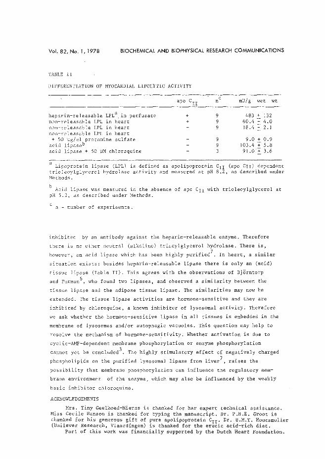
Vol. 82, No. 1, 1978 BIOCHEMICAl AND BIOPHYSICAl RESEARCH COMMUNICATIONS
TABLE II
DIFFERE~TIATION OF HYOCARDIAL LIPOLYTIC ACTIVITY
heparin-releasable LPL3 . in perfusate non-releasable LPL ln heart non-releasable LPL in heart non-releasable LPL in heart
+ 50 '"g/ml protamine sulfate acid lipaseb acid lipase + 50 WM chloroquine
apo CII
+ +
n
9 9 9
9 9 3
c mU/g wet wt
483 + 132 60.4 + 4.0 18.4 + 2. 1
9.0 + 0.9 103.4 + 5.8
91 .0 + 3.6
3 Lipoprotein lipase (LPL) is defined as apolipoprotein c 11 (apo Crr) dependent
trioleoylglycerol hydrolase activity and measured at pH 8.2, as described under Nethods.
b Acid lipase was measured in the absence of apo c11 with trioleoylglycerol at pH 5.2, as described under Methods.
c n =number of experiments.
inhibited by an antibody against the heparin-releasable enzyme. Therefore
there is no other neutral (alkaline) triacylglycerol hydrolase, There is,
however, an acid lipase which has been highly purified7
. In heart, a similar
situation exists: besides heparin-releasable lipase there is only an (acid)
tissue lipase (Table II). This agrees with the observations of BjOrntorp
and Furman6 , who found two lipases, and observed a similarity between the
tissue lipase and the adipose tissue lipase, The similarities may now be
extended. The tissue lipase activities are hormone-sensitive and they are
inhibited by chloroquine, a known inhibitor of lysosomal activity. Therefore
we ask whether the hormone-sensitive lipase in all tissues is embedded in the
membrane of lysosomes and/or autophagic vacuoles. This question may help to
r~solve the mechanism of hormone-sens~tivity. Whether activation is due to
cyclic-AMP-dependent membrane phosphorylation or enzyme phosphorylation
cannot yet be concluded3• The highly stimulatory effect of negatively charged
phospholipids on the purified lysosomal lipase from 1iver7 raises the
possibility that membrane phosphorylation can influence the regulatory mem
brane environment of the enzyme, which may also be influenced by the weakly
basic inhibitor chloroquine.
ACKNOWLEDGEMENTS
Mrs. Tiny Geelhoed-Mieras is thanked for her expert technical assistance. Miss Cecile Hanson is thanked for typing the manuscript. Dr. P.H.E. Groot is thanked for his generous gift of pure apolipoprotein c11 . Dr. U.M.T. Houtsmuller (Unilever Research, Vlaardingen) is thanked for the erucic acid-rich diet.
Part of this work was financially supported by the Dutch Heart Foundation.
Vol. 82, No. 1, 1978 BIOCHEMICAl AND BIOPHYSICAl RESEARCH COMMUNICATIONS
REFERENCES
1. Hollenberg, C.H., Raben, H.S. and Astwood, E.B. (1961) Endocrinology 68, 589-598
2. Rizack, M.A. (1961) J. Biol. Chern. 236, 657-662 3. Huttunen, J.K. and Steinberg_, D. (1971) Biochim. Biophys. Acta 239, 411-427 4. Williamson, J.R. (1964) J. Biol, Chern. 239, 2721-2729 5. Challoner, D.R. and Steinberg, D. (1965) Nature (London) 205, 602-603 6. BjOrntorp, P. and Furman, R.H. (1962) Am. J. Physiol. 203, 323-326 7. Teng, M.-H. and Kaplan, A. (1974) J, Biol. Chern, 249, 1064-1070 8, Fiedling, C,.J. (1972) Biochim. Biophys. Acta 280, 569-578 9. Kraus, R.M., Windmueller, H.G., Levy, R.I. and Fredrickson, D.S. (1973)
J, Lipid Res. 14, 286-295 10. Jansen, H., Van Zuylen-Van Wiggen, A. and Hlllsmann, hl.C. (1973) Biochem.
Biophys. Res. Commun. 55, 30-37 11. Guder, W., FrOhlich, J., Patzelt, C. and Wieland, 0. (1970) FEES Lett. 10,
215-218 12. Ashford, T.P. and Porter, K.R. (1962) J. Cell Biol. 12, 198-202 13. Deter, R.L. and De Duve, C. (1967) J. Cell Biol. 33, 437-449 14. Weismann, G. (1964) Fed. Proc. 23, 1038-1044 15. De Duve, C., De Barsy, Th., Poole, B., Trouet, A., Tulkens, P. and Van
Hoof, F. (1964) Biochem. Parmacol. 23, 2495-2531 16. Wibo, M. and Poole, B. (1974) J. Cell Biol. 63, 430-440 17. Goldstein, J.L., Brunschede, G.Y. and Brown, M.S. (1975) J. Biol. Chern. :250,
7854-7862 18. Floren, c.-H. and Nilsson, R. (1977) Biochem. Biophys. Res. Commun, 74,
520-528 19. Starn, H. and Hlllsmann, W.C. (1977) Basic Res. Cardiol. 72. 365-375 20. Laurell, S. and Tibbling, G. (1966) Clin. Chim. Acta 13, 317-322 21. Chajek, T., Friedman, G., Stein, 0. and Stein, Y. (1977) Biochim. Biophys.
Acta 488, 270-279 22. Schatz, M.C., Twu, J.-S., Pedersen, M.E., Chen, C.-H., Garfinkel, A.S. and
Borensztajn, J. (1977) Biochim. Biophys. Acta 489, 214-224 23. Rodbell, M. (1964) J. Biol. Chern. 239, 375-380 24. Belfrage, P. and Vaughan, M. (1969) J. Lipid Res. 10, 341-344 25. Abdellatif, A.M.M. and Vles, R.O. (1970) Nutr. Metabol. 12, 285-295 26. Jansen, H., Hlllsmann, W.C., Van Zuylen-Van Wiggen. A., Struijck, C.B.
and Houtsmuller, U.M.T. (1975) Biochem. Biophys. Res, Commun. 64, 747-751 27. Jansen, H., Oerlemans, M.C. and HUlsmann, W.C. (1977) Biochem. Biophys.
Res. Commun. 77, 861-867
59

Paper 4.
Erratum
The relation between fatty acid mobilization and
contractility in the isolated, perfused rat heart
H. Starn & W.C. Hlilsmann
Biochem. Biophys. Res. Cornmun. 82, 1978, 609-614
p. 612 Table II, sub~, not significant ·,..{ co:1trol

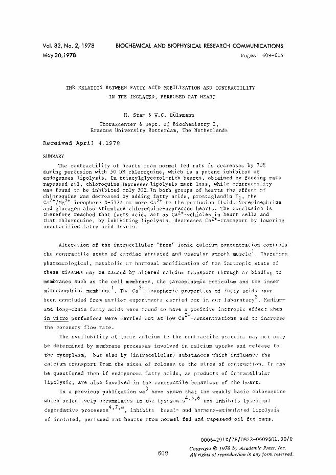
Vol. 82, No. 2, 1978
May 30,1978
BIOCHEMICAl AND BIOPHYSICAl RESEARCH COMMUNICATIONS
Pages 609-614
THE RELATION BETWEEN FATTY ACID MOBILIZATION AND CONTRACTILITY
IN THE ISOLATED, PERFUSED RAT HEART
H. Starn & W.C. Hlllsmann
Thoraxcenter & Dept. of Biochemistry I, Erasmus University Rotterdam, The Netherlands
Received April 4,1978
SUMMARY
The contractility of hearts from normal fed rats is decreased by 70% during perfusion with 50 wM chloroquine, which is a potent inhibitor of endogenous lipolysis. In triacylglycerol-rich hearts, obtained by feeding rats rapeseed-oil, chloroquine depresses lipolysis much less, while contra~tility was found to be inhibited only 30%. In both groups of hearts the effect of chloroquine was decreased by adding fatty acids, prostaglandin E1, the ca2+/Mg2+ ionophore X-537A or more ca2+ to the perfusion fluid. Norepinephrine and glucagon also stimulate chloroquine-depressed hearts. The conclusion is therefore reached that fatty acids act as ca2+-vehicles in heart cells and that chloroquine, by inhibiting lipolysis, decreases ca2+_transport by lowering unesterified fatty acid levels.
Alteration of the intracellular "free" ionic calcium concentration controls
the contractile state of cardiac striated and vascular smooth muscle 1, Therefore
pharmacological, metabolic ur hormonal modification of the inotropic state of
these tissues may be caused by altered calcium transport through or binding to
membranes such as the cell membrane, the sarcoplasmic reticulum and the inner . h . 1 1 2+ . . . . m1toc ondr1a membrane . The Ca -1onophor1c propert1es of fatty ac1ds have
been concluded from earlier experiments carried out in our laboratory2
. Medium
and long-chain fatty acids were found to have a positive inotropic effect when
in vitro perfusions were carried out at low ca 2+-concentrations and to increase
the coronary flow rate.
The availability of ionic calcium to the contractile proteins may not only
be determined by membrane processes involved in calcium uptake and release to
the cytoplasm, but also by (intracellular) substances which influence the
calcium transport from the sites of release to the sites of contraction. It may
be questioned then if endogenous fatty acids, as products of intracellular
lipolysis, are also involved in the contractile behaviour of the heart.
In a previous publication we 3 have shown that the weakly basic chloroquine
which selectively accumulates in the 4, 7,8 . h' . degradative processes , 1n 1b1ts
4 5 6 lysosomes ' ' and inhibits lysosomal
basal- and hormone-stimulated lipolysis
of isolated, perfused rat hearts from normal fed and rapeseed-oil fed rats.
609
0006-291X/78/0822-0609$01.00/0
Copyright© 1978 by Academic Press. inc. All rights of reproduction in any form reserved.
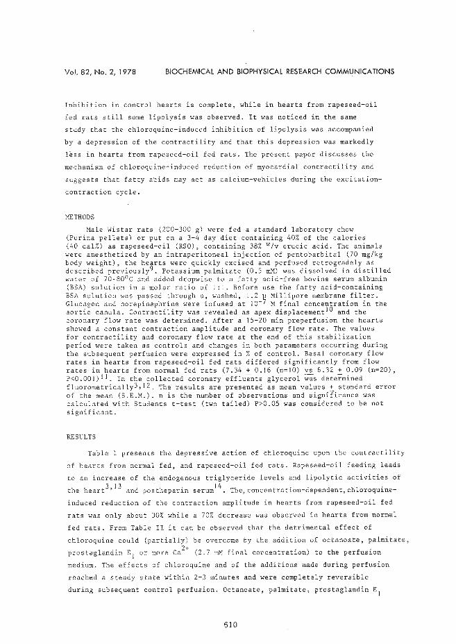
Vol. 82, No. 2, 1978 BIOCHEMICAl AND BIOPHYSICAl RESEARCH COMMUNICATIONS
Inhibition in control hearts is complete, while in hearts from rapeseed-oil
fed rats still some lipolysis was observed. It ,.,ras noticed in the same
study that the chloroquine-induced inhibition of lipolysis was accompanied
by a depression of the contractility and that this depression was markedly
less in hearts from rapeseed-oil fed rats. The present paper discusses the
mechanism of chloroquine-induced reduction of myocardial contractility and
suggests that fatty a·cids may act as calcium-vehicles during the excitation
contraction cycle.
l>JETHODS
Male Wistar rats (200-300 g) were fed a standard laboratory chow (Purina pellets) or put on a 3-4 day diet containing 40% of the calories (40 cal%) as rapeseed-oil (RSO), containing 38% w;v erucic acid. The animals were anesthetized by an intraperitoneal injection of pentobarbital (70 mg/kg body weight), the hearts were quickly excised and perfused retrogradely as described previously9. Potassium palmitate (0.5 roM) was dissolved in distilled water of 70-80°C and added dropwise to a fatty acid-free bovine serum albumin (BSA) solution in a molar ratio of 1:1, Before use the fatty acid-containing BSA solution was passed through a, washed, 1.2 )J Millipore membrane filter. Glucagon and norepinephrine were infused at Jo-7 M final concentration in the aortic canula. Contractility was revealed as apex displacement 10 and the coronary flow rate was determined. After a 15-20 min preperfusion the hearts showed a constant contraction amplitude and coronary flow rate. The values for contractility and coronary flow rate at the end of this stabilization period were taken as controls and changes in both parameters occurring during the s~bsequent perfusion were expressed in% of control. Basal coronary flow rates in hearts from rapeseed-oil fed rats differed significantly from flow rates in hearts from normal fed rats (7.34 + 0.16 (n=10) vs 6.32 + 0.09 (n=20), P<0.001)11. In the collected coronary effluents glycerol was determined fluorometrically3,12. The results are presented as mean values+ standard error of the mean (S.E.M.). n is the number of observations and significance was calculated with Students t-test (two tailed) P>O.OS was considered to be not significant.
RESULTS
Table I presents the depressive action of chloroquine upon the contractility
of hearts from normal fed, and rapeseed-oil fed rats. Rapeseed-oil feeding leads
to an increase of the endogenous triglyceride levels and lipolytic activities of
h h 3, 1 3 d . . 1 4 h . d d hl . t e, eart an posthepar~n serum . T e,concentratlon- epen ent,c _oroqulne-
induced reduction of the contraction amplitude in hearts from rapeseed-oil fed
rats was only about 30% while a 70% decrease was observed in hearts from normal
fed rats. From Table II it can be observed thaL the detrimental effect of
chloroquine could (partially) be overcome by the addition of octanoate, palmitate,
prostaglandin E1
or more ca2+ (2.7 ml:1 final concentration) to the perfusion
medium. The effects of chloroquine and of the additions made during perfusion
reached a steady state within 2-3 minutes and were completely reversible
during subsequent control perfusion. Oc~anoate, palmitate, prostaglandin E1
610
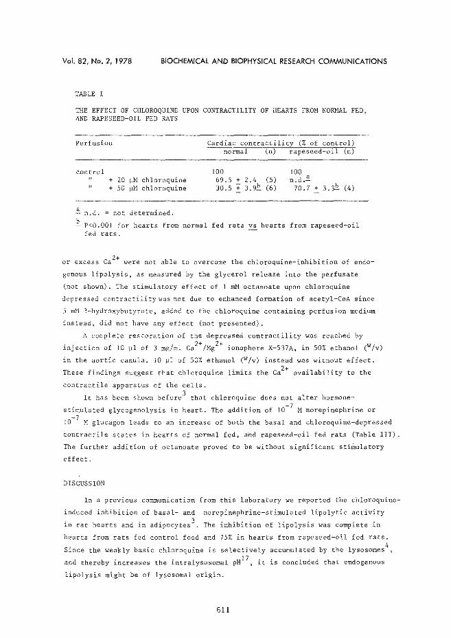
Vol. 82, No. 2, 1978 BIOCHEMICAl AND BIOPHYSICAl RESEARCH COMMUNICATIONS
TABLE I
THE EFFECT OF CHLOROQUINE UPON CONTRACTILITY OF HEARTS FROM NORMAL FED, AND RAPESEED-OIL FED RATS
Perfusion
control + 20 1-JH chloroquine + 50 1-JM chloroquine
~ n.d. =not determined.
Cardiac contractility (% of control) normal (n) rapeseed oil (n)
100 69.5 + 2.4 (5) 30.5 + 3.9E. (6)
100 n.d.~
70.7 + 3.3t (4)
~ P<O.OOI for hearts from normal fed rats vs hearts from rapeseed-oil fed rats.
or excess ca2+ were not able to overcome the chloroquine-inhibition of endo
genous lipolysis, as measured by the glycerol release into the perfusate
(not shown). The stimulatory effect of I mM octanoate upon chloroquine
depressed contractility~.o.ras not due to enhanced formation of acetyl-CoA since
5 mM 3-hydroxybutyrate, added to the chloroquine containing perfusion medium
instead, did not have any effect (not presented).
A complete restoration of the depressed contractility was reached by 2+ 2+ - -injection of 10 ~l of 3 mg/ml Ca /Mg 1onophore X-537A, 1n 50% ethanol (w/v)
in the aortic canula, 10 ~l of 50% ethanol (w/v) instead was without effect.
These findings suggest that chloroquine limits the Ca2+ availability to the
contractile apparatus of the cells.
It has been shown before3 that chloroquine does not alter hormone
stimulated glycogenolysis in heart. The addition of 10-7 M norepinephrine or -7
10 H glucagon leads to an increase of both the basal and chloroquine-depressed
contractile states in hearts of normal fed, and rapeseed-oil fed rats (Table III).
The further addition of octanoate proved to be without significant stiffiulatory
effect.
DISCUSSION
In a previous communication from this laboratory we reported the chloroquine
induced inhibition of basal- and norepinephrine-stimulated lipolytic activity
in rat hearts and in adipocyces 3 The inhibition of lipolysis was complete in
hearts from rats fed control food and 75% in hearts from rapeseed-oil fed rats. 4
Since the weakly basic chloroquine is selectively accumulated by the lysosomes
and thereby increases the intralysosomal pH 17 , it is concluded that endogenous
lipolysis might be of lysosomal origin.
611
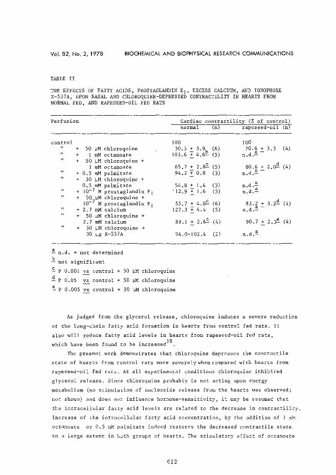
Vol. 82, No. 2, 1978 BIOCHEMICAl AND BIOPHYSICAl RESEARCH COMMUNICATIONS
TABLE II
THE EFFECTS OF FATTY ACIDS, PROSTAGLANDIN El• EXCESS CALCIUM, AND IONOPHORE X-537A, UPON BASAL AND CHLOROQUINE-DEPRESSED CONTRACTILITY IN HEARTS FROM NORMAL FED, AND RAPESEED-OIL FED RATS
Perfusion Cardiac contractility (% of control) normal (n) rapeseed oil (n)
control
" "
~ n.d.
+ SO ~M chloroquine + I mM octanoate + 50 ~M chloroquine +
I mM octanoate + 0.5 roM palmitate + 50 ~M chloroquine +
0.5 mM palmitate + Jo-7 M prostaglandin E 1 + 50 ~M chloroquine +
Jo-7 M prostaglandin E1 + 2. 7 mM calcium + 50 ~M chloroquine +
2.7 mM calcium
=
+ 50 ~M chloroquine + 30 ]Jg X-537A
not determined b
not significant
100 30.5.:: 3.9b
105.6 + 4.fr::.:-
65.7 + 2.~ 94.2 + 0.8
54.8 + I .4 '12.9 + I .6
55.7 + 4.~ I27 .3 + 4.4
89.1 + 2.6.::.
94.0-I02.4
"' p 0.001 vs control + so WM chloroquine
<l_ p o.os vs control + so wM chloroquine
".p o.oos vs control + so WM chloroquine
100 (6) 70.6 + 3.3 (4) (S)
a-n.d.-
(S) 80.6 + 2.o'! (4) (3) a-
n.d.-
(3) n.d.~ (S) n.d . .§.
(6) 83.2 + 3.2<l. (4) (S)
a-n.d.-
( 4) 90.7 + 2.:? (4)
(2) n.d.~
As judged from the glycerol release, chloroquine induces a severe reduction
of the long-chain fatty acid formation in hearts from control fed rats. It
also will reduce fatty acid levels in hearts from rapeseed-oil fed rats,
which have been found to be increased18
.
The present work demonstrates that chloroquine depresses the contractile
state of hearts from control rats more severely when compared with hearts from
rapeseed-oil fed rats. At all experimental conditions chloroquine inhibited
glycerol release. Since chloroquine probably is not acting upon energy
metabolism (no stimulation of nucleoside release from the hearts was observed;
not shown) and does not influence hormone-sensitivity, it may be assumed that
the intracellular fatty acid levels are related to the decrease in contractility.
Increase of the intracellular fatty acid concentration, by the addition of l rnM
octanoate or 0.5 mM palmitate indeed restores the decreased contractile state
to a large extent in both groups of hearts. The stimulatory effect of octanoate
612
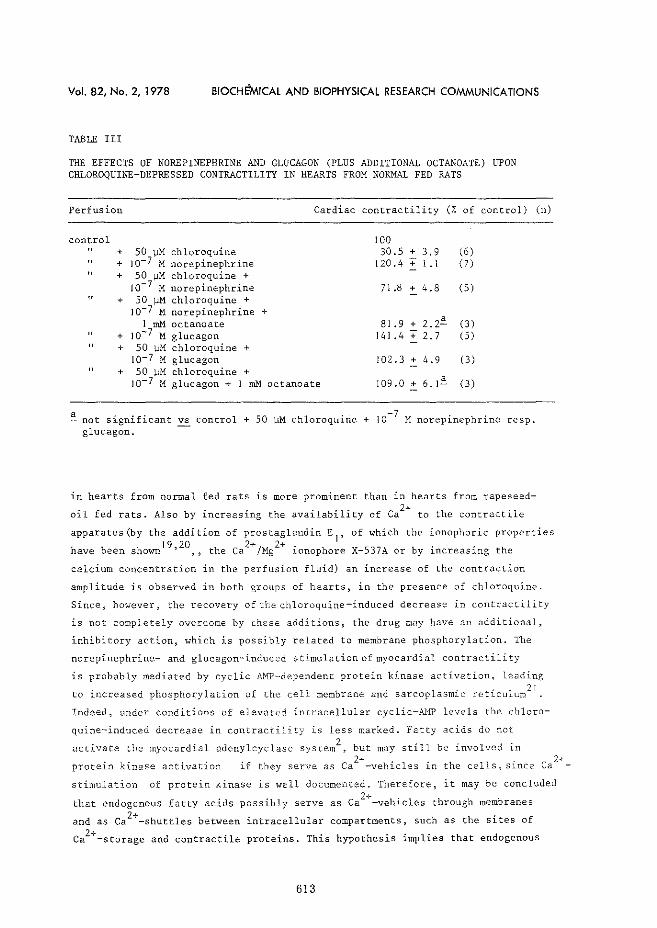
Vol. 82, No. 2, 1978 810CH~MICAl AND BIOPHYSICAl RESEARCH COMMUNICATIONS
TABLE III
THE EFFECTS OF NOREPINEPHRINE AND GLUCAGON (PLUS ADDITIONAL OCTANOATE) UPON CHLOROQUINE-DEPRESSED CONTRACTILITY IN HEARTS FROM NORMAL FED RATS
Perfusion Cardiac contractility (% of control)
control 100 + 50 ~M chloroquine 30.5 + 3.9 (6) + 10-7 M norepinephrine 120.4 + 1.1 (7)
" + 50 1-!M chloroquine + 10-7 M norepinephrine 71.8 + 4.8 (5)
" + 50 f)M chloroquine + Jo-7 M norepinephrine +
1 mM octanoate 81.9 + 2.2~ (3) + 10-7 M glucagon 141 .4 + 2.7 (5) + 50 l-IM chloroquine +
JQ-7 M glucagon 102.3 + 4.9 (3)
+ 50 l-lM chloroquine + Jo-7 M glucagon + 1 mM octanoate 109.0 + 6.1~ (3)
a not significant vs control + 50 1-!M chloroquine + 10-? M norepinephrine glucagon.
resp.
(n)
in hearts from normal fed rats is more prominent than in hearts from rapeseed
oil fed rats. Also by increasing the availability of Ca2
+ to the contractile
apparatus(by the addition of prostaglandin E1
, of which the ionophoric properties
h b 19,20 2+/ 2+ . . . ave een shown ,, theCa Mg ~onophore X-537A or by ~ncreas1.ng the
calcium concentration in the perfusion fluid) an increase of the contraction
amplitude is observed in both groups of hearts, in the presence of chloroquine.
Since, however, the recovery of the chloroquine-induced decrease in contractility
is not completely overcome by r:hese additions, the drug may have an additional,
inhibitory action, which is possibly related to membrane phosphorylation. The
norepinephrine- and glucagon-induced stimulation of myocardial contractility
is probably mediated by cyclic AMP-dependent protein kinase activation, leading
to increased phosphorylation of the cell membrane and sarcoplasmic: reticulun/ 1
Indeed, under conditions of elevated intracellular cyclic-N1P levels the chloro-
quine-induced decrease in contractility is less marked. Fatty acids do not
activate the myocardial adenylcyclase system2 , but may still be involved in
protein kinase activation ;f c 2+ h. l . h ll . c l+ ... they serve as a -ve 1.c es 1n t e ce s, s1.nce a -
stimulation of protein kinase is well documented. Therefore, it may be concluded 2+
that endogenous fatty acids possibly serve as Ca -vehicles through membranes
and as ca2+-shuttles between intracellular compartments, such as the sites of 2+ Ca -storage and contractile proteins. This hypothesis implies that endogenous
613
Vol. 82, No. 2, 1978 BIOCHEMICAL AND BIOPHYSICAL RESEARCH COMMUNICATIONS
lipolysis might be involved in the modulation (or regulation) of myocardial
contractility.
ACKNPWLEDGEMENTS
The authors wish to thank Mr. Wout Breeman for his skilled technical assistance and Hiss Cecile HansLm for typing the manuscript. Drs. F. ten Hoar and U.M.T. Houtsmuller (Unilever Research, Vlaardingen, The Netherlands) are thanked for their generous gifts of prostaglandin E 1 and the rapeseed-oil diet. The ca2+jMg2+ ionophore X-537A was a gift from Roffman-La Roche (Nutley, New Jersey, U.S.A.).
This study was financially supported by the Dutch Heart Foundation.
REFERENCES
l. Ebashi, S. (1976) Ann. Rev. Physiol, 38, 293-313 :2. Hi.ilsmann, h'.C. (1976) Basic Res. Cardiol. 71, 179-191 3. Hlilsmann, \<.1 .C. and H. Starn (1978) Biochem. Biophys. Res. Cornmun.,
accepted for publication 4. Wibo, M. and Poole, B. (1974) J. Cell. Biol. 63, 430-440 5. De Duve, C., De Barsy, Th., Poole, B., Trouet, A., Tulkens, P. and
\'an Hoof, F. (1964) Biochem. Pharmacal. 23, 2495-2531 6. Hendy, R.J., Abraham, R. and Grasso, P. (1969) J. Ultrastr. Res. 29, 485-495 7. Goldstein, J.L., Brunschede, G.Y. and Brown, M.S. (1975) J. Biol. Chern. 250,
7854-7862 S. Floren, C.-H. and Nilsson, ~. (1977) Biochem. Biophys. Res. Comrnun. 74,
520-528 9. Starn, H. and Hlilsmann, W.C. (1977) Basic Res. Cardiol. 72, 365-375
10. Starn, H. and DeJong, J.W. (1977) J. Mol. Cell. Cardiol. 9, 633-650 11 . Ten Hoor, F. and De Deckere, E., personal communication 12. Laurell, S. and Tibbling, G. (1966) Clin. Chim. Acta 13, 317-322 13. Jansen, H., Hlilsmann, W.C., Van Zuylen-Van Wiggen, A., Struijk, C.B. and
Houtsmuller, U.M.T. (1975) Biochem. Biophys. Res. Commun. 64, 747-751 14. Struijk, C.B., Houtsmuller, U.M.T., Jansen, H. and Hlilsmann, W.C. (1973)
Biochim. Biophys. Acta 296, 253-256 15. Schaffer, S.W., Safer, B., Scarpa, A. and Williamson, J.R. (1974) Biochem.
Pharmacal. 23, 1609-1617 16. Starn, H. and Hlilsmann, W.C. (1978) Basic Res. Cardiol., accepted for
publication 17. Reyngoud, D.J. and Tager, J.M. (1977) Biochim. Biophys. Acta 472, 419-449 18. Houtsmuller, U.M.T., Struijk, C.B. and VanderBeek, A. (1970) Biochim.
Biophys. Acta 218, 564-566 19. Kirtland, S.J. and Baum, H. (1972) Nature (New Biology) 236, 47-49 20. Asimakis, G.K. and Sordahl, L.A. (1977) Arch. Biochem. Biophys. 179, 200-210 21. Katz, A.M. and Repke, D.I. (1973) Am. J. Cardiol. 31, 193-201
614
Paper 5. Sephadex-induced reduction of coronary flow in the
isolated rat heart: A model for ischemic heart
disease
H. Starn & J.W. de Jong
J. Mol. Cell. Cardiol. 9, 1977, 633-650

Journal of Molecular and CellularCardirJio/I)' ( 1977) 9, 633--4;50
Sephadex-induced Reduction of Coronary Flow in the Isolated Rat Heart: A Model for Ische:mic Heart Disease*
H: STAM ANDJo W. DEJONG Department of Biochemistry I and Tlwraxcenter,
Erasmus University Rotterdam, Rotterdam, The Netherlands
(Received 12 March 1976, accepted in revised form 7 October !976)
H. STAM AND J. W, DEJoNG. Sephadex:~induced Reduction of Coronary Flow in the Isolated Rat Heart: A Model for Ischemic Heart Disease. Journal of Molecular and Cellular Cardiolr;gy (1977) 9, 633-650. Embolization of the coronary arteries with a polysaccharide (Sephadex) microsphere suspension caused a reduction in coronary flow of isolated perfused rat hearts. Obstruction by the spheres occurred in arterioles at the sub-epicardial layers of the ventrides, resulting in a whole-heart ischemia. Flow reduction was proportional to the amount ofmic:rospheres injected and ''das followed by an increase in flow. The increase in coronary flow after the initial decrease is probably due to vasodilation of the nQn-obstructed vessels, During the first 10-rnin period of this Sephadex-induced ischemia (with 0.70 mg of wicrospheres) the energy charge, ([ATPJ +-!- [ADP])/ ([ATP] + [ADP] + [AMP]), dropped from 0087 ± 0.01 to 0077 ± O.G! (fi ± s.E., P <0.005) and creatine phosphate content dropped from 34.9 ± 1.45 to 13.5 ± 0.63 (P <0.001) ~mol. g myocardial protein-1 . In the effluents of the ischemic hearts total nucleoside (adenosine, inosine, hypoxanthine) and lactate release increased indicating severe oxygen deprivation during ischemia. Mechanical performance measured as apex displacement, correlates weU with left ventricular systolic pressure. Under ischemic conditions a rapid rise in contractility during the first minute (to 129% of control values, P < 0.005) was followed by a sharp ,:! ::crease to 40%. The results of the experiments with ischemic hearts were compared W-\""1 t..~ose from hearts made hypoxic by lowering the oxygen percentage in the gas-mixture bubbling through the perfusion medium (60%, 30% and 0% oxygen). With 30% oxygen, no significant differences were measured between ischemic and hypoxic hearts with respect to contractility (apex displacement), energy charge and creatine phosphate content, whereas total nucleoside and lactate release were in the same order of magnitude. The model described here could be useful in studies of ischemia in which a low coronary flow accompanying hypoxia is essential, and of consequences of the vascular respome to embolization-induced ischemia.
KEY WoRDs: Creatine phosphate; A TP; ADP; AMP; Adenosine; Inosine; Hypoxanthine; Lactate; Coronary flow; Apex displacement; Sephadex-induced ischemia; Hypmcia.
1. Introduction
Lack of oxygen during hypoxia and ischemia is responsible for the alteration of myocardial high-energy phosphate content while cardiac function is strongly impaired [ 10, 18, 29]. In studies with isolated perfused heart preparations, hypoxia was produced by lowering the oxygen tension of the perfusate without any decrease in coronary flow. Ischemia was produced by ligation of the main branch of the left
* This investigation was supported by the Dutch Heart Foundation.
634 H. STAM AND J· W. DEJONG
coronary artery [5], by placing a one-way valve in the aortic outflow tract preventing retrograde perfusion of the coronary arteries during diastole [27, 31], by lowering of the perfusion pressure [ 14] or, in vivo, by constricting the abdominal aorta [7].
The reduced coronary flow under these ischemic conditions impairs the delivery of oxygen and energy substrates and prevents an adequate removal of metabolic products. During hypoxia as well as ischemia, metabolic breakdown products of ATP, such as adenosine, inosine and hypoxanthine, are released by the isolated perfused heart [3, 4, 16] and in vivo [7]. The increased anaerobic glycolytic flux enhances lactate production and release [31].
In the past, several investigators reported the use of embolization of the coronary arteries with microspheres of several kinds and sizes in order to produce a cardiagenic shock [J, 19, 33, 37]. Other studies dealt with the coronary vascular response after occlusion of a coronary artery or coronary embolization with spheres [13, 25]. The reduction in coronary flow after embolization and some circulatory, biochemical and pathological consequences were studied by Bing et al. [6, 34].
The abovementioned studies were performed in closed- or open-chested animals (dogs, calves), while Monroe et al. [ 26] first studied embolization in the isolated, supported dog heart.
In the study presented here, rat hearts were perfused retrogradely and reduction of coronary flow was induced by embolization of the coronary arteries with Sephadex G-25 microspheres. The ischemic state induced by embolization was characterized and the alterations in cardiac function, high-energy phosphate content of the myocardial tissue and release of nucleosides and lactate from the heart following embolization were compared with results obtained under hypoxia. The observed changes in cardiac function, high-energy phosphate content, nucleoside and lactate release after Sephadex addition indicate that ischemia induced by embolization of the coronary arteries may be useful to create the combination of hypoxia and decreased coronary flow, as is observed in ischemic heart disease.
2. Materials and Methods
Male Wistar rats (220 to 250 g) were fed ad libitum, anesthetized with an intraperitoneal injection of sodium pentobarbital (70 mg. kg-1 body weight, Abbott, Saint-Remy sur _,_6;,.v:re, France). Prior to the cannulation procedure they were heparinized with 500 iu Thromboliquine (Organon, Oss, The Netherlands)o The hearts were perfused retYogradely at 37°C and pH 7A with a 1nodified Tyrode solution [24]) equilibrated vrith 95o/0 02 + 5~·~ C02~ according to the Langendorff technique. The hea:rts vv.ere at 300 pulses. m.in-1~ 2 ms duration and 0.2 rnA. The perfusion pressure was l 00 em Contractility was measured as apex displacement (AD), using a :tfe-v\llett·-Packard transducer (7-DCDT-100), fitted with a core of tampon steel (l .3 g), ,_.v}Lich was hooked to the apex of the heart [ 41] 0
SEPHADEX-INDUCED ISCHEMIA IN ISOLATED HEART 635
Left ventricular pressure was measured by inserting a 19-gauge needle through the apex of the heart into the left ventricle, connected with a Statham pressure transducer (model P23Gb). Pressure and displacement were recorded simultaneously on a SE Oscillograph 3006/DL. Hypoxia was induced by substituting the oxygen in the medium by nitrogen. Final oxygen percentages were 60, 30 and O%.
Reduction of coronary flow was induced by injecting Sephadex G-25 fine microspheres (particle size 20 to 80 p.m), suspended in oxygenated perfusion medium, into the lumen of the cannula. Hypoxia and embolization took place after 30 min of control perfl1sion.
At regular intervals, eflluent was collected in liquid nitrogen and stored at -20°C. At the end of the perfusion beating rats hearts were clamped between aluminium blocks at the temperature of liquid nitrogen [ 40] and extracted as des~ribed by DeJong [9], The myocardial tissue extracts were stored at -20°C. All deterffiinations in extracts and effluents were carried out within 48 h after clamping the heart. Creatine phosphate, ATP, ADP and AMP were determined according to standard enzymatical procedures [ 21] on a Gilford 2400 recording spectrophotometer. Values for AMP were corrected for AMP-like material in the NADH (Boehringer, Mannheim). Adenosine, inosine and hypoxanthine were estimated with an Aminco DW-2, double-beam spectrophotometer. Lactate was determined enzymatically on an AutoAnalyzer H (Technicon, Tarry Town, New York) according to Apstein et al. [2]. Protein was measured by the biuret method [ 15]. All chemicals were analytical grade.
For microscopic examination whole hearts were rapidly frozen in isopentane cooled to -150°C with liquid nitrogen [23], 5 min after embolization with 0.70 mg of Sephadex microspheres. Sect,~ns, from different parts of the myocardium, at a thickness of 6 to 10 tJ.ffi were cut on a cryostat-microtome. The cross-sections were stained with hematoxilin-eosin (HE). For histological demonstration of glycogen in the tissue staining was carried out with the periodic acid-Schiff (PAS) reaction. Due to the fixation and staining technique the Sephadex microspheres were subjected to shrinkage. Results are given in mean values ± s.E.," and significance was calculated with Student's I-test. P>0.05 was considered to be not si'l"nificant.
3. Results
Apex displacement as a marker for contractility
Measurement of the apex displacement of the beating heart can be regarded as the registration of isotonic contraction [ 24]. Apex displacement, so, is a parameter for contractility. Figure l illustrates the similarity of the changes in the apex displacement and left ventricular systolic pressure (L VSP) during anoxia and embolizationinduced reduction of coronary flow (ischemia: see next section). Immediately after
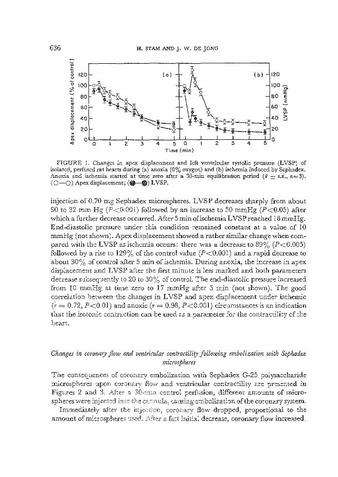
636 H. STAMAND J. W. DEJONG
"§ - 2 c
120 ( 0) - (b) - 120 0 u 0\ - o-O 0 100 - 100-~ !-~2'sk \Q, ~
~ 0 :z: c 80 80 E
E e '!-! '2,~ 60 E 60 - '! 0. w '! "' u 0 40
- ~~·-~ ll!,,)-H-<:i-'2 40 > 0. ...)
• ~ 20 1- Q-Q !-~-!-!-!- 20 X
I I I I I I w L I _!_ I 5 0 ~ 0
"' 0 2 3 4 5 0 2 3 4 rime (m-in)
FIGURE 1. Changes in apex displacement and left ventricular systolic pressure (LVSP) of isolated, perfused rat hearts during (a) anoxia (0% oxygen) and (b) ischemia induced by Sephadex. l\.noxia and ischemia started at time zero after a 30-min equilibration period (X ± s.i., n=3). ( 0-0) Apex displacement; (-II) LVSP.
injection of 0.70 mg Sephadex microspheres. LVSP decreases sharply from about 90 to 32 mm Hg (P<O.OOI) followed by an increase to 50 mmHg (P<0.05) after which a further decrease occurred. After 5 min of ischemia L VSP reached 18 mmHg. End~diastolic pressure under this condition remained constant at a value of 10 mmHg (not shown). Apex displacement showed a rather similar change when compared with the LVSP as ischemia occurs: there was a decrease to 89% (P<0.005) followed by a rise to 129% of the control value (P<O.OOl) and a rapid decrease to about 30%) of control after 5 min of ischemia. During anoxia, the increase in apex displacement and L VSP after the first minute is less marked and both parameters decrease subsequently to 20 to 30% of control. The end-diastolic pressure increased from !0 mmHg at time zero to 17 mmHg after 5 min (not shown). The good correlation between the changes in L VSP and apex displacement under ischemic (i- = 0.72, P<O.OI) and anoxic (r = 0.98, P<O.OOI) circumstances is an indication that the isotonic contraction can be used as a parameter for the contractility of the heart.
Changes in coronary flow and ventricular contractility following embolization with Sephadex microspheres
The consequences of coronary embolization with Sephadex G-25 polysaccharide microspheres upon co:ronar; fl_m,v and ventricular contractility are presented in Figures 2 and 3 .. After a 30-.min control perfusion~ different amounts of micro-spheres were injected in.:-:o the causing embolization of the coronary system.
Immediately a±l:e:r the coronary flow dropped, proportional to the amount of microsphe:res usedo .i;,.fte:r a fast initial decrease, coronary flow increased.
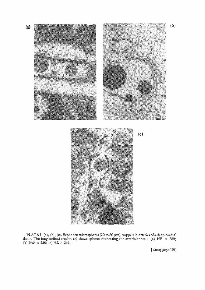
(al (b)
(c)
PLATE l. (a), (b), (c). Sephadex microspheres (20 to 80 fLID) trapped in arteries of sub-epicardial tissue. The longitudinal section (c) shows spheres dislocating the arteriolar wall. (a) HE X 280; (b) PAS X 350; (c) HE X 245.
[facing page 636]

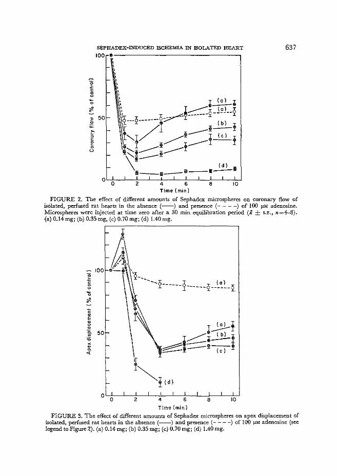
SEPHADEX-INDUCED ISCHEMIA IN ISOLATED HEART
100;~------------------------------,
' t-' \ ' ' t- .. '
Time (min)
637
FIGURE 2. The effect of different amounts of Sephadex microspheres on coronary flow of isolated, perfused rat hearts in the absence (---) and presence (-- - -) of 100 !J.M adenosine. Microspheres were injected at time zero after a 30 min equilibration period (X ± S.E., n=4--8). (a) 0.14mg; (b) 0.35 mg, (c) 0.70 mg; (d) 1.40 mg.
100 ' :g '2-._ 0 ---2---- ~ 2 (a l 0 -- ------ ------2. u -0
l ~ • ~ ~
E ~
I~! u 0
0. 50 ·" -----=•~! ~ :ei::::=!-( c-)-! X ~ ~
"' ~ ~!tdl
0 0 2 4 6 8 10
Time {min)
FIGURE 3. The effect of different amounts of Sephadex microspheres on apex displacement of isolated, perfused rat hearts in the absence (---) and presence (--- -) of 100 !J.M adenosine (see legend to Figure~). (a) 0.14 mg; (b) 0.35 mg; (c) 0.70 mg; (d) 1.40 mg.
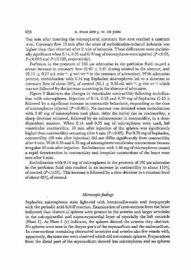
638 H. STAMAND J. W. DEJONG
Ten min after inserting the microspheres; coronary flow rate reached a constant level. Coronary flow lO min after the onset of embolization-induced ischemia was higher than that observed after 2 min of ischemia. These differences were statistically significant when 0.14, 0.35 and 0. 70 mg ofmicrospheres were injected (P<0.05, P<0.025 and P<0.025, respectively).
Perfusion in the presence of !00 fLM adenosine in the perfusion fluid caused a severe increase in coronary flow (6.40 ± 0.61 during control in the absence, and 22.15 ± 0.27 ml. min-1 . g wet wt-1 in the presence of adenosine). With adenosine present, embolization with 0.14 mg Sephadex microspheres led to a decrease in coronary flow of about 50% of control (10.1 ± 0.50ml. min-1. g wet wt-1 which was not followed by the increase occurring in the absence of adenosine.
Figure 3 illustrates the changes in ventricular contractility following embolization with microspheres. Injection of 0.14, 0.35 and 0.70 mg of Sephadex G-25 is followed by a significant increase in contractile behaviour, responding to the dose ofmicrospheres injected (P<O.OO!). No increase was detected when embolization with 1.40 mg of microspheres took place. After the initial rise in contractility, a sharp decrease occurred, followed by an enhancement in contractility, in a dosedependent manner. When 0.14 and 0.35 mg of microspheres were used, left ventricular contractility, 10 min after injection of the spheres was significantly higher than contractility occurring after 4 min (P<0.05). For 0. 70 mg ofSephadex, contractility (10 min after injection) did not differ significantly from contractility after 4 min. With 0.35 and 0. 70 mg of microspheres ventricular contractions became irregular !0 min after injection. Embolization with 1.40 mg of microspheres caused a rapid deterioration in contractility and irregular contractions of the heart were seen after 4 min.
Embolization with 0.14 mg of microspheres in the presence of l 00 fLM adenosine in the perfusion fluid also resulted in an increase in contractility to about ll 0% of control (P<O.Ol). This increase is followed by a slow decrease to a constant level of about 85% of control.
Microscopic findings
Sephadex microspheres stain light-red with hematoxilin-eosin and deep-purple with the periodic acid-Schiff reaction. Examination of cross-sections from the heart indicated that clusters of spheres were present in the arteries and larger arterioles in the sub-epicardial and supra-myocardial layer of especially the left ventricle (Plate l). As Plate l (c) indicates, the spheres distend the arteries they obstruct. No spheres were seen in the deeper parts of the myocardium and the endocardium. In cross-sections containing obstructed arterioles and arteries also few vessels with, apparently, the same size were observed which did not contain spheres. Preparations from the distal part of the myocardium showed less microspheres and no spheres
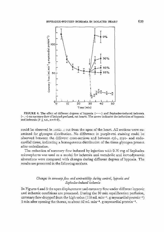
SEPHADEX~INDUCED ISCHEMIA IN ISOLATED HEART
·~ -~ e roo ~
0
~ 0 u 0 ~
E ~
;-c
E
"E so ~ 0
I: 0 c 0
5 u
---! 0%
Time {min}
639
FIGURE 4. The effect of different degrees of hypoxia (--) and Sephadex·induced ischemia (-.-)on coronary flow of isolated perfused, rat hearts. The arrow indicates the induction of hypoxia and ischemia (X± s.E., n=4-8).
could be observed in .cectic .. 3 cut from the apex of the heart. All sections were examined for glycogen distribution. No difference in purple-red staining could be observed between the different cross-sections and between epi-, myo- and endocardial tissue, indicating a homogeneous distribution of the tissue glycogen present after embolization.
The reduction of coronary flow induced by injection with 0. 70 mg of Sephadex microspheres was used as a model for ischemia and metabolic and hemodynamic alterations were compared with changes during different degrees of hypoxia. The results are presented in the following sections.
Changes in coronary flow and contractility during control, hypoxia and Sephadex-induced ischemia
In Figures 4 and 5 the apex displacement and coronary flow under different hypoxic and ischemic conditions are presented. During the 30 min equilibration perfusion, coronary flow dropped from the high value ( 118 ml. min -1. g myocardial protein -1) 5 min after opening the thorax, to about 52 ml. min-1. g myocardial prptein-1.
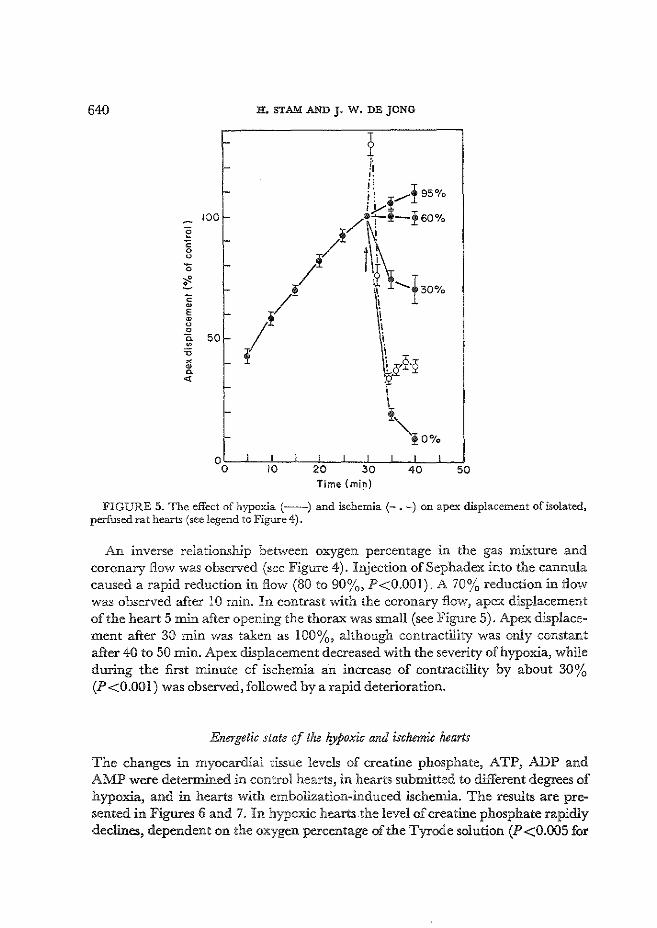
640 H, STAMAND J• W. DEJONG
:- 1 f-
i• ·I .. 'I
- I. ! 0 i I !,.....- 95 ~o 1)/
100 f- 1/tO-!oo% e i-" t j; 0
u
'0 i-,I! p_/ It ~----± 30% !.. t-c w ( \ E t-Q 0
I \ g6·2
0 c. 50 t--~ ~
X -0 0. ~l ~ <(
I ! "!O%
0 I I I I I I I 0 10 20 30 40 50
Time (min)
FIGURE 5. The effect of hypoxia(--) and ischemia(-.-) on apex displacement of isolated, perfused rat hearts (see legend to Figure 4).
An inverse relationship between oxygen percentage i.rt the gas wixture and coronary flow was observed (see Figure 4). Injection ofSephadex into the cannula caused a rapid reduction in flow (SO to 90%, P<O.OOJ). A 70% reduction in flow was observed after 10 min" In contrast with the coronary flow, apex displacement of the heart 5 min after opening the thorax was small (see Figure 5). Apex displacement after 30 min was taken as 100%, although contractility was onJy constant after 40 to 50 min. Apex displacement decreased with the severity of hypoxia, while during the first minute of ischemia an increase of contractility by about 30% (P <0.00 l) was observed, followed by a rapid deterioration.
Energetic state of the hypoxic and ischemic hearts
The changes in myocardial tissue levels of creatine phosphate, ATP, ADP and AMP were determined in control hearts, in hearts submitted to different degrees of hypoxia, and in hearts with embolization-induced ischemia. The results are presented in Figures 6 and 7. In hypoxic hearts the level of creatine phosphate rapidly declines, dependent on the oxygen percentage of the Tyrode solution (P<0.005 for

0 E 10 "-
SEPHADEX-INDUCED ISCHEMIA IN ISOLATED HEART
I b l
o'--L~~~~-L~,~~~L-~_J--~~~~~~r,J-~,~~-fl~~~~ 0 10 30 40 50 0 10 30 40 50 Time (min)
641
FIGURE 6. Creatine-phosphate (a) and ATP (b) content in hypoxic(---) and ischemic(-.-) rat hearts (see legend to Figure 4).
Time (min}
FIGURE 7. ADP (a) and AMP (b) contents in hypoxic(---) and ischemic(-.-) rat hearts (see legend to Figure 4).
all degrees of hypoxia compared with the control value), The decrease in tissue levels of ATP is less rapid than creatine phosphate breakdown, but still significant (P<0,025 for all degrees of hypoxia),
The decrease in tissue ATP levels is accompanied by a rise in ADP and AiviP contents during the different degrees of hypoxia (P<0.05 for ADP and P<O.Ol for AMP), The energy charge of the myocardial tissue, ([ATP] + 7 [ADP])/([ATP] + [ltDPJ + [AMP]), decreased from 0,87 ± 0,01 with fully oxygenized medium to 0,66 ± 0,22 under anoxic conditions (P<0,005), During the first lO min of
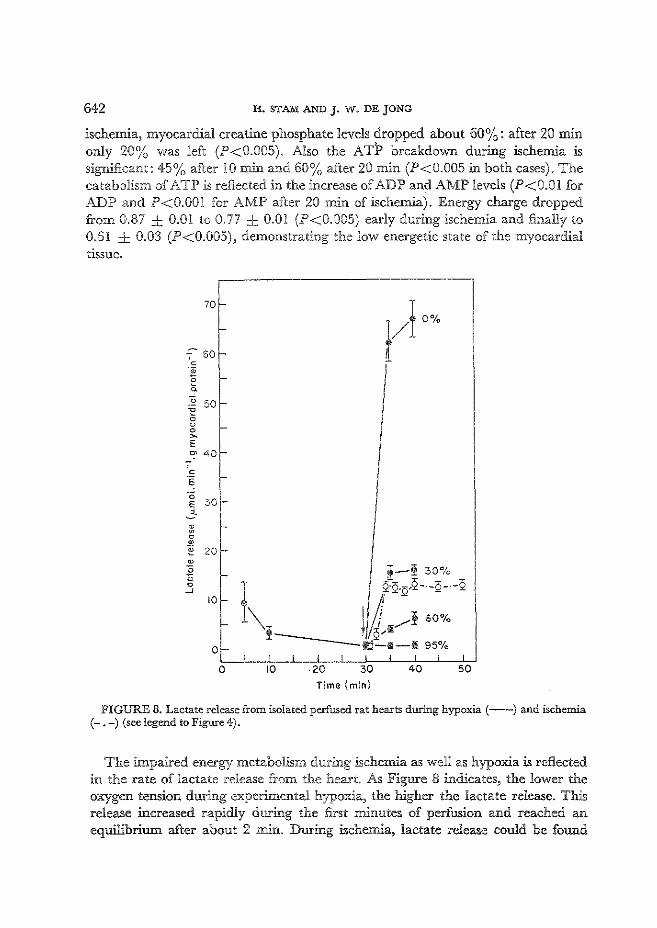
642 H. STAMAND J· W. DEJONG
ischemia, myocardial creatine phosphate levels dropped about 60%: after 20 min only 20% was left (P<0.005). Also the ATP breakdown during ischemia is significant: 45% after 10 min and 60% after 20 min (P<0.005 in both cases). The catabolism of ATP is reflected in the increase of ADP and AMP levels (P<O.Ol for ADP and P<O.OOl for AMP after 20 min of ischemia). Energy charge dropped from 0.87 ..:_ 0,01 to 0.77 ± 0.0! (P<0.005) early during ischemia and finally to 0.61 ± 0.03 (P<0.005), demonstrati11g the low energetic state of the myocardial tissue.
70
7 60 c •• 0 0.
~ 50 0 u 0 > E c:' 40
~ 0 ® e 20
10
0
0 10 20 30 40 50 Time {min)
FIGURE 8. Lactate release from isolated perfused rat hearts during hypoxia (--) and ischemia (-. -) (seelegend to Figure 4).
The impaired energy metabolism during ischemia as well as hypoxia is reflected in the rate of lactate release from the heart. As Figure 8 indicates, the lower the oxygen tension during experi.menta! hypoxia, the higher the lactate release. This release increased rapidly during the first minutes of perfusion and reached an equilibrium after about 2 min. During ischemia, lactate release could be found
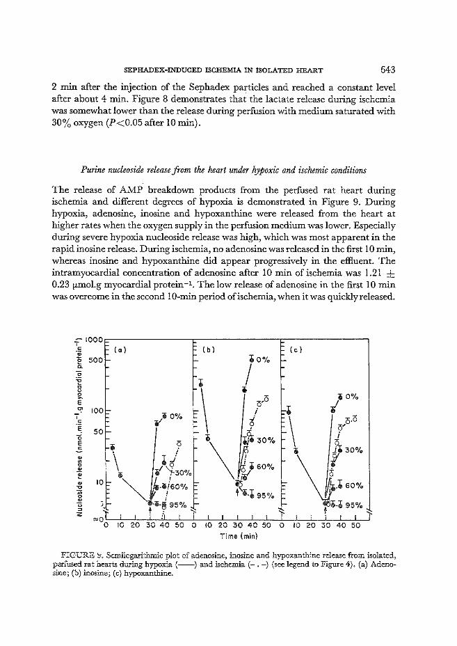
SEPHADEX-INDUCED ISCHEMIA IN ISOLATED HEART 643
2 min after the injection of the Sephadex particles and reached a constant level after about 4 min. Figure 8 demonstrates that the lactate release during ischemia was somewhat lower than the release during perfusion with medium saturated with 30% oxygen (P<0.05 after 10 min).
Purine nucleoside release from the heart under hypoxic and ischemic conditions
The release of AMP breakdown products from the perfused rat heart during ischemia and different degrees of hypoxia is demonstrated in Figure 9. During hypoxia, adenosine, inosine and hypoxanthine were released from the heart at higher rates when the oxygen supply in the perfusion medium was lower. Especially during severe hypoxia nucleoside release was high, which was most apparent in the rapid inosine release. During ischemia, no adenosine was released in the first 10 min, whereas inosine and hypoxanthine did appear progressively in the effluent. The intramyocardial concentration of adenosine after 10 min of ischemia was 1.21 ± 0.23 fLmol.g myocardial protein-1 . The low release of adenosine in the first 10 min was overcome in the second 10-min period of ischemia, when it was quickly released.
=-1000~-------------.~------------~--------------. ' c ·;:; E soo 0.
~ 100
50
10
(a) (b) (c)
4io%
iO%
I
I •\ i icl'
i \ c/j.o e T~ oO% -d._
i\ ~ / \ ., i f" 30%
iO "i ~30% .~· 60% \j· ""' .• ,!;60% ~ ;~·· 60%
""' I •·h5% il·li·!!l 95% ! i.$ 95%
,.0l I I t il I J I I I I I 1 I I I I I J 0 10 20 30 40 50 0 10 20 30 40 50 0 10 20 30 40 50
Tlme (min)
FIGURE ~. Semilogarithmic plot of adenosine, inosine and hypoxanthine release from isolated, perfused rat hearts during hypoxia (---) and ischemia(-.-) (see legend to Figure 4). (a) Adenosine; (b) inosine; (c) hypoxanthine.

644. H. STAMAND J. W. DEJONG
TABLE l. Correlations between metabolic and fWlctional changes in isolated perfused rat hearts during Sephadex-induced ischemia and different degrees of hypoxia
Coronary flow: hypoxia* ischemiat
Apex displacement hypoxia* ischemiat
vs Energy charge
0.98 0.95
Correlation coefficient (r) vs Creatine vs Adenosine vs Lactate phosphate release release
0.94 0.97
0.94 -0.16t
-0.98 -0.94
* 10 min after subjecting rat hearts to different degrees of hypoxia all parameters shown were determined and correlated to each other.
t Perfused rat hearts were made ischemic by injecting Sephadex microspheres into the coronary circulation system. At regular intervals during ischemia the parameters shown were determined and correlated.
:j: Not significant, all other correlations P < 0.001.
In Table !, some correlations between metabolic and functional changes during ischemia and hypoxia are presented. During ischemia as well as during hypoxia, high-energy phosphate content, energy charge and lactate release correlate well with contractility. Coronary flow and adenosine release correlate significantly during hypoxia, while no correlation is found during ischemia.
4. Discussion
Pressure development in the left ventricle is often used as a parameter for the mechanical performance of the heart [17, 20]. In this study, the isotonic contraction, apex displacement was taken as marker for cardiac function. Evidence is presented that the apex displacement correlates well with the L VSP under ischemic and anoxic conditions. Measurement of the apex displacement is simple and the trauma caused by connecting the tampon steel to the apex of the heart seems less compared to the insertion of a needle or left atrial appendectomy prior to balloon catheterization [ 35]. The investigations presented here describe a model for the induction of ischemia by embolization of the coronary arteries by Stphadex microspheres with a particle size of 20 to 80 f!.m. The reduction of coronary flow caused by embolization is dependent on the amount of microspheres used. This implicates that the higher the amount of microspheres injected the greater the number of vessels obstructed by the spheres.
Obstruction occurs at the level of the coronary arteries and larger arterioles in the sub-epicardial layers of, especially, the left ventricle. So, epicardial flow, the major contributor of total coronary flow probably is affected especially by embolization.

SEPHADEX-INDUCED ISCHEMIA IN ISOLATED HEART 645
The presence of non-obstructed arteries indicates that flow reduction is not uniform. The arterial muscular wall is well developed and these vessels play an important role in the circulatory mechanism. Vasomotion is controlled by the many sympathic fibres ending in the wall musculature and by vasoactive metabolites. In the isolated, perfused heart preparation, autonomic influences can be excluded. A second regulatory possibility is found in the precapillary sphincters, a small muscular cuff at the origin of capillaries and met-arterioles, the tone of which regulates fluid entrance to the capillary bed. The sphincter muscle-tone is sensitive to local chemical influences.
Cardiac metabolic activity and coronary flow rate are very well related and metabolites have been suggested to mediate coronary flow during different physiological and pathophysiological states. Berne and Rubio [ 3] reported the potent vasodilator adenosine as possible mediator of coronary blood flow.
The increase in coronary flow following the initial decrease after injection of the microspheres is ai;>sent in the presence of adenosine. It may be concluded that this increase is caused by coronary dilation of the non-obstructed vessels [ 13]. The coronary vascular response is less marked when more arterioles are obstructed after injection of a larger amount of microspheres. It is emphasized that coronary flow in embolized hearts, in the presence of adenosine, is still abo'lt three times higher than flow in embolized (ischemic) hearts perfused without adenosine. Obviously, in the presence of exogenous adenosine a greater number of capillaries is open for perfusion. Also ventricular contractility recovered from the low level 4 min after different degrees of embolization, probably by a partial re-establishment of energy met~bolism due to the increase in coronary flow rates.
Several models for experimental ischemia in isolated heart preparations have been described [5-7, 14, 27, 31, 39]. The reduction of retrograde aortic perfusion as developed by Neely et al. [17] is often used in studies of biochemical and hemodynamic changes during the acute phase of whole heart ischemia. In this model, however, the reduction of coronary flow is an all or nothing effect in response to closure of the aortic outflow tract and the consequences of cor'!nary vasodilation, occurring after the onset of ischemia cannot be studied. in this preparation.
Embolization with microspheres reduces epicardial flow in a dose-dependent manner and the vascular response (vasodilation) to ischemia is only absent when an "overdose" ofmicrospheres is used. With 0. 70 mg ofSephadex microspheres not all the arterioles are obstructed. The glycogen, still present after 5 min of ischemia is distributed homogeneously over the myocardial tissue.
If embolization would cause local ischemia, differences in tissue glycogen staining between ischemic and non-ischemic tissue would be expected. Since this was not the case it can be concluded that, although flow reduction induced by microsphere embolization seemed not uniform, a whole heart ischemia is induced after injection of 0. 70 mg ofmicrospheres.
A substantial number of our experiments was carried out in which coronary
646 H. STAMAND J. W. DEJONG
flow was reduced to 70% of control. Rovetto et·a[. [ 31] reduced the flow through isolated hearts to 90% using an one-way valve in the aortic outflow tract and of DeJong et al. [8] with a 74% reduction of the flow through the anterior descending coronary artery in open-chest pigs.
The changes in myocardial function and metabolism during ischemia ( embolization with 0. 70 nig ofmicrospheres) were compared with changes of metabolism and function in rat hearts subjected to different degrees of hypoxia.
The increase in contractility (apex displacement) immediately after insertion of the microspheres is probably due to increased myocardial levels of adenosine 3', 5' -cyclic monophosphate mediated by catecholamines [II, 30, 36]. When 1.40 mg ofmicrospheres are injected contractility remains at a 100% level for I min before a rapid deterioration takes place. The absence of an increase seen when an "over~ dose" ofmicrospheres was used is probably caused by a fast improvement of energy metabolism, overruling the proposed catecholamine effect. The rise in contractility during hypoxia is far less marked which does not implicate any physiological importance. The abrupt interruption of coronary flow by Sephadex is more likely to give rise to increased cAMP levels than the more gradual induction of hypoxia. As can be seen from Figures 4 to 9 the anoxic period during part of the operation required for the cannulation of the aorta, and mounting of the heart in the perfusion apparatus, is responsible for the differences in mechanical and biochemical parameters between 5 and 30 min of the equilibration period. After this period the heart is assumed to have recovered from anoxia during surgery. The metabolic effects of the impaired coronary flow after Sephadex injection are drastic. Myocardial creatine phosphate and ATP levels decline and the increase in ADP and AMP contents are a reflection of this ATP-breakdown. The lactate release from ischemic heart indicates a severe degree of hypoxia accompanying ischewJa. The decline in lactate release in working rat hearts after about 12 min of ischemia mentioned by Neely et al. [27], indicating a restriction of glycolytic flux, was not seen during our ischemic conditions where a steady-state lactate release was observed. This may be a consequence of the increased coronary flow after about 10 min of ischemia. The importance of coronary flow rates on the glycolytic flux has been reported in 2.
recent study ofN eely et at. [ 28]. Inhibition of the glucose utilization occurs at lower flow rates. The 70% reduction of flow in non-working hearts (this study) did not have an effect on the enhanced glycolysis.
The high rate of ATP breakdown is manifested in the release of purine nucleosides. AMP is degraded to adenosine by 5' -nucleotidase, localized Lc the cell membrane [ 4]. Adenosine present :b_ the interstitial fluid gives rise to vasodilation of the coronary arterioles [ 32]. No adenosine is released during the first period of ischemia although it is formed intracdlularly. The low coronary flow and the high rate of adenosine deamination [9] to inosine probably are responsible for this lack in adenosine release. Fu:rtherrno.re~ no adenosine deaminase activity could be found in the effluents (not shown). We were able to demonstrate appreciable amounts
SEPHADEX~INDUCED ISCHEMIA IN ISOLATED HEART 647
of intramyocardial adenosine after 10 min of ischemia. Adenosine deamination could be insufficient and as a result adenosine leaks out of the heart. Under hypoxic conditions an increase in coronary flow and the fall of contractility is seen to be dependent on the amount of oxygen in the perfusate. Creatine phosphate and A TP were also broken down at a rate dependent on the oxygen supply. This rapid catabolism elevated ADP and AMP levels, and is reflected in the release of adenosine, inosine and hypoxanthine.
The dear correlation between adenosine release and coronary flow during different degrees of hypoxia indicates the vasodilatory effect of this nucleoside (Table I) [4]. The higher glycolytic rate is responsible for lactate release from the heart and also is dependent on the oxygen supply. The lower rate oflactate release during ischemia compared with hypoxia (30% oxygen) and anoxia (0% oxygen) illustrate& the lower glucose supply and the inadequate catabolite removal due to the lower coronary flow.
The mechanical performance during perfusion with 60% oxygen and 30% oxygen parallels the decreasing myocardial ATP-Ievels (see Figures 6 and 7). During anoxia (0% oxygen), the fall in apex displacement is more severe than ATP breakdown, indicating that factors other than tissue levels of creatine phosphate and ATP are rate limiting under severe hypoxic conditions [ 12]. The clear negative correlation between lactate release and the apex displacement during hypoxia as well as during ischemia (Table l) may be an indication that lactate, by decreasing the intracellular pH, plays a role in the deterioration of contractility. However, Lai and Scheuer [ 20] concluded that a fall in intracellular pH was not responsible for early mechanical damage during hypoxia (20% oxygen). Certainly the effects of high lactate concentrations on cardiac function could be important since Marrannes et al. [22] and Wissner [39] reported some effects of lactate upon the excit,.bility of cardiac Purkinje fibres during metabolic acidosis.
The irregular contractions of the heart after 10 min of ischemia and anoxia could thus be caused by lactate. However, hydrogen-calcium .competiti0n concerning actine-myosine interaction [ 18, 19] and calcium release from intracellular stores [ 38] have been proposed as factors responsible for the deterioration of cardiac contractility during hypoxia and ischemia.
Comparing the consequences of Sephadex-induced ischemia and hypoxia, it can be concluded that changes in metabolism and function induced by hypoxia (30% oxygen) and embolization-induced ischemia are in the same order of magnitude for myocardial energy charge, creatine-phosphate content and the release oflactate, inosine and hypoxanthine. Adenosine release is present during hypoxia, but absent in the first ischemic period. In this model for acute, whole heart ischemia the vascular response to the impaired metabolism is retained. In conclusion, a model for ischemic heart disease can be obtained with Sephadex-induced reduction of coronary flow.
648 H. STAMAND J· W. DEJONG
Acknowledgements
The authors acknowledge the skilled technical assistance of Mrs E. M. EysbroekSipman and Mr W. Breeman. Professor W. C. Hulsmann is thanked for advice. Dr W. Ruitenbeek is thanked for assistance in the microscopic study and Mrs C. Viool for typewriting the manuscript. The cooperation of Mr H. L. van der Wiel in the initial stage of this investigation is appreciated.
REFERENCES
1. AGREss, C. M., RosENBERG, M.J.,JACOBS, H. I., BINDER, M.J., ScHNEIDERMAN, A. & CLARK, W. G. Protracted shock in the dosed-chest dog following coronary embolization with graded microspheres. American Journal of Physiology 170, 536-549 (1952).
2. APsTEIN, C. S., PuCHNER, E. & BRACHFELD, N. Improved automated lactate determination. Analytical Biochemistry 38,20-34 (1970).
3. BERNE, R. M. & RuBIO, R. Acute coronary occlusion: early changes that induce coronary dilatiOn and the development of collateral circulation. American Journal of Cardiology 24, 776-781 (1969).
4. BERNE, R. M. & RuBIO, R. Adenine nucleotide metabolism in the heart. Circulation Research 34 and 35, Suppl. 3, III-, 109-120 (1974).
5. BESTER, A. J., BASJUSZ, E. & LocHNER. A. Effect of ischaemia and infarction on the metabolism and function of the isolated, perfused rat heart. Cardiovascular Research 6, 284-294 (1972).
6. BING, R. J., CAsTELLANOs, A., GRADEL, E., LuPTON, C. & SIEGEL, A. Experimental myocardial infarction: circulatory, biochemical and pathological changes. American Journal of the Medical Sciences 232, 533-554 ( 1956).
7. DEGENRING, F. H., RUBIO, R. & BERNE, R. M. Adenine nucleotide metabolism during cardiac hypertrophy and ischemia in rats. Journal of Molecular and Cellular Cardiology 7, 105-113 (1975).
8. DEjoNG,]. W., REMME, W.J. & VERDOuw, P. D. Myocardial arteriovenous difference in carbohydrates, purine nucleosides and electrolytes following occlusion and release of pig coronary artery. American Journal of Cardiology 37, 130 ( 1976) (Abstract).
9. DE JoNG, J. W. Phosphorylation and deamination of adenosine by the isolated, perfused rat heart. Biochimica et biophysica acta 286,252-259 (1972).
10. DHALLA, N. S., YATES, J. C., WALZ, D. A., McDoNALD, V. A. & 0L'3oN, R. E. Correlation between changes in the endogenous energy stores and myocardial function due to hypoxia in the isolated perfused rat heart. Canadian Journal of Physiology and Pharmacology 50, 333-345 (1972).
II. EPSTEIN, S. E., LEVEY, G. S. & SKELTON, C. L. Adenyl cyclase and cyclic AMP: Biochemical links in the regulation of myocardial contractility. Circulation 43, 437-450 (1971).
12. GUDBJARNASON, S., :MATHEs, P. & RAVENS, K. G. Functional compartmentation of ATP and creatine phosphate in heart muscle. Journal of Molecular and Cellular Cardiology 1, 325-339 (1970).
13. HERZBERG, R. H., RUBIO, R. & BERNE, R. M. Coronary occlusion and embolization: effect of blood flow in adjacent arteries. American Journal of Physiology 210, 169-175 (1966).
SEPHADEX-INDUCED ISCHEMIA IN ISOLATED HEART 649
14. HOuGH, F. S. & GEVERS, W. Catecholamine release as mediator of intracellular enzyme activation in ischaemic perfused rat hearts. South African Medical Journal 49, 538--543 (1975).
15. jACOBs, E. E., jAcoB, M., SANADI, D. R. & BRADLEY, L. B. Uncoupling of oxidative phosphorylation hy cadmium ions.Joumal if Biological Chemistry 223, 147-153 (1956).
16. KAToru, M. & BERNE, R. M. Release of adenosine from anoxic hearts: Relationship to coronary flow. Circulation Research 19, 420-425 ( 1966).
17. KATz, A. M. & HEcHT, H. M. The early "pump" faihrre of the ischemic heart. American Journal qf Medicine 4 7, 497-502 ( 1969).
18. KATz, A. M. Effects ofischemia on the contractile processes of heart muscle. American Journal if Cardiology 32,456-460 (1973).
19. LAFARGE, C. G., C.4RR, J. E., CoLEMAN, S. & BERNHARD, W. F. The maintenance of circulatory competence during chronic microsphere-induced myocardial failure. Journal of Tharacic and Cardiovascular Surgery 64, 652-658 ( 1972).
20. LAI, F. & ScHEUER, J. Early changes in myocardial hypoxia: Relations between mechanical function~ pH and intracellular compartmental metabolites. Journal of Molecular and Cellular Cardiology 7, 289-303 ( 1975),
21. LAMPRECHT, W., STEIN, P., HEINZ, F. & WEISSER, H. In Methoden der enzymatischen Ana/yse (2nd ed.), H, U, Bergmeyer, Ed. pp, 1729-1733, 2024-2033, 2051-2056. Weinheim/Bergstrasse, Germany: Verlag Chemie (1970).
22. MA.R:R.ANNEs, R., DE HEMPTINNE, A. & LEUSEN, I. Influence oflactate on the electrical activity of cardiac Purkyn6 fibres in condition of metabolic acidosis. Archives inter~ nationales du physiologie et de biochimie 83, 605-606 ( !97 5) (Abstract).
23. MEIJER, A. E. F. H. Semipermeable membranes for improving the histochemical demonstration of enzyme activities in tissue sections. Histochemie 30, 31-39 (1972).
24. MEYLER, F. L., BonE, C. & 0FFERIJNS, F. G. J. A simple method for the recording of the contractions of the isolated rat's heart, if necessary, together with the electro~ cardiogram. Archives internationales du physiologie et de biochimie 66, 303-308 (1958).
25. MocHas, C. B., LEHAN, f'. H., ~;LDE'NURSTEL, H. A., CAsANEGRA, P. & KoRoXENIDxs, G. Coronary vascular reactivity following arterial versus arteriolar obstruction. Clinical Research 12, 190 (1964) (Abstract),
26, MoNROE, R. G,, LAFARGE, G G., GAMBLE, W. J., KuMAR, A. E, & iMANAsEK, F.J. Left ventricular performance and coronary flow after coronary embolization with plastic microspheres. Journal of Clinical investigation 50, 1656-1665 ( 1971).
27. NEELY,j. R., RoVEno, M.J,, 1;\THITMER,J. T. & MoRGAN, H. E: Effects ofischemia on fu.."lction and metabolism of the isolated working rat heart. American Journal of Physiology 225, 651-658 ( 1973).
28. NEELY, J. R., WHITMER,]. T. & ROVETTO, M.J. Effect of coronary flow on glycolytic flux and intracellular pH iniso]ated rat hearts. Circulation Research 31, 733-741 (1975).
29. OLSON, R. E., DHALLA, N. S. & SUN, C. N. Changes in energy stores in the hypoxic heart. Cardiology 56, 114-124 (1971-2),
30. RABINoWITz, B., PARMLEY, "\V. W., KLIGEJRMAN, M., NoRMAN; J., FuJIMURA, S., C:a:rnA, S. & MATLOFF, J. M. Iviyoca:rdial and plasma !evels of adenosine 3': 5'-cyclic phosphate: Studies in experimental myocardial ischemJa. Chest6B, 69-74 (1975).
31. RoVETTo, M.J., VVHITMER,J. T. & NEELY,]. R. Comparison of the effects of anoxia and whole heart ischemia on carbohydrate utilization in isolated working rat hearts. Circulation Research 32,699-711 (1973).
32. RUBIO, R., WIEDMEIER, V. T, & llERNE, R, M. Relationship hetween coronary flow and adenosine production and release. Journal of Molecular and Cellular Cardiology 6, 561-566 (1974).
650 H. STAMAND J· W. DEJONG
33. SATAVA, R. M. & McGooN, D. C. Cardiac a:ssist with an intraventricular balloon. Journal qf Thoracic and Cardiovascular Surgery 61, 78fJ--787 ( 1974).
34. SIEGEL, A. & BING, R. J. Plasma enzyme activity in myocardial infarction in dog and man. Proceedings qfthe Society for Experimental Biology and Medicine 91,604--£07 (1956).
35. SMITHEN, C., CimrsTODOULou, J., KILLIP, T. & BRACHFELD, N. Metabolic and hemodynamic consequences of mannitol following myocardial anoxia. American Journal of Physiology 229,847-352 (1975).
36. SOBEL, B. E. & MAYER, E. Cyclic adenosine monophosphate and cardiac contractility. Circulation Research 32, 407-414 (1973).
37. WEsT,j. W., KoBAYASHI, T. & ANDERSON, F. S. Effects of selective coronary embolization on coronary blood flow and coronary sinus blood oxygen saturation in dogs. Circulation Research 10,722-738 (1962).
38. WILLIAMSON, J. R., SAFER, B., RicH, T., ScHAFFER, S. & KoBAYASHI, K. Effects of acidosis on myocardial contractility and metabolism. Acta medica scandinavica 587, 95-lll (1976).
39. WISSNER, S. B. The effect of excess lactate upon the excitability of the sheep Purkinje fiber. Journal of Electrocardiology 1, 17-26 ( 197 4).
40. WoLLENBERGER, A., RisTAu, 0. & ScHOFFA, G. Eine einfache Technik der extrem schnellen Abki.ihlung gr6sserer Gewebestticke. Pjliigers Archiu fiir die gesamte Physiologic des Menschen und der Tieren 270, 399-412 (!960).
41. ZIMMERMAN, A. N. E. Substraat-geinduceerde contractiliteitsremming en de "calciwnparadox" bij het gelsoleerde doorstroomde rattehart. Academi' Thesis, pp. 21-32. Amsterdam. (1965).
Paper 6. Effect of glucose on ~~-catabolite release during
fatty acid perfusion in normal and ischemic rat
hearts
H. Starn & W. Breernan
Life Sciences, accepted for publication

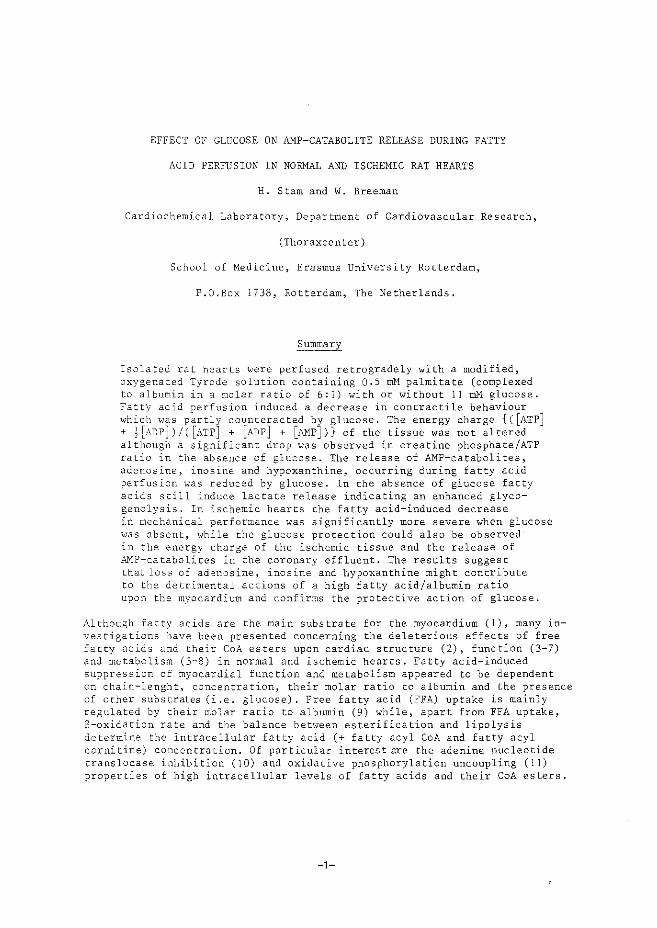
EFFECT OF GLUCOSE ON AMP-CATABOLITE RELEASE DURING FATTY
ACID PERFUSION IN NORMAL AND ISCHEMIC RAT HEARTS
H. Starn and W. Breeman
Cardiochemical Laboratory, Department of Cardiovascular Research,
(Thoraxcenter)
School of Nedicine, Erasmus University Rotterdam,
P.O.Box 1738, Rotterdam, The Netherlands.
Summary
Isolated rat hearts were perfused retrogradely with a modified, oxygenated Tyrode solution containing Q.S mM palmitate (complexed to albumin in a molar ratio of 6:1) with or without 11 mM glucose. Fatt)' acid perfusion induced a decrease in contractile behaviour which >vas partly counteracted by glucose. The energy charge {C[ATP] + j[ADP]J/( [ATP] + [ADP] + [&'1P]Jl of the tissue was not altered although a significant drop was observed in creatine phosphate/ATP ratio in the absence of glucose. The release of AMP-catabolites, adenosine, inosine and hypoxanthine, occurring during fatty acid perfusion was reduced by glucose. In the absence of glucose fatty acids still induce lactate release indicating an enhanced glycogenolysis. In ischemic hearts the fatty acid-induced decrease in mechanical performance was significantly more severe when glucose "'as absent, while the glucose protection could also be observed in the energy charge of the ischemic tissue and the release of ~1P-catabolites in the coronary effluent. The results suggest that loss of ad~nosine, inosine and hypoxanthine might contribute to the detrimental actions of a high fatty acid/albumin ratio upon the myocardium and confirms the protective action of glucose.
Although fatty acids are the main substrate for the myocardium (1), many investigations have been presented concerning the deleterious effects of free fatty acids and their CoA esters upon cardiac structure (2), function (3-7) and metabolism (3-8) in normal and ischemic hearts. Fatty acid-induced suppression of myocardial function and metabolism appeared to be dependent on chain-lenght, concentratiOn, their molar ratio to albumin and the presence of other substrates (i.e. glucose), Free fatty acid (FFA) uptake is mainly regulated by their molar ratio to albumin (9) while, apart from FFA uptake, S-oxidation rate and the balance between esterification and lipolysis determine the intracellular fatty acid (+ fatty acyl CoA and fatty acyl carnitine) concentration. Of particular interest are the adenine nucleotide translocase inhibition (10) and oxidative phosphorylation uncoupling (1 1) properties of high intracellular levels of fatty acids and their CoA esters.
-1-
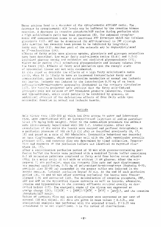
These actions lead to a decrease of the cytoplasmatic ATP/ADP ratio. The decrease in cytoplasmatic ATP levels may be buffered by'the creatine kinase reaction. A decrease in creatine phosphate/ATP ratios during perfusion with a high acid/albumin ratio has been presented (8). The enhanced cytoplas-m:Jtic ADP concentration leads to an increased AMP formation which may initiate glycolysis (12). AtlP may be deaminated (by AMP-deaminase) to IMP although this reaction has recently been proven to be inhibited by long-chain btty acyl Co.:\ ( 13). Another part of the molecule may be dephosphorylated by 5 1 -nucleotidase (14). Effect~ of fatty acids upon glucose uptake, glycolysis and glycogen metabolism have heen describ2d. Lo"' molar fatty acid-albumin ratios (0.6:1 and 1.2:1) inhibited glucose uptake and oxidation and abolished glycogenolysis (15). HL;.ller molar ratios (3:1) stimulated glycogenolysis and lactate release from titc \teart (16). Effects of fatty acid perfusion upon the energic state Ll:'" m~'ocardial tissue are not \Vell documented. Lt this paper He describe the effects of a high fatty acid/albumin ratio (b:l), h'hen it is likely to have an increased intracellular fatty acid cc'HLt!ntration, upon lactate and nucleotide metabolism of normal and ischemic r~:t he~Hts. Ischemia \vas induced by the introduction 0.70 mg of an inert ~L;lysac.::"lride-microsphere suspension (Sephadex) in the coronary circulation ( 17). Tht: r0sults presented here indicate that the fatty acid-induced .;lycl~gcnlllysis and release of AMP breakdown products (adenosine, inosine and hyp'-lxanthine), hlhich could partially be inhibited by glucose, is additional evid~ncc of the deleterious effects of free fatty acids upon myocardial function in normal and ischemic hearts.
Methods
:!ale 1.,'istar rats (200-300 g) ,,,hich had free access to water and laboratory c!1'-'H, \•'ere anest~etized v.'ith an intraperitoneal injection of sodium pentobarbital (70 :ng/kg body v.'eight). Prior to the cannulation procedure the animals ~>'ere intravenously heparinized with 500 IX. tromboliquine. After the cannulation of the aorta the hearts were excised and perfused retrogradely at a perfusio:1 pressure of 100 em H
2o (10 kPa) as described previously (8, 17,
18) and paced at a rate of 300 beats/min. Contractile behaviour was recorded as apex displacement, which correlates well with the left ventricular systolic pressure ( 17), and coronary flow was determined by timed collection. Preparation and contents of the perfusion buffers was identical as reported else'·'·:-:ere (8, 17) . . Hter a stabilization perfusion period of 30 min with glucose-containing perfusion bu£fer the hearts were perfused with a modified Tyrode buffer containing 0.5 ITO! po~assium palmitate complexed to fatty acid free bovine serum albumin (BSA) in 2 :nolar ratio of 6:1 with or without 11 ~~glucose. After the subsequent 1,~ min perfusion, when the coronary flow rate and apex displacement has reached equilibrium, 0.70 mg of polysaccharide-microspheres (Sephadex G-25, particle size 20-80 ·Jm) suspended in the proper buffer was injected in the aortic cannula. Ischemic perfusion lasted 20 min. At the end of each perfusion period (3G, 40 and 60 min after starting perfusion) the hearts were freezecla:nped (19) and extracted (20), The determination of creatine phosphate, ATP, ADP and AHP in the myocardial tissue extracts as well as lactate, adenosine, inosine and hypoxanthine in the coronary effluents occurred exactly as described before (17). The energetic state of the tissue was expressed as energy charge (21), {([ATP] +! [ADP])/C[ATP] + [ADPJ +[AMP])}, and the creatine phosphate/ATP ratio. \'alues of coronary flov.' and apex displacement were expressed as per cent of control (30 min value). All data are given in mean values+ S.E.M., and statistical analysis 1vas performed with the unpaired t-test. P > 0.05 was co:>.sidered to be not significant. n is the number of·observations.
-2-
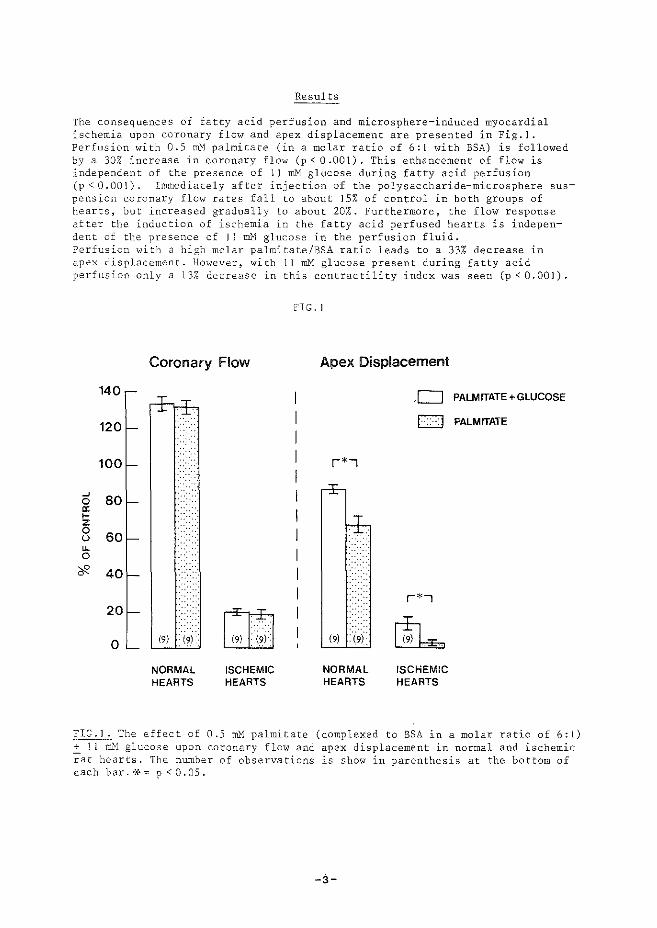
Results
The consequences of fatty acid perfusion and microsphere-induced myocardial ischemia upon coronary flow and apex displacement are presented in Fig.]. Perfusion with 0.5 mM palmitate (in a molar ratio of 6:1 with BSA) is followed by a 30% increase in coronary flow (p < 0 .001). This enhancement of flow is independent of the presence of l I rnM glucose during fatty acid perfusion (p < 0.001). Immediately after injection of the polysaccharide-microsphere suspension coronary flow rates fall to about 15% of control in both groups of hearts, but increased g-radually to about 20%. Furthermore, the flow response after the induction of ischemia in the fatty acid perfused hearts is independent of the presence of II Illi'1 glucose in the perfusion fluid. Perfusion with a high molar palmitate/BSA ratio leads to a 33% decrease in apex displacement. Ho'ivever, with 11 rnM glucose present during fatty acid perfusion only a 13% decrease in this contractility index was seen (p < 0.001).
FIG. I
Coronary Flow Apex Displacement
140 D PALMITATE +GLUCOSE
120 EJ PALMITATE
100 r*>
-' 80 0 a: 1-z 0
60 u u. 0
'if' 40
r*-, 20
1:1~1 0 19) (9) ~ NORMAL ISCHEMIC NORMAL ISCHEMIC HEARTS HEARTS HEARTS HEARTS
FIG.l. The effect of 0.5 ~'1 palmitate (complexed to BSA in a molar ratio of 6:1) ~ 1 1 ~1 glucose upon coronary flow and apex displacement in normal and ischemic rat hearts. The number of observations is show in parenthesis at the bottom of each bar.*"" p<O.OS.
-3-
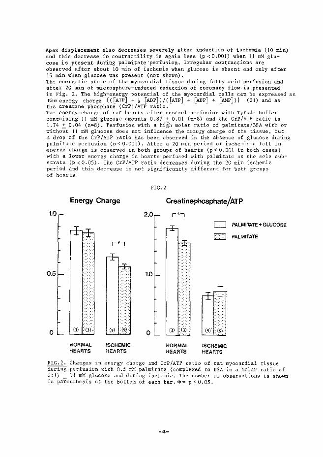
Apex displacement also decreases severely after induction of ischemia (10 min) and this decrease in contractility is again less (p <0.001) when 11 mM glucose is present during palmitate ·perfusion. Irregular contractions are observed after about 10 min of ischemia when glucose is absent and only after 15 min when glucose was present (not shown). The energetic state of the myocardial tissue during fatty acid perfusion and after 20 min of microsphere-induced reduction of coronary flow· is presented in Fig. 2. The high-energy potential of the myocardial cells can be expressed as the energy charge {([ATP] + j [ADP])/C[ATP] + [ADP] +[AMP])} (21) and as the creatine phosphate (CrP)/ATP ratio. Tpe energy charge of rat hearts after control perfusion with Tyrode buffer containing 11 mM glucose amounts 0.87 + O.OI (n=B) and the CrP/ATP ratio is I .74 + 0.04 {n=8). Perfusion with a hiih molar ratio of palmitate/BSA with or witho~t II mM glucose does not influence the energy charge of the tissue, but a drop of the CrP/ATP ratio has been observed in. the absence of glucose during palmitate perfusion (p < 0.001). After a 20 min period of ischemia a fall in energy charge is observed in both groups of hearts (p < O.OOI in both cases) with a lower energy charge in hearts perfused with palmitate as the sole substrate {p <0.05). The CrP/ATP ratio decreases during the 20 min ischemic period and this decrease is not significantly different for both groups of hearts.
Energy Charge
1.0
0
NORMAL HEARTS
r*•
ISCHEMIC HEARTS
FIG. 2
2.0
0
Creatinephosphate/ATP
r*l
NORMAL HEARTS
D PALMITATE+ GLUCOSE
CJ PALMITATE
(9)" :19):
ISCHEMIC HEARTS
FIG.2. Changes in energy charge and CrP/ATP ratio of rat myocardial tissue during perfusion with 0.5 mM palmitate {complexed to BSA in a molar ratio of 6:l) ~II mM glucose and during ischemia. The number of observations is shown in parenthesis at the bottom of each bar.*= p < 0 .OS,
-4-
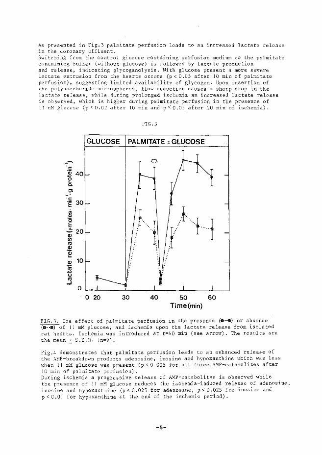
As presented in Fig.3 palmitate perfusion leads to an increased lactate release in the coronary effluent. Switching from the control glucose containing perfusion medium to the palmitate containing buffer (without glucose) is followed by lactate production and release, indicating glycogenolysis. With glucose present a more severe lactate extrusion from the hearts occurs (p < 0.05 after 10 min of palmitate perfusion), suggesting limited availability of glycogen. Upon insertion of the polysaccharide microspheres, flow reduction causes a sharp drop in the lactate release, .while during prolonged ischemia an increased lactate release is observed, which is higher during palmitate perfusion in the presence of 11 ml'1 glucose (p < 0.02 after 10 min and p < 0.05. after 20 min of ischemia).
~~
'c ·a; 40 e a. Ol
'"' E 30
"' ~ E "- 20 -cv (/)
"' cv ~ ~
cv 10 -"' -u "' ..J
0
FIG.3
GLUCOSE PALMITATE ±GLUCOSE
0
f- .......___
-
f· .. ···1_ -
-
~ -en I
_l: 0 20 30 40
~ l.
I/ lJ
I
50 Time(min)
I
60
-
-
-
-
FIG.3. The effect of palmitate perfusion in the presence (8-8) or absence (m .. ll) of 11 m}1 glucose, and ischemia upon the lactate release from isolated rat hearts. Ischemia was introduced at t=40 min (see arrow). The results are the mean.! S.E.H. (n=9).
Fig.4 demonstrates that palmitate perfusion leads to an enhanced release of the AMP-breakdown products adenosine, inosine and hypoxanthine which was less when 11 Illi'1 glucose was present (p < 0.005 for all three AMP-catabolites after 10 min of palmitate perfusion). During ischemia a progressive release of AMP-catabolites is observed while the presence of 1 I mM glucose reduces the ischemia-induced release of adenosine, inosine and hypoxanthine (p < 0.025 for adenosine, p < 0.025 for inosine and p < 0.01 for hypoxanthine at the end of the ischemic period).
-5-
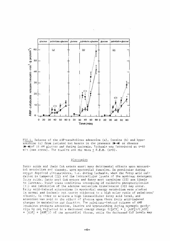
glucose palmitate,.glucose glucose palmitate±glucose g'ucose palmitate:glucose
(c) -
-
-
. -
jJJ .· ~--
i!if .,d' l I I
0 20 30 40 50 60 0 20 30 40 so 60 0 20 30 40 50 60
Timecmin)
FIG.~. Release of the M1P-catabolites adenosine (a), inosine (b) and hypo-;zanthine (c) from isolated rat hearts in the presence (......._.) or absence (W··m)of l 1 rnM glucose and during ischemia. Ischemia was introduced at t=40 min (see arrow). The results are the mean+ S.E.M. (n=9).
Discussion
Fatty acids and their CoA esters exert many detrimental effects upon myocardial metabolis~ and thereby, upon myocardial function. In particular during oxygen deprived circumstances, i.e. during ischemia, when the fatty acid oxidation is hampered (22) and the intracellular levels of the membrane detergent fatty acids, fatty acyl CoA esters and fatty acyl carnitine (23) are likely to increase. Under these conditions uncoupling of oxidative phosphorlylation (l 1) and inhibition of the adenine nucleotide translocator (10) may occur. Fatty acid-induced alterations in myocardial energy metabolism 1:vere studied in normal and ischemic rat hearts subjected to a high molar ratio of palmitate/ albumin, in order to achieve a high intracellular fatty acid level, and attention·was paid to the effect of glucose upon these fatty acid-induced changes in metabolism and function. The palmitate-induced release of AMP breakdown products adenosine, inosine and hypoxanthine during normoxic perfusiC?.n is not reflected in a decreased energy charge {([ATP] + i [ADP])/([ATP] + ~ADP] + [AMP])} of the myocardial tissue, 'vhile the decreased CrP levels may
-6-
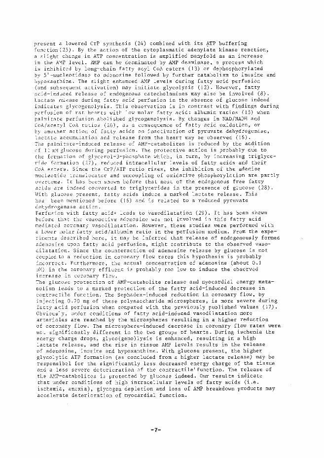
present a lowered CrP synthesis (24) combined with its ATP buffering function (25), By the action of the cytoplasmatic adenyl~te kinase reaction, a slight change in ATP concentration is amplified manyfold as an increase ln the AMP level. Al'1P can be deaminated by AMP deaminase, a process which is inhibited by long-chain fatty acyl CoA esters (13) or dephosphorylated by 5 1 -nucleotidase to adenosine followed by further catabolism to inosine and hypoxanthine. The slight enhanced AMP levels during fatty acid perfusion (and subsequent activation) may initiate glycolysis (12). However, fatty acid-induced release of endogenous catecholamines may also be involved (8). Lactate n.;l.-oase du,ring fatty acid perfusion in the absence of glucose indeed indicates glycogenolysis. This observation is in contrast with findings during perfusion of rat hearts with low molar fatty acid: albumin ratios (15) when palmitate perfusion abolished glycogenolysis. By changes in NAD/NADH and CoA/acetyl CoA ratios (26), as a consequence of fatty acid oxidation, or by another action of fatty acids on inactivation of pyruvate dehydrogenase, lactate accumulation and release from the heart may be observed (16). The palmitate-induced release of AMP-catabolites is reduced by the addition (Jf II m.\f glucose during perfusion. The protective action is probably dUe to the form<J.tion of glycerol-3-phosphate which, in turn, by increasing triglyceride formation (27), reduced intracellular levels of fatty acids and their CoA esters. Since the CrP/ATP ratio rises, the inhibition of the adenine nucleotide translocator and uncoupling of oxidative phosphorylation are partly overcome. It has been shown before that most of the endogenous free fatty 2c:ids are indeed converted to triglycerides in the presence of glucose (28) . \\lith glucose present, fatty acids induce a marked lactate release. This has been mentioned before (16) and is related to a reduced pyruvate dehydrogenase action. Perfusion i>.'ith fatty acids leads to vasodilatation (29). It has been shown before that the vasoactive adenosine was not involved in this fatty acid mediated coronary vasodilatation. However, these studies were performed with a lO'i,•er Tiolar fatty acid/albumin ratio in the perfusion medium. From the experiments described here, it may be inferred that release of endogenously formed adenosine upon fatty acid perfusion, might contribute to the observed vasodilatation. Since the counteraction of adenosine release by glucose is not coupled to a reduction in coronary flow rates this hypothesis is probably incorrect. Furthermore, the actual concentration of adenosine (about 0.1 ]J)!) in the coronary effluent is probably too low to induce the observed increase in coronary flo'"· The glucose protection of AMP-catabolite release and myocardial energy metabolism leads to a marked protection of the fatty acid-induced decrease in contractile function. The Sephadex-induced reduction in coronary flow, by injecting 0.70 mg of these polysaccharide microspheres, is more severe during fatty acid perfusion when compared with the previously published values (17). Obviously, under conditions of fatty acid-induced vasodilatation more arterioles are reached by the microspheres resulting in a higher reduction of coronary flow. The microsphere-induced decrease in coronary flow rates were not significantly different in the two groups of hearts. During ischemia the energy charge drops, glyco(geno)lysis is enhanced, resulting in a high lactate release, and the rise. in tissue AMP levels results in the release of adenosine, inosine and hypoxanthine. With glucose present, the higher glycolytic ATP formation (as concluded from a higher lactate release) may be responsibel for the significantly less decreased energy charge of the tissue and a less severe deterioration of the contractile 1 function. The release of the A}fP-catabolites is protected by glucose indeed. Our results indicate that under conditions of high intracellular levels of fatty acids (i.e. ischemia, anoxia), glycogen depletion and loss of AMP breakdown products may accelerate deterioration of myocardial function.
-7-
Acknowledgements
The skillful technical assistance of Miss M. Stewart is acknowledged. Prof. W.C. Hlllsmann is thanked for advice and Miss J.J. v.d. Kleijn for type-writing the manuscript. Financial support was obtained from the Dutch Heart Foundation.
References
I. J.R. NEELY and H. MORGAN, Ann. Rev. PhysioL 36 413-459 (1974). 2. J. DE LEIRIS and D. FEUVRAY, J. Mol. Cell."Cardiol. 9 365-373 (1977). 3. L.H: OPIE. Nature 227 1055-1056 (1970). -4. A.H. HENDERSON, R.~GRAIG, R. GORLIN, and E.H. SONNENBLICK, Cardiovasc.
Res. 4 446-472 (1970). 5. D.R. CHALLONER and D.R. STEINBERG, Am. J. Physiol. lll 897-902 (1966). 6. A.F. WILLEBRANDS, H.F. TER WELLE, and S.J.A. TASSERON, J. Mol. Cell.
Cardiol. 5 259-273 (1973). 7. J.M .. J. LAMERS and W.C. HULSMANN, J. Mol. Cell. Cardiel. 9, 343-346 (1977). 8. H. STAMand W.C. HULSMANN, Basic Res. Cardiel. 73 208 219 (1978). 9. A. SPECTOR, Progr. Biochern. Pharmacal. val~. p~ 130-176 (Karger, Basel
( 1971) . 10. E. SHRAGO, Life Sciences 1l J-6 (1978). 11. W.C. HULSMANN, W.B. ELLIOT, and E.C. SLATER, Biochim. Biophys. Acta
39 267-276 (1960). 12. ~A. NEWSHOLME, Cardiology 56 22-34 (1971/1972). 13. A.SKLADANOWSKI, K. KALETHA, and M. ZYDOWO, Int. J. Biochem. 9 43-47
(1978). 14. R. RUBIO, R.M. BERNE, and J.G. DOBSON, Jr., Am. J. Physiol. 225 938-953
(1973). 15. M.G. Grass III, E.S. McCaskill, and J.C. SHIPP, J. Appl. Physiol. ~
87-91 (1970). 16. O.WIELAND, H.V. 'FUNCKE, and G. LtiFFLER, FEES Letters 15 295-298 (1971). i7. H.STAM and J.W. DEJONG, J. Mol. Cell. Cardiol. 9 633-650 (1977). 18. H. STAMand W.C. HDLSMM~N, Basic Res. Cardiel. 72 365-375 (1977). 19. A. WOLLENBERGER, 0. RISTAU, and G. SCHOFFA, PfUgers Archiv 270 399-412
(1960). -20. J.W. DE JONG, Biochim. Biophys. Acta 286 252-259 (1972). 21. D.E. ATKINSON, Biochemistry 7 4030 4034 (1968). 22. J. BREMER and A.J. WOJTCZAK, Biochim. Biophys. Acta 280 515-530 (1972). 23. A.L. SHUG, J.H. THOMSEN, J.D. FOLTS, N. BITTAR, M.I.JZLEIN, J.R. KOKE~ and
P.J. HUTZ, Arch. Biochem. Biophys. 187 25-33 (1978). 24. V.A. SAKS, N,V, LIPINA, N.V. LYULIN~G.B. CHERNOUSOVA, R. FET!ER, V.N.
SMIRNOV, and E.I. CHAZOV, Biochemistry 41 1191-1199 (1976). 25. H.R. SCHOLTE, Biochim. Biophys. Acta 30s-413-427 (1973). 26. M.S. OLSON, S.C. DENNIS, C.A. ROUTH, and M.S. DE BUYSERE, Arch. Biochem.
Biophys. 187 l21-13l (.1978). 27. A. LOCHNER, J,L.N" KOTZE, and W. GEVERS, J. Mol. Cell. Cardiel. 8 575-584
(1976). 28. 0. STEIN and Y. STEIN, J, Cell, Biol. 36 63-77 (!968) 29. W.C. HDLSMANN, Basic Res, CarCiol. 71 179-!91 (1976).
-a-