L.J. Boersma, radiotherapeut-oncoloog MUMC+, MAASTRO ... radiotherapeur en de marges_L.J... ·...
28
De radiotherapeut en de marges L.J. Boersma, radiotherapeut-oncoloog MUMC+, MAASTRO clinic Maastricht
Transcript of L.J. Boersma, radiotherapeut-oncoloog MUMC+, MAASTRO ... radiotherapeur en de marges_L.J... ·...
De radiotherapeut en de marges
L.J. Boersma, radiotherapeut-oncoloog
MUMC+, MAASTRO clinic
Maastricht
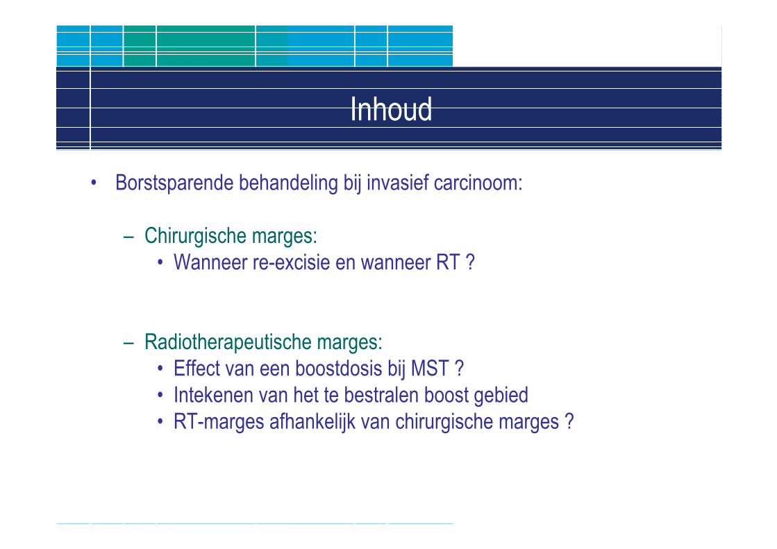
Inhoud
• Borstsparende behandeling bij invasief carcinoom:
– Chirurgische marges:
• Wanneer re-excisie en wanneer RT ?
– Radiotherapeutische marges:
• Effect van een boostdosis bij MST ?
• Intekenen van het te bestralen boost gebied
• RT-marges afhankelijk van chirurgische marges ?
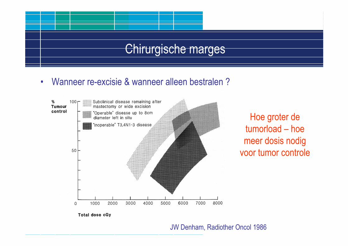
Chirurgische marges
• Wanneer re-excisie & wanneer alleen bestralen ?
JW Denham, Radiother Oncol 1986
Hoe groter de
tumorload – hoe
meer dosis nodig
voor tumor controle
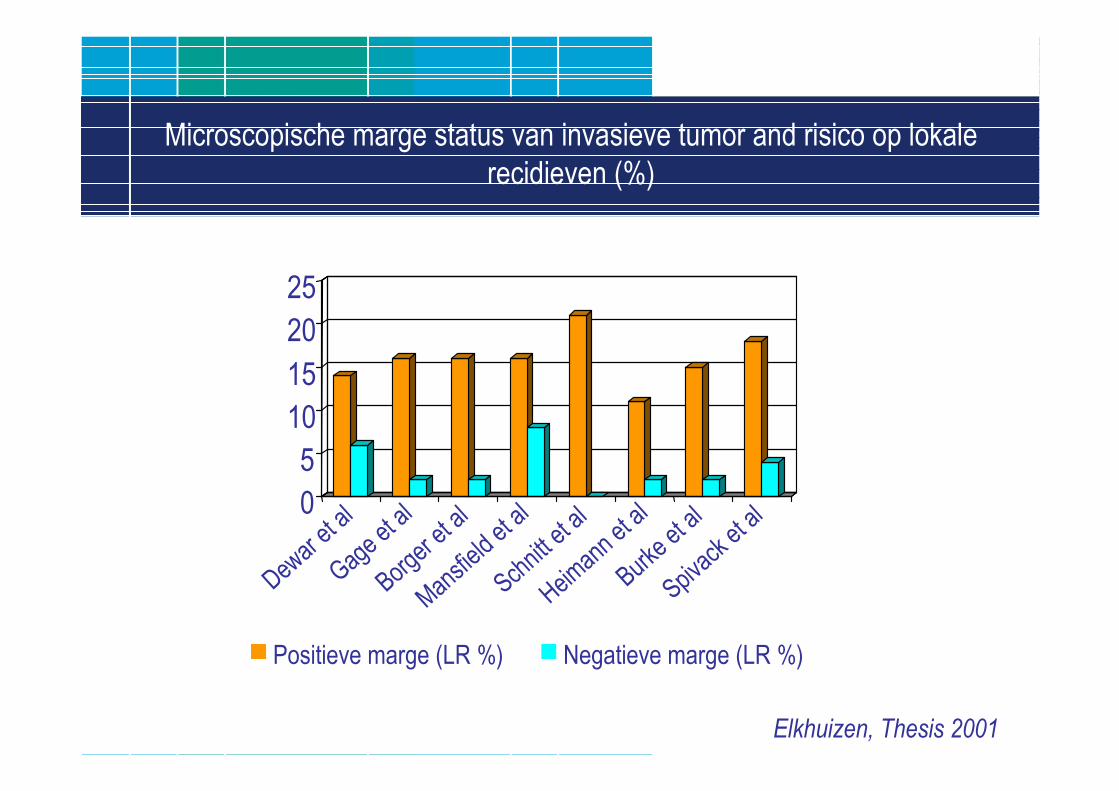
Microscopische marge status van invasieve tumor and risico op lokale
recidieven (%)
0
5
10
15
20
25
Dewar et al
Gage et al
Borger et al
Mansfield et al
Schnittet al
Heimann et al
Burke et al
Spivacket al
Positieve marge (LR %) Negatieve marge (LR %)
Elkhuizen, Thesis 2001
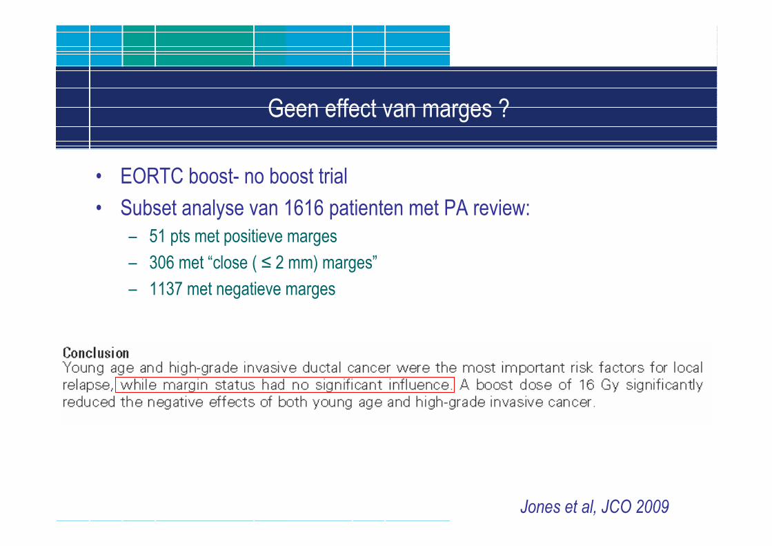
Geen effect van marges ?
• EORTC boost- no boost trial
• Subset analyse van 1616 patienten met PA review:
– 51 pts met positieve marges
– 306 met “close ( ≤ 2 mm) marges”
– 1137 met negatieve marges
Jones et al, JCO 2009
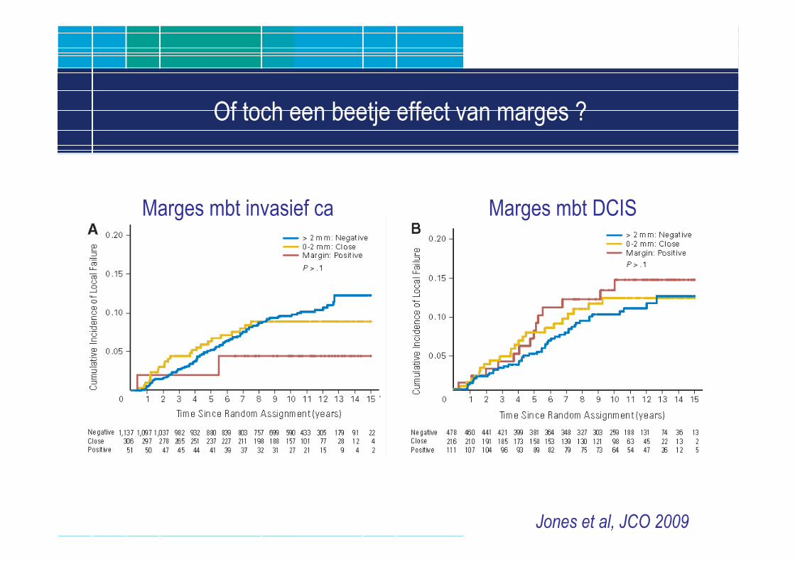
Of toch een beetje effect van marges ?
Jones et al, JCO 2009
Marges mbt invasief ca Marges mbt DCIS
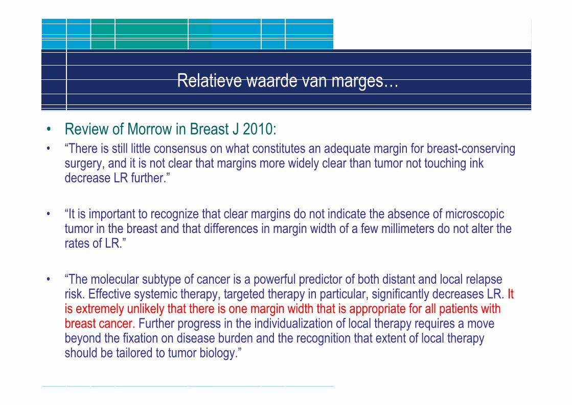
Relatieve waarde van marges…
• Review of Morrow in Breast J 2010:• “There is still little consensus on what constitutes an adequate margin for breast-conserving
surgery, and it is not clear that margins more widely clear than tumor not touching inkdecrease LR further.”
• “It is important to recognize that clear margins do not indicate the absence of microscopictumor in the breast and that differences in margin width of a few millimeters do not alter the rates of LR.”
• “The molecular subtype of cancer is a powerful predictor of both distant and local relapserisk. Effective systemic therapy, targeted therapy in particular, significantly decreases LR. Itis extremely unlikely that there is one margin width that is appropriate for all patients withbreast cancer. Further progress in the individualization of local therapy requires a move beyond the fixation on disease burden and the recognition that extent of local therapyshould be tailored to tumor biology.”
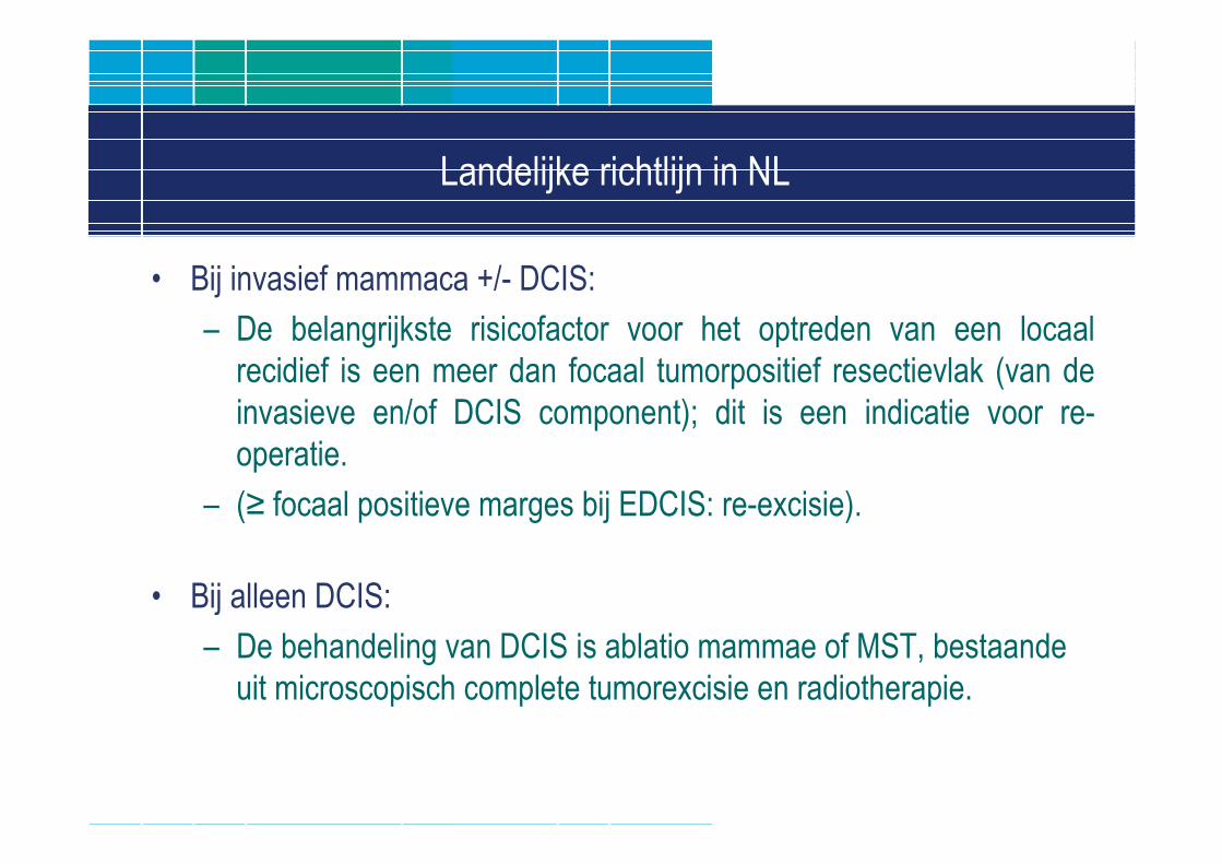
Landelijke richtlijn in NL
• Bij invasief mammaca +/- DCIS:
– De belangrijkste risicofactor voor het optreden van een locaal
recidief is een meer dan focaal tumorpositief resectievlak (van de
invasieve en/of DCIS component); dit is een indicatie voor re-
operatie.
– (≥ focaal positieve marges bij EDCIS: re-excisie).
• Bij alleen DCIS:
– De behandeling van DCIS is ablatio mammae of MST, bestaande
uit microscopisch complete tumorexcisie en radiotherapie.

Inhoud
• Borstsparende behandeling bij invasief carcinoom:
– Chirurgische marges:
• Wanneer re-excisie en wanneer RT ?
– Radiotherapeutische marges:
• Effect van een boostdosis bij MST ?
• Intekenen van het te bestralen boost gebied
• RT-marges afhankelijk van chirurgische marges ?
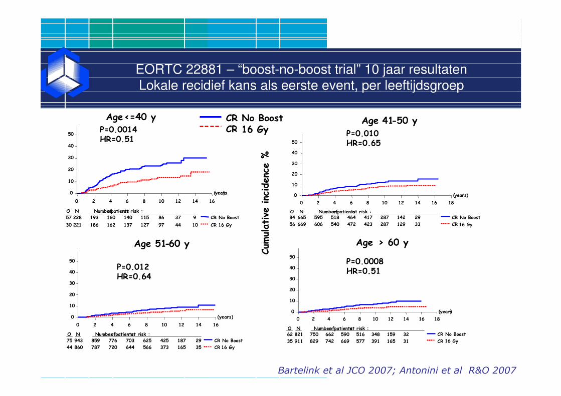
EORTC 22881 – “boost-no-boost trial” 10 jaar resultatenLokale recidief kans als eerste event, per leeftijdsgroep
Bartelink et al JCO 2007; Antonini et al R&O 2007
CR No BoostCR 15 GyP=0.0014
HR=0.51
(years)
0 2 4 6 8 10 12 14 16
0
10
20
30
40
50
O N Numberof patientsat risk :
57 228 193 160 140 115 86 37 9
30 221 186 162 137 127 97 44 10
CR No Boost
CR 15 Gy
Age<=40 y
(years)
0 2 4 6 8 10 12 14 16 18
0
10
20
30
40
50
O N Numberof patientsat risk :84 665 595 518 464 417 287 142 29
56 669 606 540 472 423 287 129 33
CR No Boost
CR 15 Gy
Age 41-50 y
P=0.012 HR=0.64
(years)
0 2 4 6 8 10 12 14 16
0
10
20
30
40
50
O N Numberof patientsat risk :
75 943 859 776 703 625 425 187 29
44 860 787 720 644 566 373 165 35
CR No Boost
CR 15 16 Gy
Age 51-60 y
P=0.0008 HR=0.51
(years)
0 2 4 6 8 10 12 14 16 18
0
10
20
30
40
50
O N Numberof patientsat risk :62 821 750 662 590 516 348 159 32
35 911 829 742 669 577 391 165 31
CR No Boost
CR 15 Gy
Age > 60 y
P=0.010 HR=0.65
16 Gy
16 Gy 16 Gy
16 Gy
Cum
ulative
inc
idenc
e %
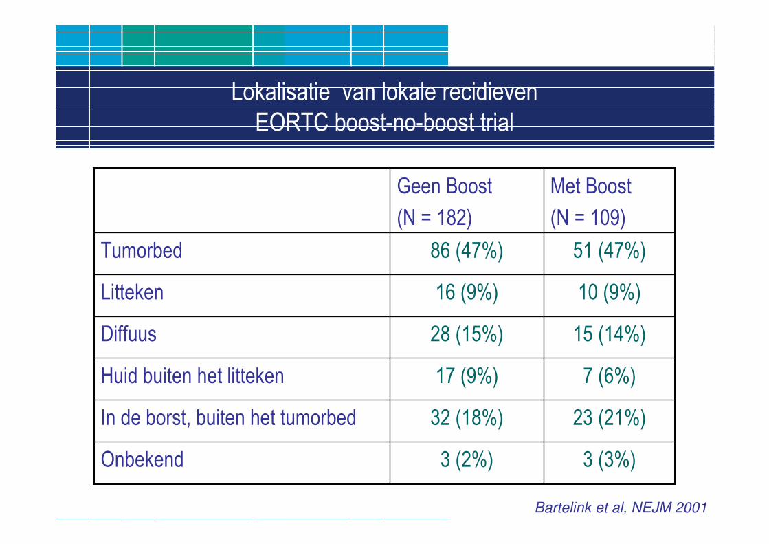
Lokalisatie van lokale recidieven
EORTC boost-no-boost trial
3 (3%)3 (2%)Onbekend
23 (21%)32 (18%)In de borst, buiten het tumorbed
7 (6%)17 (9%)Huid buiten het litteken
15 (14%)28 (15%)Diffuus
10 (9%)16 (9%)Litteken
51 (47%)86 (47%)Tumorbed
Met Boost
(N = 109)
Geen Boost
(N = 182)
Bartelink et al, NEJM 2001
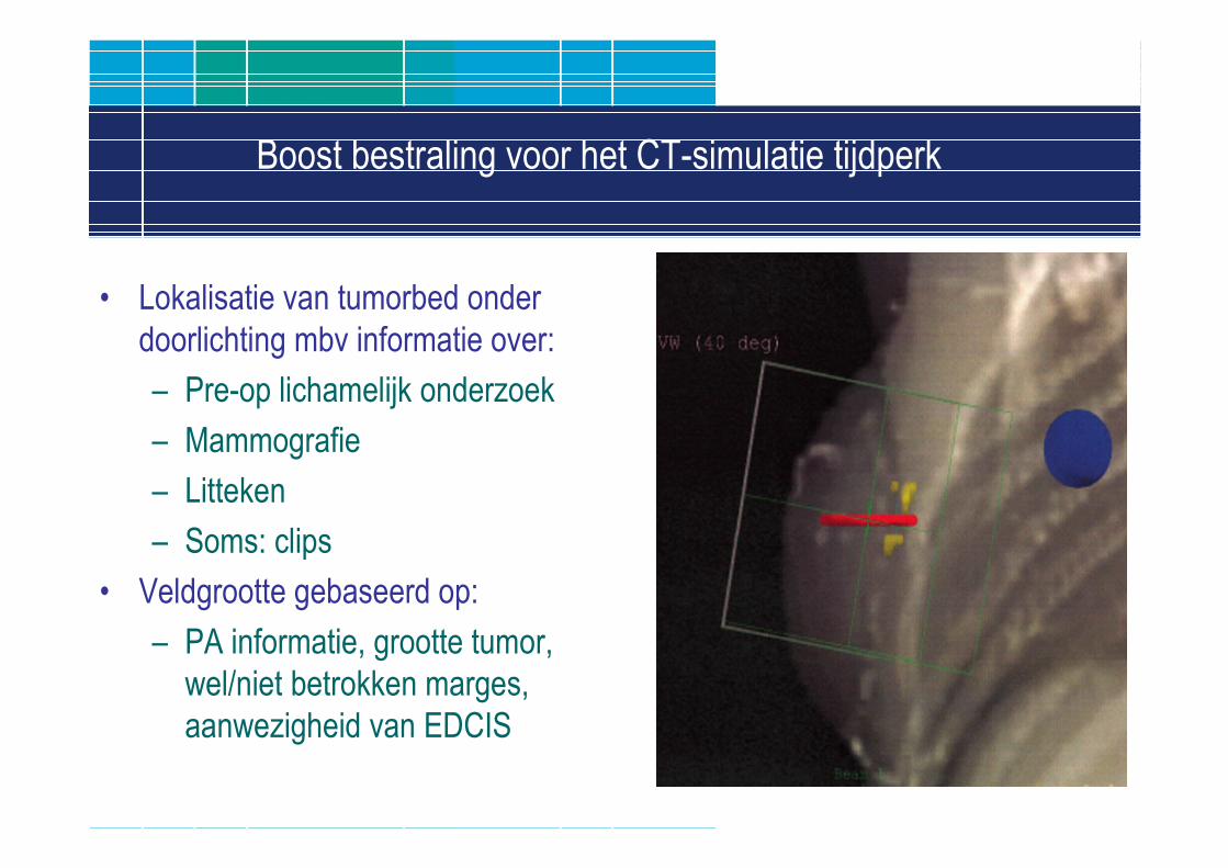
Boost bestraling voor het CT-simulatie tijdperk
• Lokalisatie van tumorbed onder
doorlichting mbv informatie over:
– Pre-op lichamelijk onderzoek
– Mammografie
– Litteken
– Soms: clips
• Veldgrootte gebaseerd op:
– PA informatie, grootte tumor,
wel/niet betrokken marges,
aanwezigheid van EDCIS
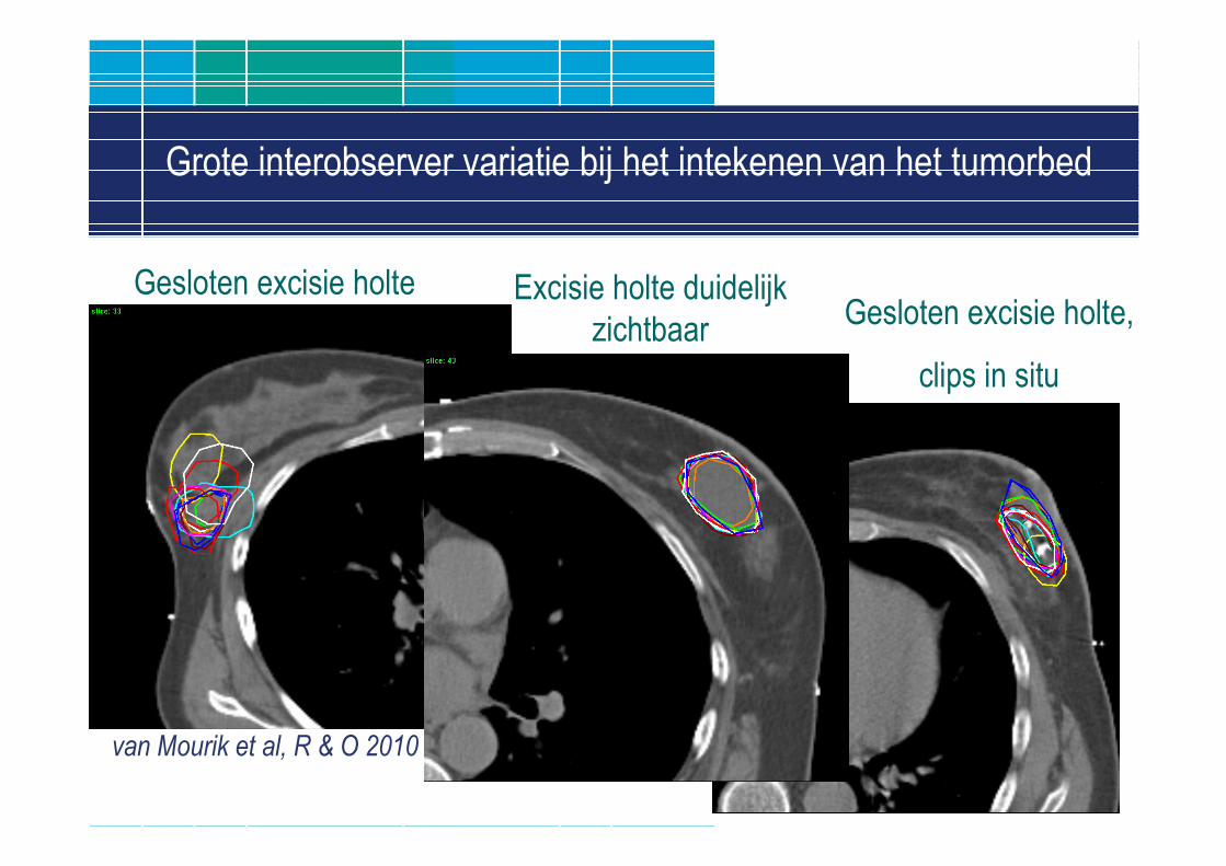
Grote interobserver variatie bij het intekenen van het tumorbed
Excisie holte duidelijk
zichtbaar
van Mourik et al, R & O 2010
Gesloten excisie holte,
clips in situ
Gesloten excisie holte
Strategieën om de interobserver variatie te verbeteren
• Gebruik van clips.
• Gebruik van pre-operatieve beeldvorming in RT houding.
Slide courtesy Ph. Poortmans
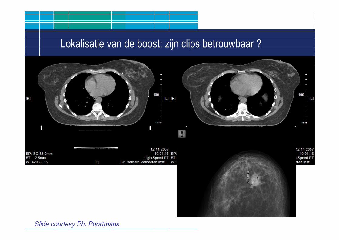
Lokalisatie van de boost: zijn clips betrouwbaar ?
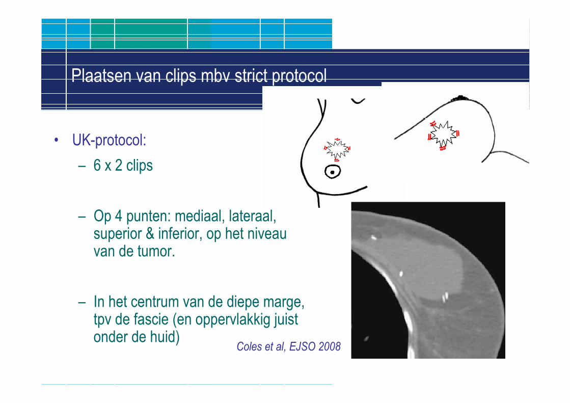
Plaatsen van clips mbv strict protocol
• UK-protocol:
– 6 x 2 clips
– Op 4 punten: mediaal, lateraal, superior & inferior, op het niveauvan de tumor.
– In het centrum van de diepe marge, tpv de fascie (en oppervlakkig juistonder de huid)
Coles et al, EJSO 2008
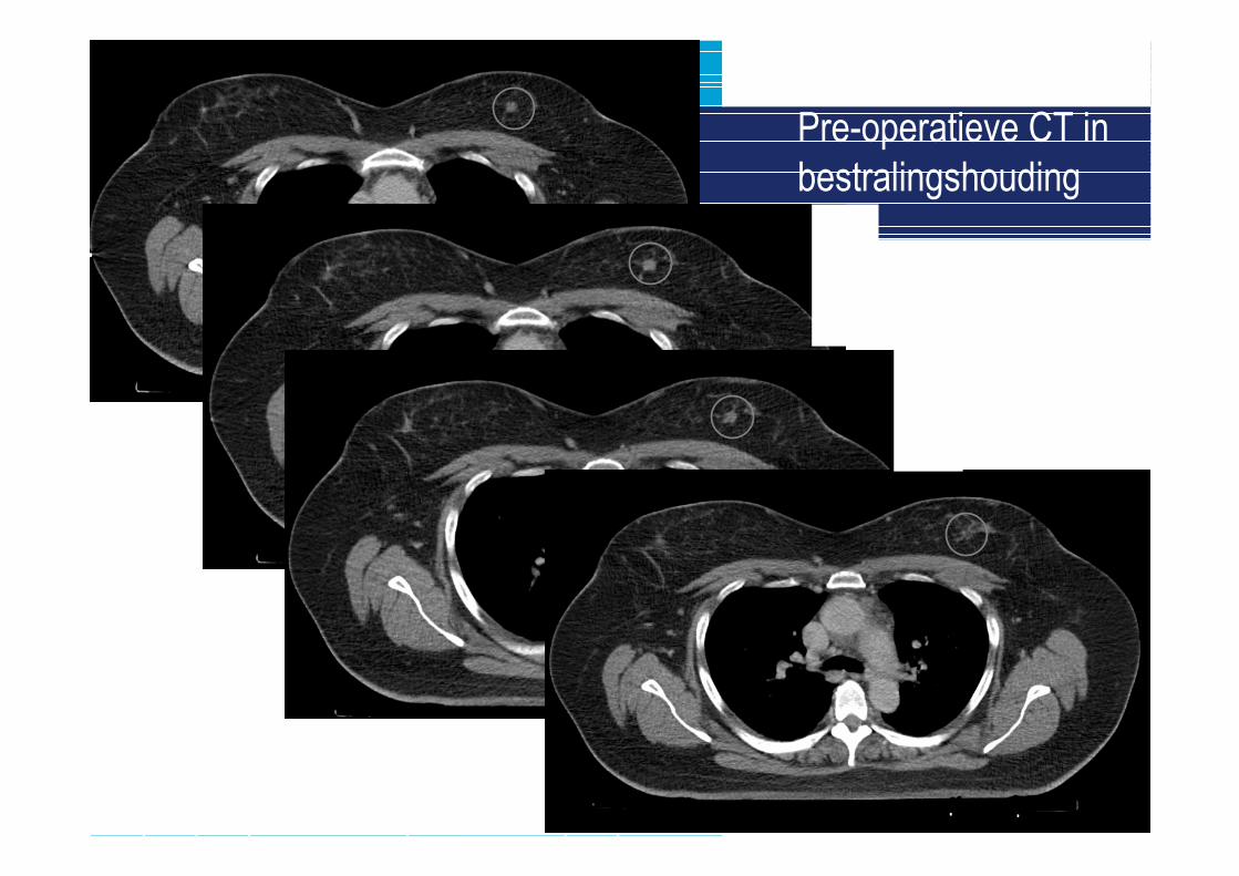
Casus met pre-op CT infoPre-operatieve CT in
bestralingshouding
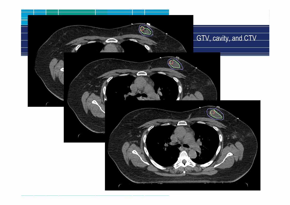
GTV, cavity, and CTV
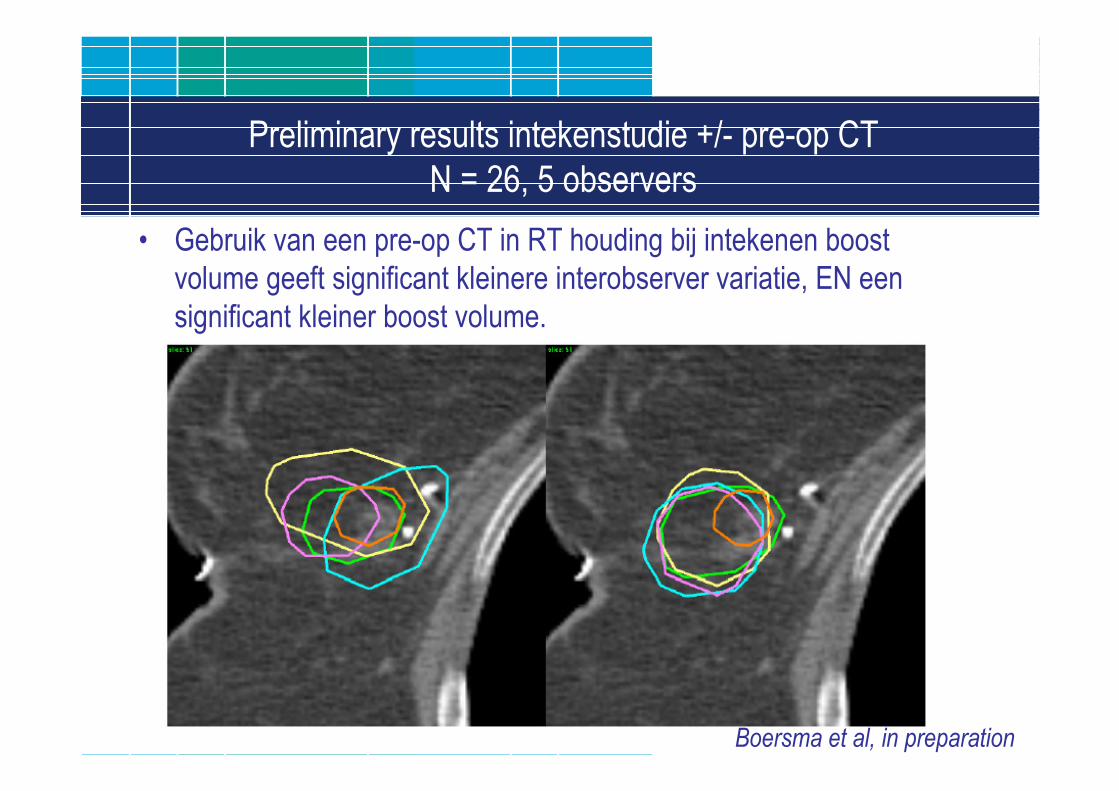
Preliminary results intekenstudie +/- pre-op CT
N = 26, 5 observers
• Gebruik van een pre-op CT in RT houding bij intekenen boost
volume geeft significant kleinere interobserver variatie, EN een
significant kleiner boost volume.
Boersma et al, in preparation
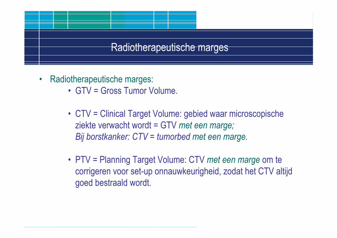
Radiotherapeutische marges
• Radiotherapeutische marges:
• GTV = Gross Tumor Volume.
• CTV = Clinical Target Volume: gebied waar microscopische
ziekte verwacht wordt = GTV met een marge;
Bij borstkanker: CTV = tumorbed met een marge.
• PTV = Planning Target Volume: CTV met een marge om te
corrigeren voor set-up onnauwkeurigheid, zodat het CTV altijd
goed bestraald wordt.
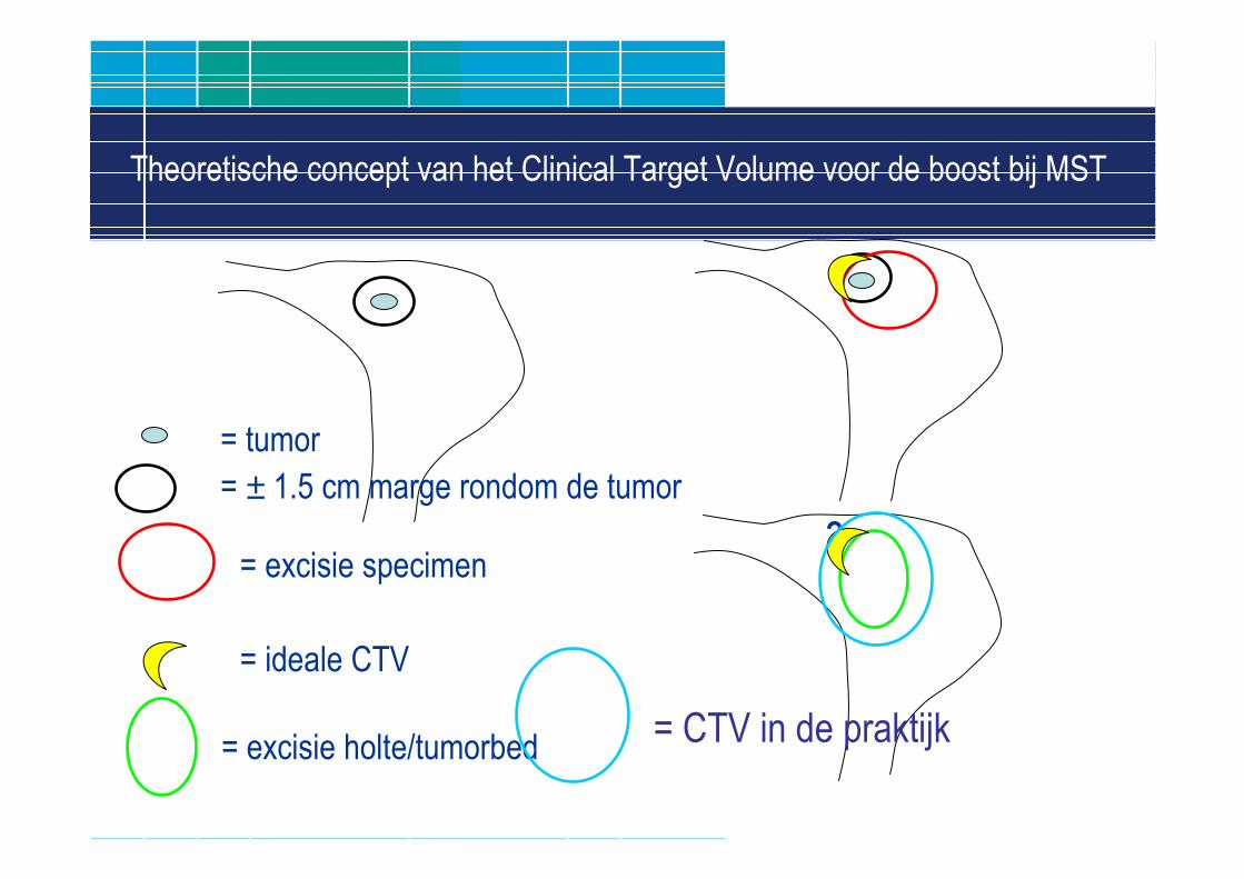
Theoretische concept van het Clinical Target Volume voor de boost bij MST
= tumor
= ± 1.5 cm marge rondom de tumor
= ideale CTV
= excisie specimen?
= excisie holte/tumorbed= CTV in de praktijk
1
2
3
4
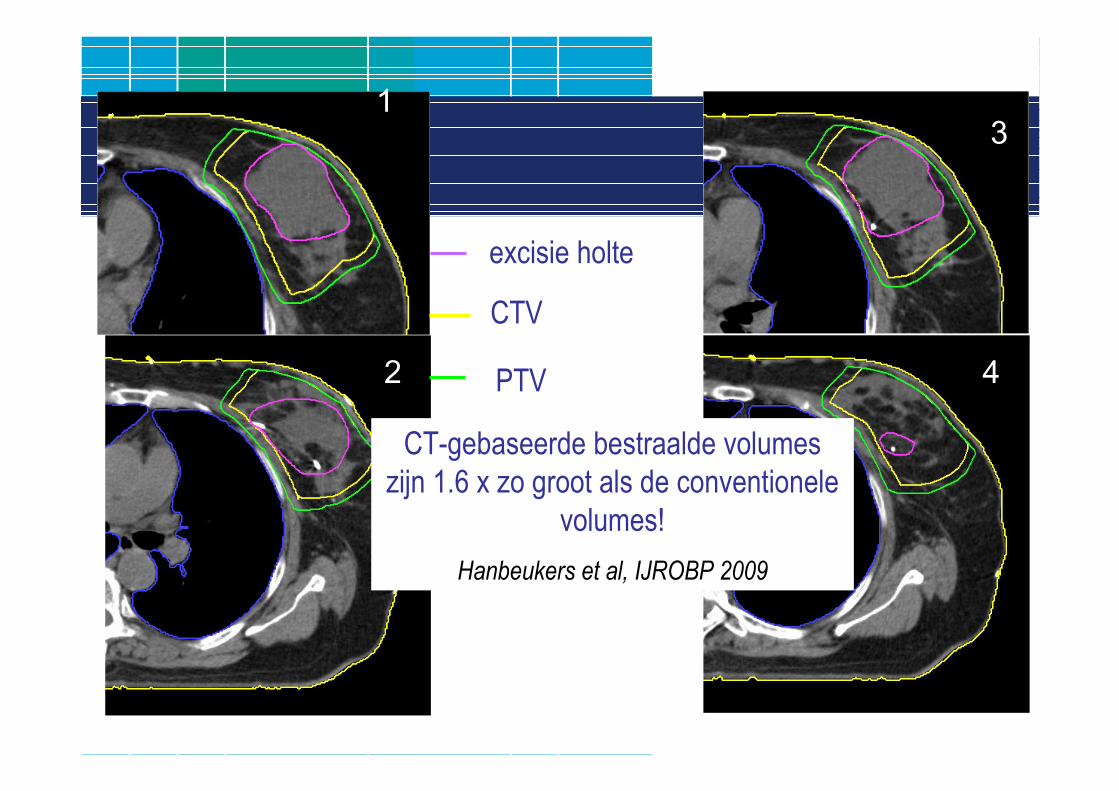
excisie holte
CTV
PTV
CT-gebaseerde bestraalde volumes
zijn 1.6 x zo groot als de conventionele
volumes!
Hanbeukers et al, IJROBP 2009
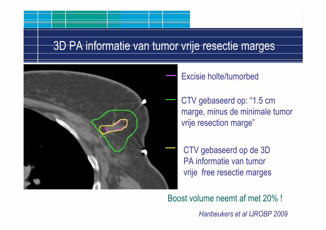
3D PA informatie van tumor vrije resectie marges
Excisie holte/tumorbed
CTV gebaseerd op: “1.5 cm
marge, minus de minimale tumor
vrije resection marge”
CTV gebaseerd op de 3D
PA informatie van tumor
vrije free resectie marges
Boost volume neemt af met 20% !
Hanbeukers et al IJROBP 2009
Enkele conclusies
Chirurgische marges:
• Bij invasief ca is re-excisie is alleen nodig in geval van > focaalpositieve marges, ( of bij ≥ focaal positieve marges van EDCIS).
• Beleid bij positieve marges zou wellicht – in de toekomst - medemoeten afhangen van andere tumorkarakteristieken.
Radiotherapeutische marges:
• Er is grote interobserver variatie bij het intekenen van het tumorbed.
• CT gebaseerde boost volumes zijn groter dan vroeger.
• Strategieën om interobserver variatie en/of volume te verkleinen:– Plaatsen van clips volgens vast landelijk protocol.
– Gebruik van pre-operatieve CT scan in RT houding.
– Verwerken van kennis over 3D chirurgische marges in het boostvolume.
Dank voor uw aandacht !
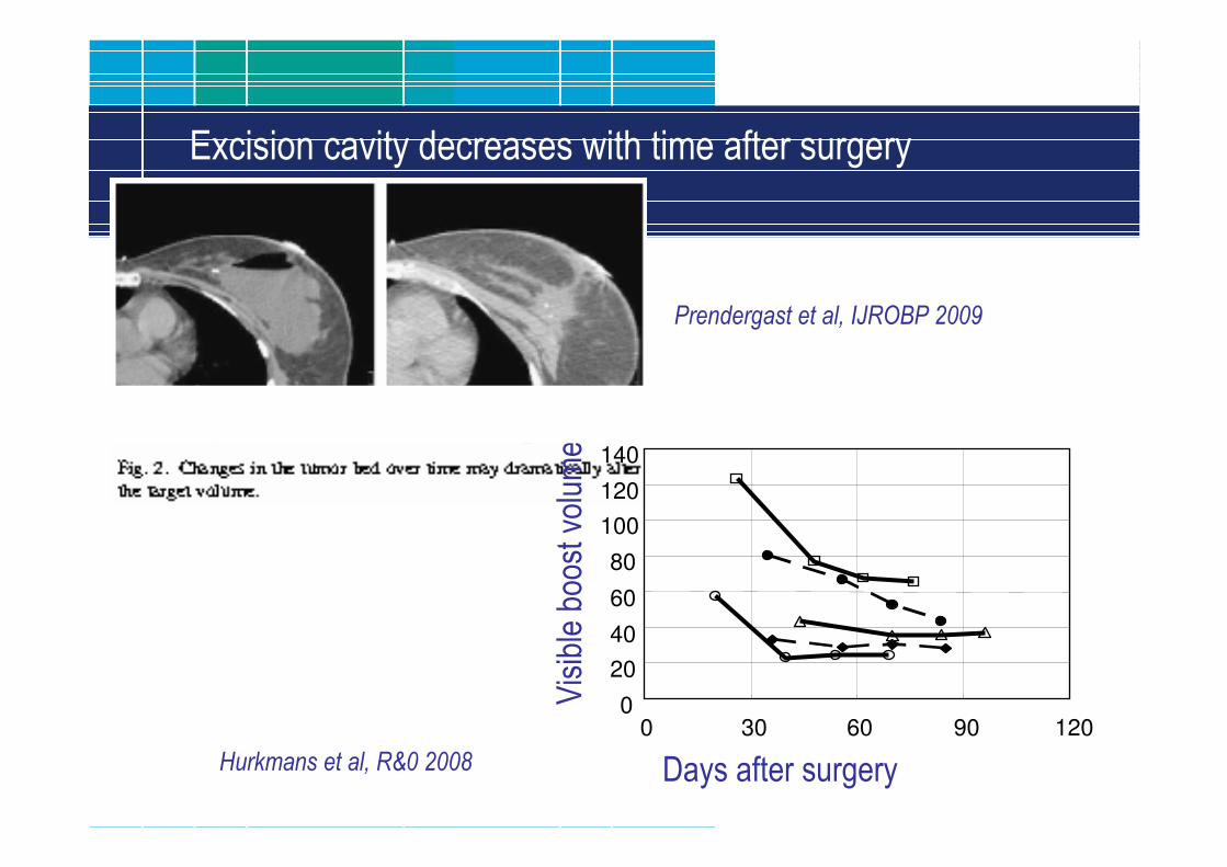
Excision cavity decreases with time after surgery
Prendergast et al, IJROBP 2009
Hurkmans et al, R&0 2008
0
20
40
60
80
100
120
140
0 30 60 90 120
Days after surgery
Visible boost volume (cc)
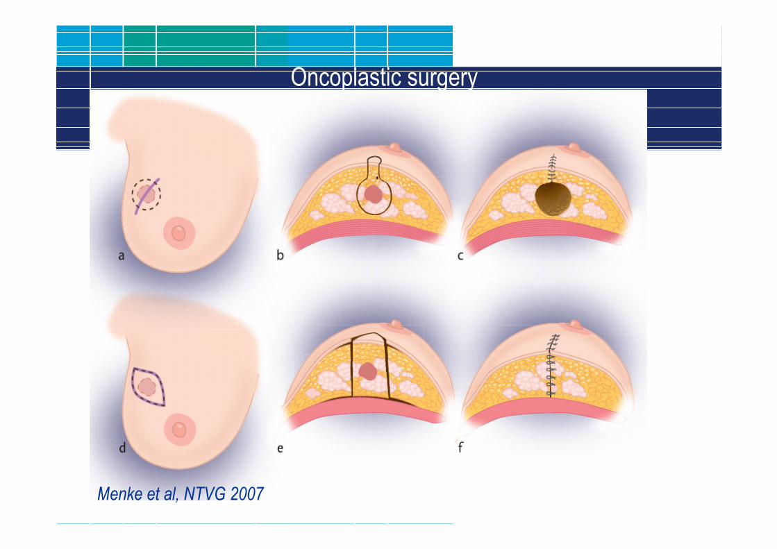
Oncoplastic surgery
Menke et al, NTVG 2007
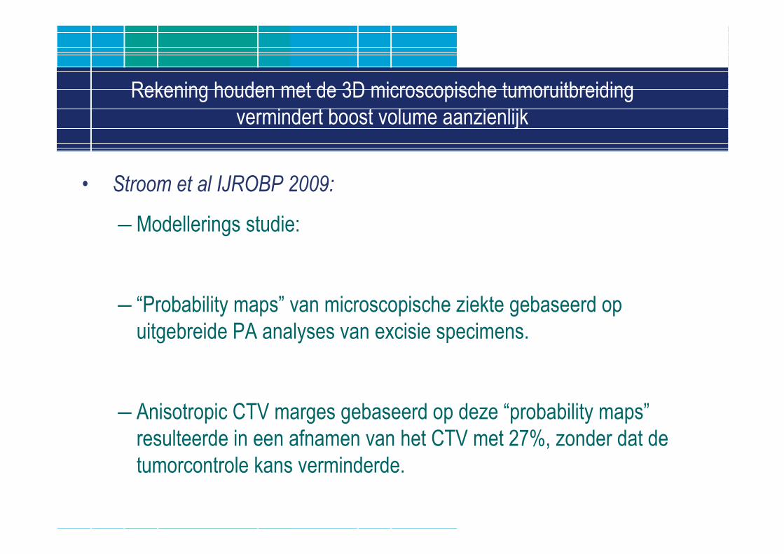
Rekening houden met de 3D microscopische tumoruitbreiding
vermindert boost volume aanzienlijk
• Stroom et al IJROBP 2009:
―Modellerings studie:
―“Probability maps” van microscopische ziekte gebaseerd op
uitgebreide PA analyses van excisie specimens.
―Anisotropic CTV marges gebaseerd op deze “probability maps”
resulteerde in een afnamen van het CTV met 27%, zonder dat de
tumorcontrole kans verminderde.