EAE Recommendations - Contrast echocardiography: evidence ...
22
EAE RECOMMENDATIONS Contrast echocardiography: evidence-based evidence based recommendations by European Association fEh di h of Echocardiography Roxy Senior, Harald Becher, Mark Monaghan, Luciano Agati, Jose Zamorano, Jean Louis Vanoverschelde, and Petros Nihoyannopoulos European Journal of Echocardiography (2009) 10 194–212 European Journal of Echocardiography (2009) 10, 194–212 ©European Association of Echocardiography 2009
Transcript of EAE Recommendations - Contrast echocardiography: evidence ...
EAE RECOMMENDATIONSContrast echocardiography:
evidence-basedevidence basedrecommendations by European Association
f E h di hof Echocardiography
Roxy Senior, Harald Becher, Mark Monaghan, Luciano Agati, Jose Zamorano, Jean Louis Vanoverschelde, and g , , ,
Petros NihoyannopoulosEuropean Journal of Echocardiography (2009) 10 194–212European Journal of Echocardiography (2009) 10, 194–212©European Association of Echocardiography 2009
European Journal of Echocardiography (2009) 10, 194–212
European Journal of Echocardiography (2009) 10, 194–212
European Journal of Echocardiography (2009) 10, 194–212
European Journal of Echocardiography (2009) 10, 194–212
European Journal of Echocardiography (2009) 10, 194–212
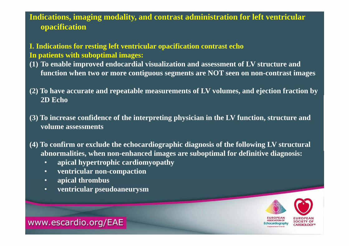
Indications, imaging modality, and contrast administration for left ventricular opacification
I. Indications for resting left ventricular opacification contrast echoIn patients with suboptimal images:(1) To enable improved endocardial visualization and assessment of LV structure and ( ) p
function when two or more contiguous segments are NOT seen on non-contrast images
(2) To have accurate and repeatable measurements of LV volumes, and ejection fraction by 2D Echo
(3) To increase confidence of the interpreting physician in the LV function, structure and volume assessments
(4) To confirm or exclude the echocardiographic diagnosis of the following LV structural b li i h h d i b i l f d fi i i di iabnormalities, when non-enhanced images are suboptimal for definitive diagnosis:• apical hypertrophic cardiomyopathy• ventricular non-compaction
i l th b• apical thrombus• ventricular pseudoaneurysm
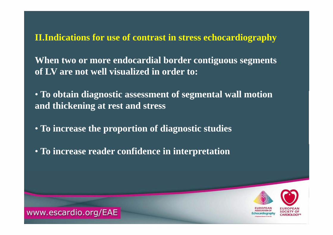
II Indications for use of contrast in stress echocardiographyII.Indications for use of contrast in stress echocardiography
When two or more endocardial border contiguous segmentsg gof LV are not well visualized in order to:
• To obtain diagnostic assessment of segmental wall motionand thickening at rest and stress
• To increase the proportion of diagnostic studies
• To increase reader confidence in interpretation
European Journal of Echocardiography (2009) 10, 194–212
European Journal of Echocardiography (2009) 10, 194–212
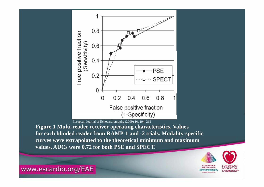
Figure 1 Multi-reader receiver operating characteristics. Valuesfor each blinded reader from RAMP-1 and -2 trials. Modality-specific
European Journal of Echocardiography (2009) 10, 194–212
y pcurves were extrapolated to the theoretical minimum and maximumvalues. AUCs were 0.72 for both PSE and SPECT.
European Journal of Echocardiography (2009) 10, 194–212
European Journal of Echocardiography (2009) 10, 194–212
European Journal of Echocardiography (2009) 10, 194–212
European Journal of Echocardiography (2009) 10, 194–212
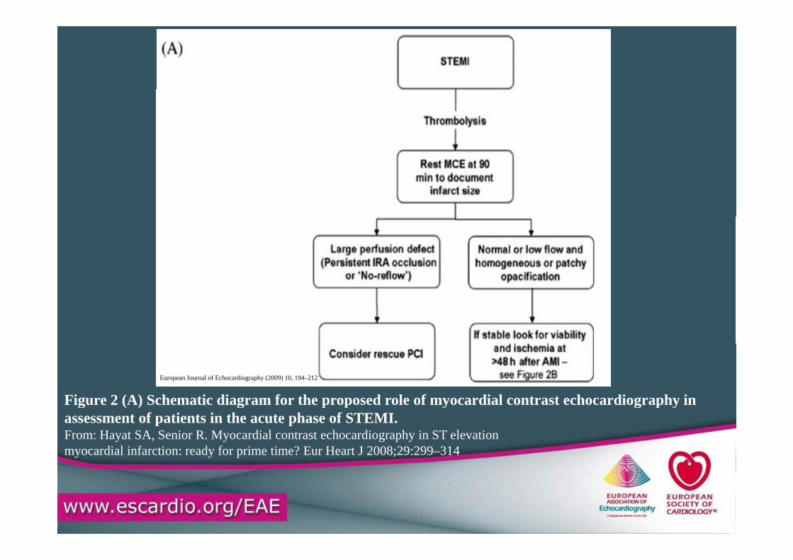
Figure 2 (A) Schematic diagram for the proposed role of myocardial contrast echocardiography in
European Journal of Echocardiography (2009) 10, 194–212
g ( ) g p p y g p yassessment of patients in the acute phase of STEMI.From: Hayat SA, Senior R. Myocardial contrast echocardiography in ST elevationmyocardial infarction: ready for prime time? Eur Heart J 2008;29:299–314
European Journal of Echocardiography (2009) 10, 194–212
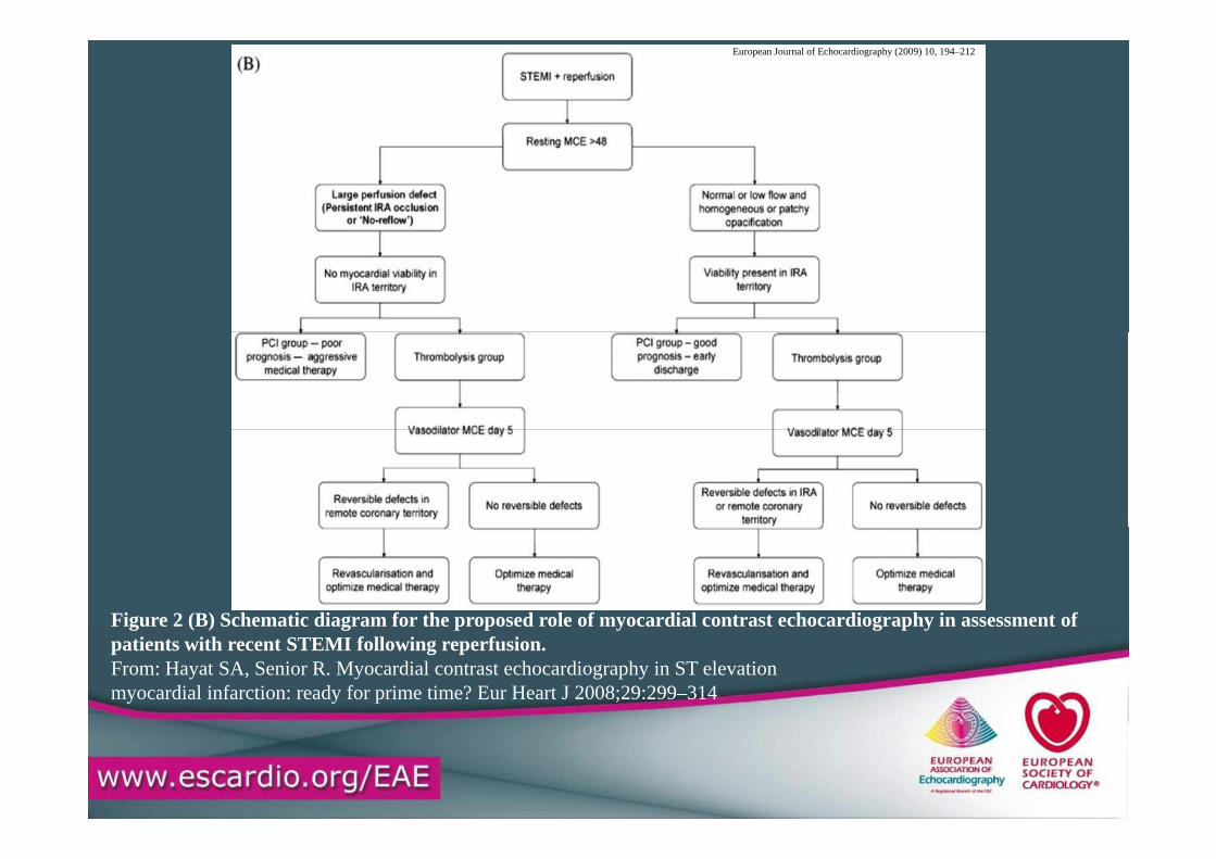
Figure 2 (B) Schematic diagram for the proposed role of myocardial contrast echocardiography in assessment ofFigure 2 (B) Schematic diagram for the proposed role of myocardial contrast echocardiography in assessment of patients with recent STEMI following reperfusion.From: Hayat SA, Senior R. Myocardial contrast echocardiography in ST elevation myocardial infarction: ready for prime time? Eur Heart J 2008;29:299–314
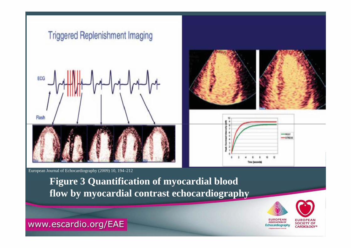
Figure 3 Quantification of myocardial bloodEuropean Journal of Echocardiography (2009) 10, 194–212
Figure 3 Quantification of myocardial bloodflow by myocardial contrast echocardiography
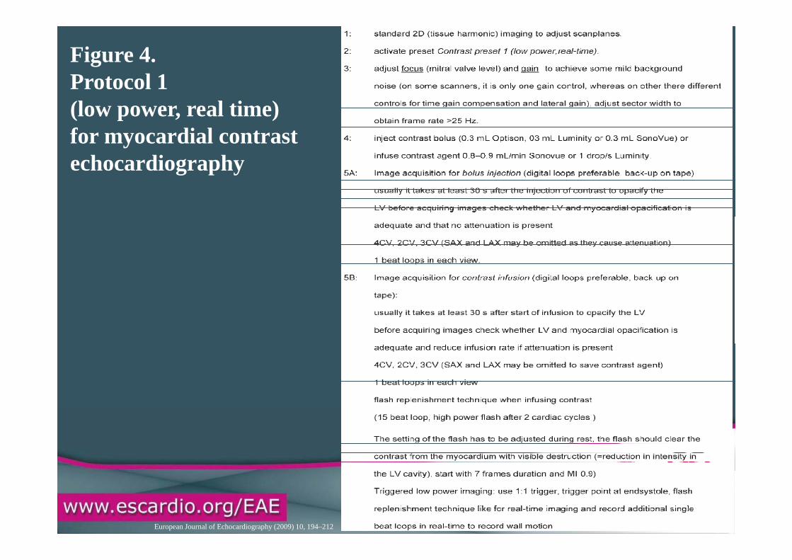
Figure 4. Protocol 1o oco(low power, real time) for myocardial contrast echocardiography
European Journal of Echocardiography (2009) 10, 194–212
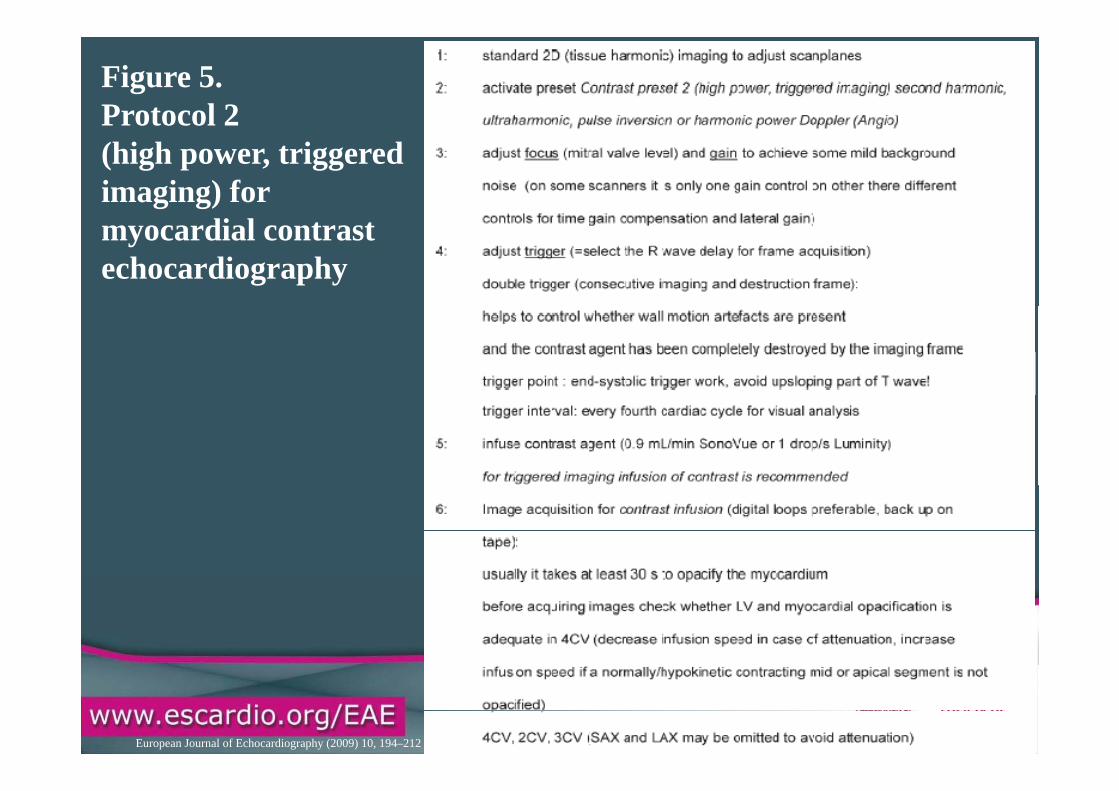
Figure 5. Protocol 2(high power, triggeredimaging) for myocardial contrast echocardiography
European Journal of Echocardiography (2009) 10, 194–212
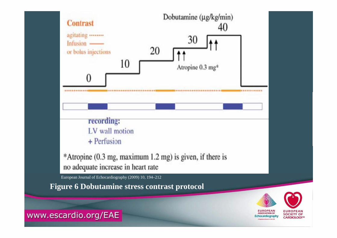
Figure 6 Dobutamine stress contrast protocolEuropean Journal of Echocardiography (2009) 10, 194–212
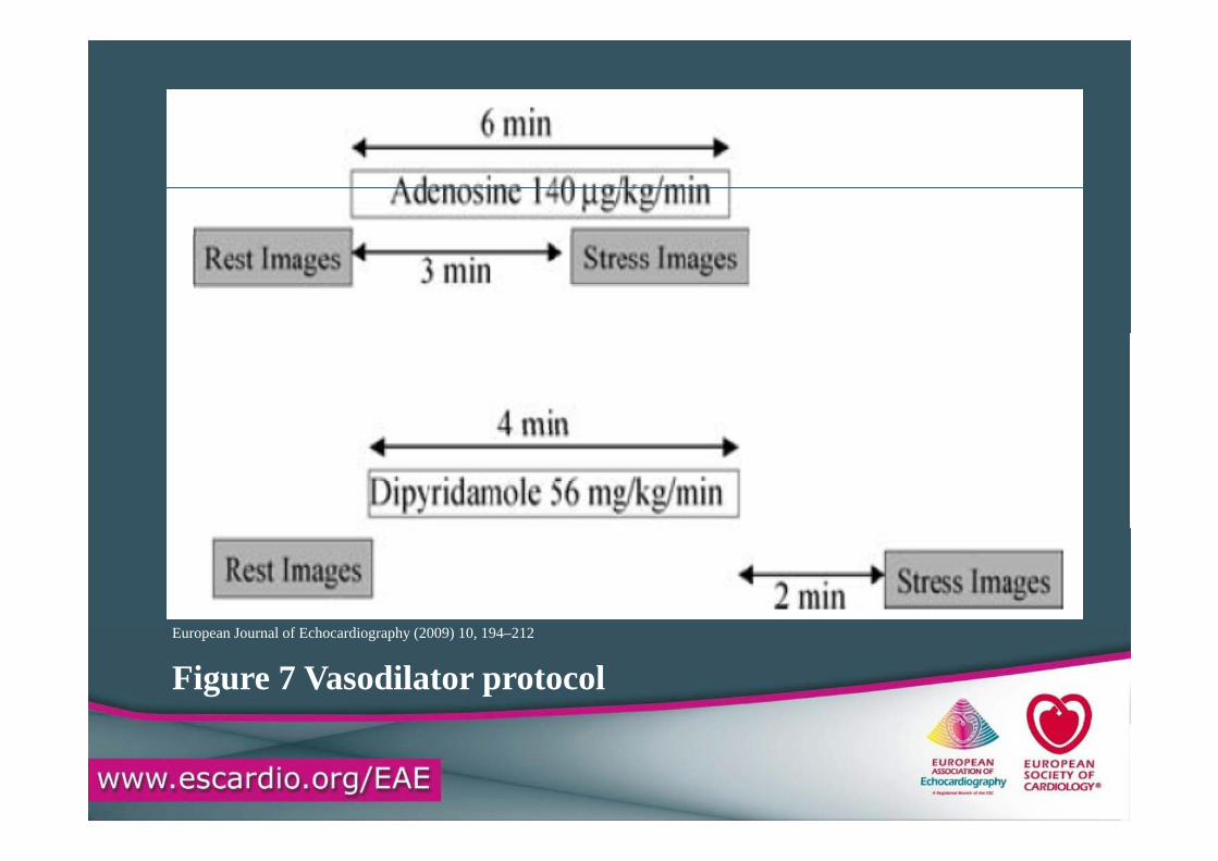
Figure 7 Vasodilator protocolEuropean Journal of Echocardiography (2009) 10, 194–212