Dente de Roots 1
59
roots inter nation al magazi ne of endodontology _case study Multiple vs. One-Step Apexification _scientific paper Control and Elimination of Endodontic Infection _overview Successful Mechanical Root Canal Conditioning 1 2006 issn 1616-6345 Vol. 1 • Issue 1/2006
-
Upload
vinisha-pandey -
Category
Documents
-
view
231 -
download
0
Transcript of Dente de Roots 1

8/13/2019 Dente de Roots 1
http://slidepdf.com/reader/full/dente-de-roots-1 1/58
rootsinternational magazine ofendodontology
_case study
Multiple vs.One-Step Apexification
_scientific paperControl and Elimination
of Endodontic Infection
_overview
Successful MechanicalRoot Canal Conditioning
1
2006
issn 1616-6345 Vol. 1 • Issue 1/2006

8/13/2019 Dente de Roots 1
http://slidepdf.com/reader/full/dente-de-roots-1 2/58
I 03
roots _ editorial I
roots1_2006
_It has finally arrived! In your hands you hold the first edition of roots, the international magazine of
endodontology. In the future we hope to offer scientists, endodontic specialists, and those generally inter-
ested in the subject a forum within its pages four times a year.
In spite of the increasing number of publications for our profession, there is not one trade publication that
concerns itself with the exchange of information between science and the practice of different specialized
disciplines. This is not at all unfortunate as on the one hand, the variety of systems existing today is often
confusing, and on the hand research frequently does not take into consideration the problems actually found
in the practice.
Direct lines of communication between scientist and practitioners can result from the establishment
of our journal and create a new impetus for innovation and for the practice. Given the upswing of endodon-
tology in the last years, we would like roots to close the gap in the spectrum of available publications and
create a place for shared debates. It allows for a greater exchange between its readership, and particularly
the Internet community created by Dr. Ken Serota at www.roots.com, to which we want to grant a worthy
new home within these pages.
roots will begin with this international English edition, soon to be followed by a country-specific edition
for the USA. Editions for North America, South America, Europe and Asia will also follow.
Our goals include counting those in general practice as well as scientists among our list of published
authors. The scientific side will be handled by Mr. Professor Benjamin Brisenio, whom I would first cordially
like to thank for his commitment.
I am eager to see your contributions and look forward to many years of lively debates and interestingcontributions from endodontic’s research side as well as its practical side.
Respectfully Yours,
Dr. Karl Behr
Editor-in-Chief
Germany
Dear Colleagues,
Dr. Karl Behr
Editor-in-Chief
8/13/2019 Dente de Roots 1
http://slidepdf.com/reader/full/dente-de-roots-1 3/58
04 I
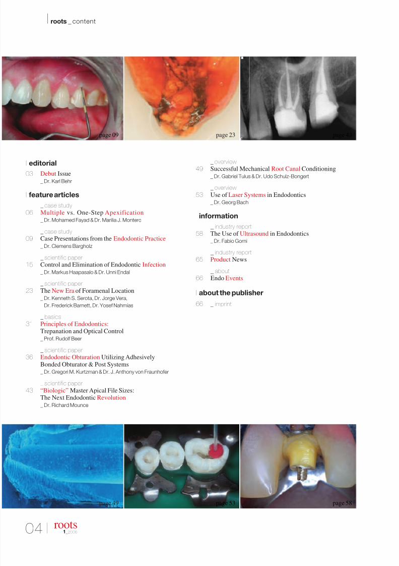
I roots _ content
roots1_2006
I editorial
03 Debut Issue_ Dr. Karl Behr
I feature articles_ case study
06 Multiple vs. One-Step Apexification_ Dr. Mohamed Fayad & Dr. Marilia J. Montero
_ case study
09 Case Presentations from the Endodontic Practice_ Dr. Clemens Bargholz
_ scientific paper
15 Control and Elimination of Endodontic Infection_ Dr. Markus Haapasalo & Dr. Unni Endal
_ scientific paper
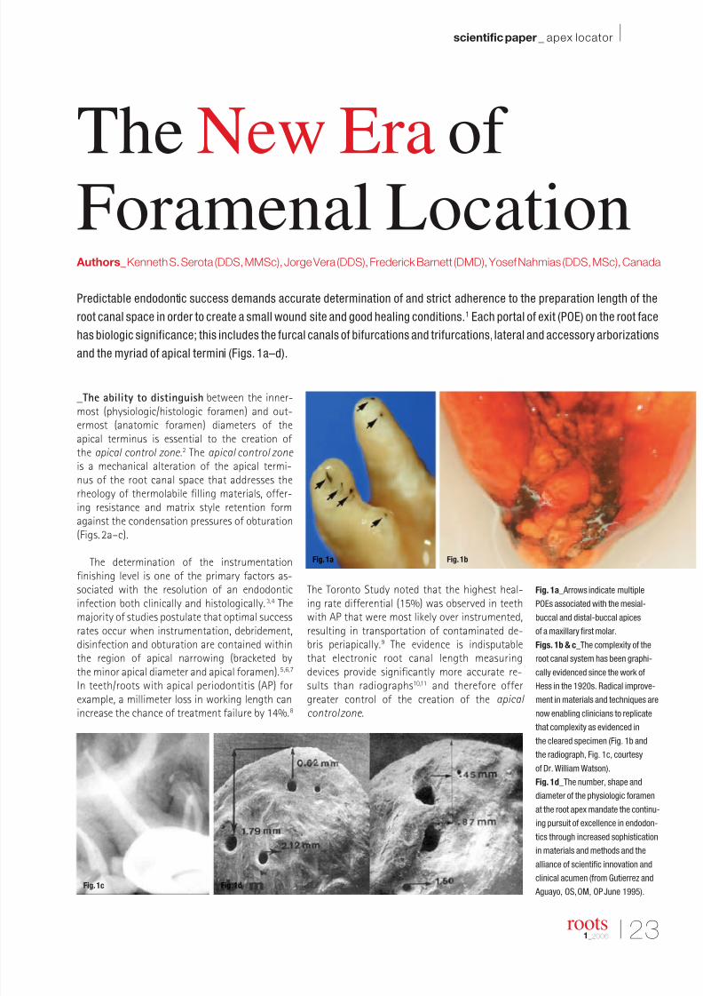
23 The New Era of Foramenal Location
_ Dr. Kenneth S. Serota, Dr. Jorge Vera,Dr. Frederick Barnett, Dr. Yosef Nahmias
_ basics
31 Principles of Endodontics:Trepanation and Optical Control_ Prof. Rudolf Beer
_ scientific paper
36 Endodontic Obturation Utilizing AdhesivelyBonded Obturator & Post Systems_ Dr. Gregori M. Kurtzman & Dr. J. Anthony von Fraunhofer
_ scientific paper
43 “Biologic” Master Apical File Sizes:
The Next Endodontic Revolution_ Dr. Richard Mounce
_ overview
49 Successful Mechanical Root Canal Conditioning_ Dr. Gabriel Tulus & Dr. Udo Schulz-Bongert
_ overview
53 Use of Laser Systems in Endodontics_ Dr. Georg Bach
I information
_ industry report
58 The Use of Ultrasound in Endodontics_ Dr. Fabio Gorni
_ industry report
65 Product News
_ about
66 Endo Events
I about the publisher
66 _ imprint
page 09 page 23 page 43
page 49 page 53 page 58
8/13/2019 Dente de Roots 1
http://slidepdf.com/reader/full/dente-de-roots-1 4/58
06 I
I case study _ apexification
_Root development commences after the comple-tion of the enamel formation. Pulp vitality is requiredfor root development to take place. Any factor thatwill sacrifice pulp vitality, such as trauma, caries, anddental anomalies (dens evaginatus and ivaginatus),may lead to the arrest of the physiological processof root formation.
Immature pulpless teeth present special problemsin meeting the objectives of nonsurgical endodontictherapy. Immature pulpless teeth have thin, short di-
vergent walls in the apical third, which
makes normal development of an apicalstop and obtaining an optimal apical sealimpossible. This leads to an inability toconfine the filling material to the canalspace. The most commonly used tech-nique is inducing apical closure by for-mation of an apical stop using calciumhydroxide as an intracanal medicament.This process is known as apexification.
A new approach is placing a biologi-cally acceptable material in the apicalportion of the root canal, thus formingan apical barrier; followed by filling the
root canal with gutta-percha and sealer.This procedure has been called one-stepapexification.
_Multiple-visit apexification
Induction of apical closure has beenthe most widely used approach to treat-ing open apex. Kaiser1 first introducedthe use of calcium hydroxide mixed withcamphorated monoparachlorophenol(CMCP) to induce apical closure. Thetechnique was popularized later in1966 by Frank2, who described a step-by-step technique and four types of
apical closure. Calcium hydroxide can be mixed witha number of different substances (CMCP, distilled wa-ter, sterile saline, anesthetic solutions and recentlychlorohexidine) to induce apical closure. The rela-tively good success rate of this procedure has beenattributed to one or more of the following properties:(a) the high pH, (b) the calcium ion, (c) the hydroxylion, and (d) the antibacterial effect.
However, the property that actually promotes themechanism for the calcific bridge formation is notknown. From the previous literature, the most impor-
tant factors in achieving apexification seem to bethorough debridment of the root canal (to remove allnecrotic pulp tissue) and sealing the tooth (to preventthe ingress of bacteria and substrate).
The usual time required to achieve apexificationwith conventional calcium hydroxide treatment is6 to 24 months (the average is 1 year ± 7 months).Factors that lead to increased treatment time are thepresence of a radiolucent lesion, inter-appointmentsymptoms, and loss of the external seal with rein-fection of the canal.
During this time, the patient is recalled at 3-monthintervals. If any signs or symptoms of reinfection or
pathology occur during this phase of the treatment,the canal is recleaned and refilled with the calciumhydroxide paste. The patient is recalled until radi-ographic evidence of apexification has become ap-parent.
Determination of the extent of the apical closureis often difficult to ascertain. Radiographic interpre-tation of apical closure may be misleading. It must beremembered that the dental radiograph is a two-di-mensional picture of a three-dimensional object. Thefaciolingual aspect of the root canal is usually the lastto become convergent apically as the root develops.Therefore, it is possible to have a dental radiographshowing an apically convergent root canal while inthe faciolingual plane the root canal is divergent.
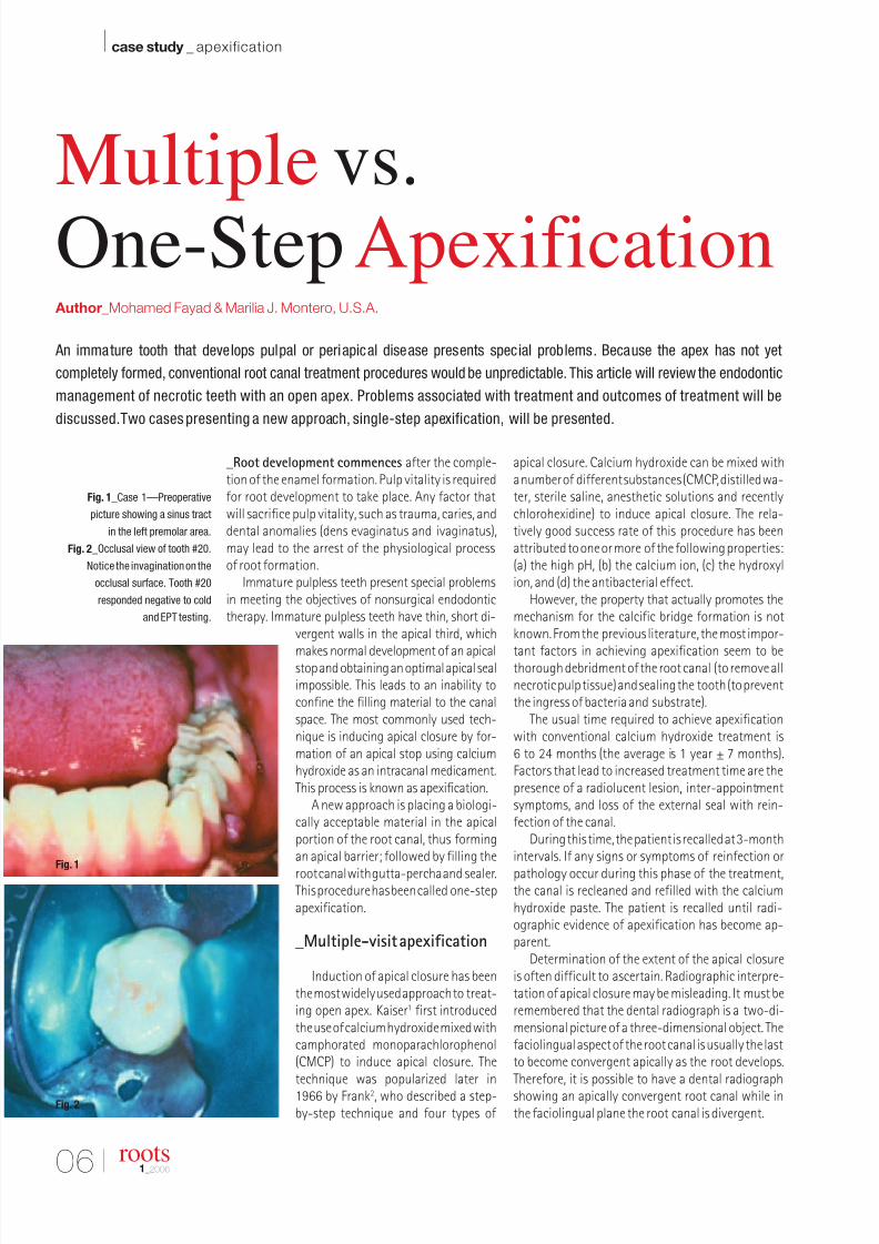
Fig. 1_Case 1—Preoperative
picture showing a sinus tract
in the left premolar area.
Fig. 2_Occlusal view of tooth #20.
Notice the invagination on the
occlusal surface. Tooth #20
responded negative to cold
and EPT testing.
roots1_2006
Multiple vs.
One-StepApexification Author_Mohamed Fayad & Marilia J. Montero, U.S.A.
An immature tooth that develops pulpal or periapical disease presents special problems. Because the apex has not yet
completely formed, conventional root canal treatment procedures would be unpredictable. This article will review the endodontic
management of necrotic teeth with an open apex. Problems associated with treatment and outcomes of treatment will be
discussed.Two cases presenting a new approach, single-step apexification, will be presented.
Fig. 1
Fig. 2
8/13/2019 Dente de Roots 1
http://slidepdf.com/reader/full/dente-de-roots-1 5/58
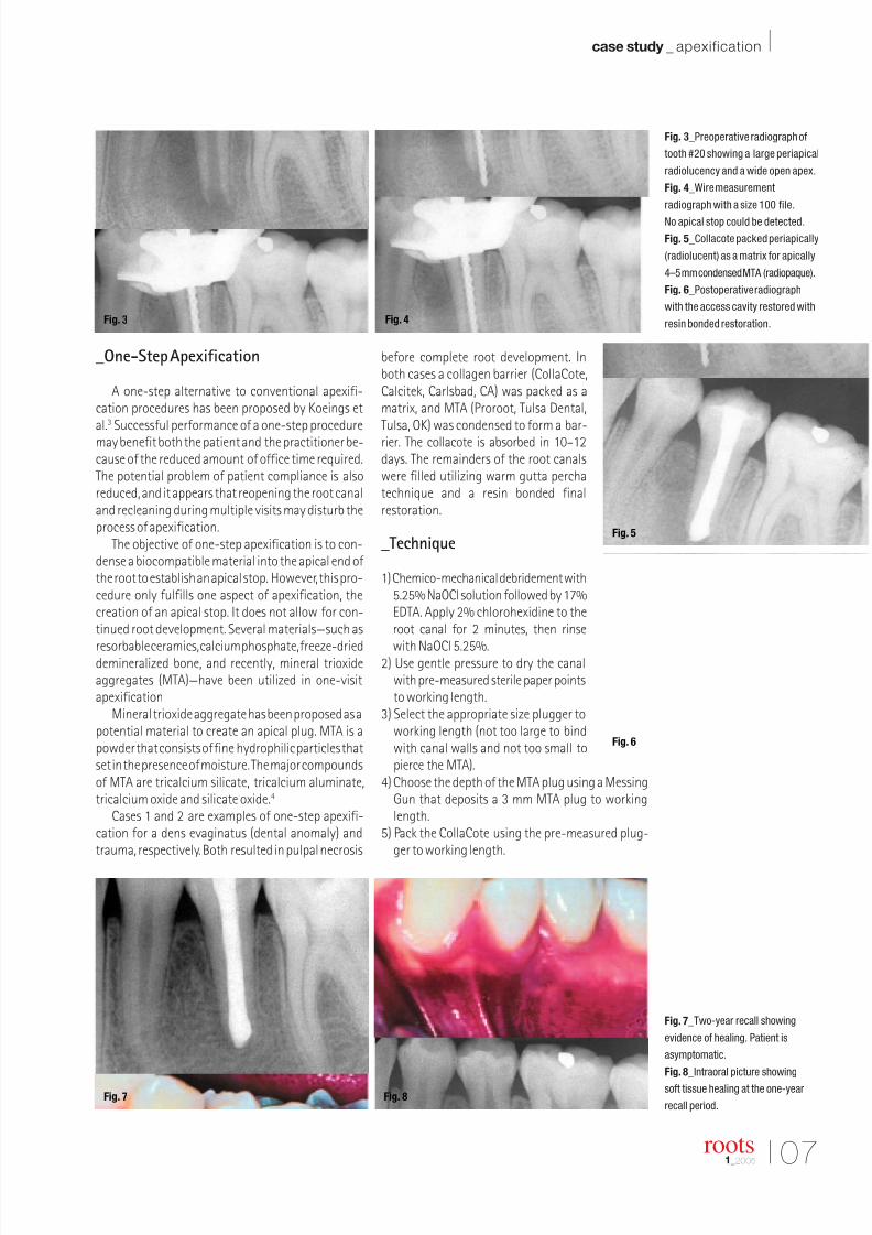
Fig. 3_Preoperative radiograph of
tooth #20 showing a large periapical
radiolucency and a wide open apex.
Fig. 4_Wire measurement
radiograph with a size 100 file.
No apical stop could be detected.
Fig. 5_Collacote packed periapically
(radiolucent) as a matrix for apically
4–5 mm condensed MTA (radiopaque).
Fig. 6_Postoperative radiograph
with the access cavity restored with
resin bonded restoration.
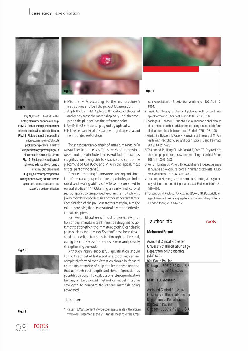
Fig. 7_Two-year recall showing
evidence of healing. Patient is
asymptomatic.
Fig. 8_Intraoral picture showing
soft tissue healing at the one-year
recall period.
I 07
case study _ apexification I
roots1_2005
_One-Step Apexification
A one-step alternative to conventional apexifi-cation procedures has been proposed by Koeings etal.3 Successful performance of a one-step proceduremay benefit both the patient and the practitioner be-cause of the reduced amount of office time required.The potential problem of patient compliance is alsoreduced, and it appears that reopening the root canaland recleaning during multiple visits may disturb theprocess of apexification.
The objective of one-step apexification is to con-dense a biocompatible material into the apical end of the root to establish an apical stop. However, this pro-cedure only fulfills one aspect of apexification, thecreation of an apical stop. It does not allow for con-tinued root development. Several materials—such as
resorbable ceramics, calcium phosphate, freeze-drieddemineralized bone, and recently, mineral trioxideaggregates (MTA)—have been utilized in one-visitapexification
Mineral trioxide aggregate has been proposed as apotential material to create an apical plug. MTA is apowder that consists of fine hydrophilic particles thatset in the presence of moisture. The major compoundsof MTA are tricalcium silicate, tricalcium aluminate,tricalcium oxide and silicate oxide.4
Cases 1 and 2 are examples of one-step apexifi-cation for a dens evaginatus (dental anomaly) andtrauma, respectively. Both resulted in pulpal necrosis
before complete root development. Inboth cases a collagen barrier (CollaCote,Calcitek, Carlsbad, CA) was packed as amatrix, and MTA (Proroot, Tulsa Dental,Tulsa, OK) was condensed to form a bar-rier. The collacote is absorbed in 10–12days. The remainders of the root canalswere filled utilizing warm gutta perchatechnique and a resin bonded finalrestoration.
_Technique
1) Chemico-mechanical debridement with5.25% NaOCl solution followed by 17%EDTA. Apply 2% chlorohexidine to theroot canal for 2 minutes, then rinse
with NaOCl 5.25%.2) Use gentle pressure to dry the canal
with pre-measured sterile paper pointsto working length.
3) Select the appropriate size plugger toworking length (not too large to bindwith canal walls and not too small topierce the MTA).
4) Choose the depth of the MTA plug using a MessingGun that deposits a 3 mm MTA plug to workinglength.
5) Pack the CollaCote using the pre-measured plug-ger to working length.
Fig. 3 Fig. 4
Fig. 5
Fig. 6
Fig. 7 Fig. 8
8/13/2019 Dente de Roots 1
http://slidepdf.com/reader/full/dente-de-roots-1 6/58
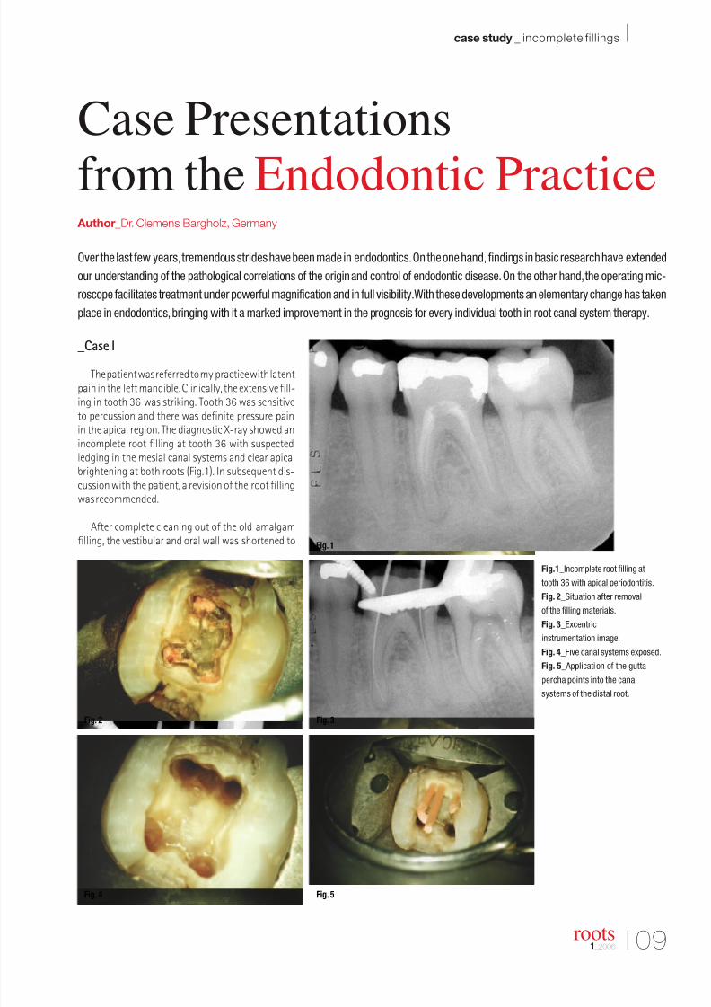
Fig. 9_Case 2—Tooth #9 with a
history of trauma and necrotic pulp.
Fig. 10_Picture through the operating
microscope showing periapical tissue.
Fig. 11_Picture through the operating
microscope showing Collacote
packed periapically as a matrix.
Periapical radiograph verifying MTA
placement in the apical 3–4 mm.
Fig. 12_Postoperative radiograph
showing a dense fill with control
in apical plug placement.
Fig.13_Six month postoperative
radiograph showing a dense fill with
apical control and reduction in thesize of the periapical lesion.
08 I
I case study _ apexification
6) Mix the MTA according to the manufacturer’sinstructions and load the pre-set Messing Gun.
7) Apply the 3 mm MTA plug to the orifice of the canaland gently tease the material apically until the stop-per on the plugger is at the reference point.
8) Verify the 3 mm apical plug radiographically.9) Fill the remainder of the canal with gutta percha and
resin bonded restoration.
These cases are an example of immature roots. MTAwas utilized in both cases. The success of the previouscases could be attributed to several factors, such asmagnification (being able to visualize and control theplacement of CollaCote and MTA in the apical, mostcritical part of the canal).
Other contributing factors are cleansing and shap-ing of the canals; superior biocompatibility, antimic-
robial and sealing ability of MTA as documented inseveral studies.5, 6, 7, 8 Obtaining an early final coronalseal compared to temporized teeth in the multiple visit(6–12 months) procedures is another important factor.Combination of the previous factors may play a majorrole in increasing the success rate of necrotic teeth withimmature apices.
Following obturation with gutta-percha, restora-tion of the immature teeth must be designed to at-tempt to strengthen the immature teeth. Clear plasticposts such as the Luminex System® have been devel-oped to allow light transmission throughout the canal,curing the entire mass of composite resin and possibly
strengthening the root.Although highly successful, apexification should
be the treatment of last resort in a tooth with an in-completely formed root. Attention should be focusedon the maintenance of pulp vitality in these teeth sothat as much root length and dentin formation aspossible can occur. To evaluate one-step apexificationfurther, a standardized method or model must bedeveloped to compare the various materials beingadvocated. _
_Literature
1.Kaiser HJ.Management of wide open apex canals with calcium
hydroxide. Presented at the 21st Annual meeting of the Amer-
ican Association of Endodontics, Washington, DC, April 17,
1964.
2.Frank AL. Therapy of divergent pulpless tooth by continued
apical formation.J Am dent Assoc.1966; 72:87–93.
3.Koeings JF, Heller AL, Brilliant JD, et al: Induced apical closure
of permanent teeth in adult primates using a resorbable form
of tricalcium phosphate ceramic.J Endod 1975; 102–106.
4.Giuliani V, Baccetti T, Pace R, Pagavino G. The use of MTA in
teeth with necrotic pulps and open apices. Dent Traumatol
2002; 18:217–221.
5.Torabnejad M, Hong CU, McDonald F, Ford TR. Physical and
chemical properties of a new root-end filling material.J Endod
1995; 21:349–353.
6.Koh ET,Torabnejad M,Ford TR,et al.Mineral trioxide aggregate
stimulates a biological response in human osteoblasts. J. Bio-
med Mater Res 1997; 37: 432–439.
7.Torabnejad M, Hong CU, Pitt-Ford TR, Ketterling JD. Cytotox-
icity of four root-end filling materials. J Endodon 1995; 21:489–492.
8.Torabnejad M,Rastegar AF,Kettring JD,Ford TR. Bacterial leak-
age of mineral trioxide aggregate as a root-end filling material.
J. Endod 1999; 21:109–112.
roots1_2006
Mohamed Fayad
Assistant Clinical Professor
University of Illin ois at ChicagoDepartment of Endodonti cs(M/C 642)
801 South PaulinaChicago IL 60612-7 212, U.S.A.
E-mail: [email protected]
Marilia J. Montero
Assistant Clinical Professor
University of Illin ois at ChicagoDepartment of Pediatrics
801 South Paulina
Chicago IL 60612-7 212, U.S.A.
_author info roots
Fig. 9
Fig. 10 Fig. 11
Fig. 12
Fig. 13
8/13/2019 Dente de Roots 1
http://slidepdf.com/reader/full/dente-de-roots-1 7/58
I 09
case study _ incomplete fill ings I
roots1_2006
_Case I
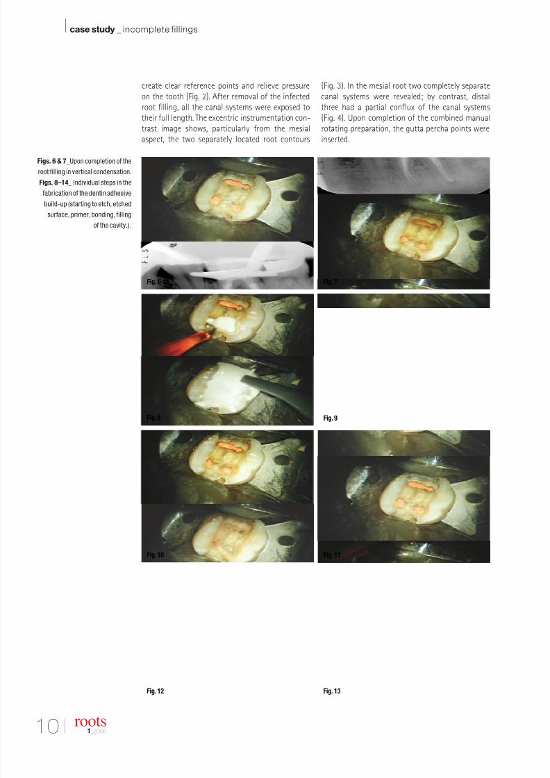
The patient was referred to my practice with latentpain in the left mandible. Clinically, the extensive fill-ing in tooth 36 was striking. Tooth 36 was sensitiveto percussion and there was definite pressure painin the apical region. The diagnostic X-ray showed anincomplete root filling at tooth 36 with suspectedledging in the mesial canal systems and clear apicalbrightening at both roots (Fig.1). In subsequent dis-cussion with the patient, a revision of the root fillingwas recommended.
After complete cleaning out of the old amalgamfilling, the vestibular and oral wall was shortened to
Fig. 2
Fig. 4 Fig. 5
Fig. 1
Fig. 3
Case Presentations
from the Endodontic Practice Author_Dr. Clemens Bargholz, Germany
Over the last few years,tremendous strides have been made in endodontics.On the one hand, findings in basic research have extended
our understanding of the pathological correlations of the origin and control of endodontic disease.On the other hand,the operating mic-
roscope facilitates treatment under powerful magnification and in full visibility.With these developments an elementary change has taken
place in endodontics,bringing with it a marked improvement in the prognosis for every individual tooth in root canal system therapy.
Fig.1_Incomplete root filling at
tooth 36 with apical periodontitis.
Fig. 2_Situation after removal
of the filling materials.
Fig. 3_Excentric
instrumentation image.
Fig. 4_Five canal systems exposed.
Fig. 5_ Application of the gutta
percha points into the canal
systems of the distal root.
8/13/2019 Dente de Roots 1
http://slidepdf.com/reader/full/dente-de-roots-1 8/58
10 I
I case study _ incomplete fill ings
create clear reference points and relieve pressureon the tooth (Fig. 2). After removal of the infectedroot filling, all the canal systems were exposed totheir full length. The excentric instrumentation con-trast image shows, particularly from the mesial
aspect, the two separately located root contours
(Fig. 3). In the mesial root two completely separatecanal systems were revealed; by contrast, distalthree had a partial conflux of the canal systems(Fig. 4). Upon completion of the combined manualrotating preparation, the gutta percha points were
inserted.
Figs. 6 & 7_Upon completion of the
root filling in vertical condensation.
Figs. 8–14_ Individual steps in the
fabrication of the dentin adhesive
build-up (starting to etch, etched
surface, primer, bonding, filling
of the cavity.).
roots1_2006
Fig. 6
Fig. 8
Fig. 10
Fig. 12 Fig. 13
Fig. 11
Fig. 9
Fig. 7
8/13/2019 Dente de Roots 1
http://slidepdf.com/reader/full/dente-de-roots-1 9/58
I 11
case study _ incomplete fill ings I
roots1_2006
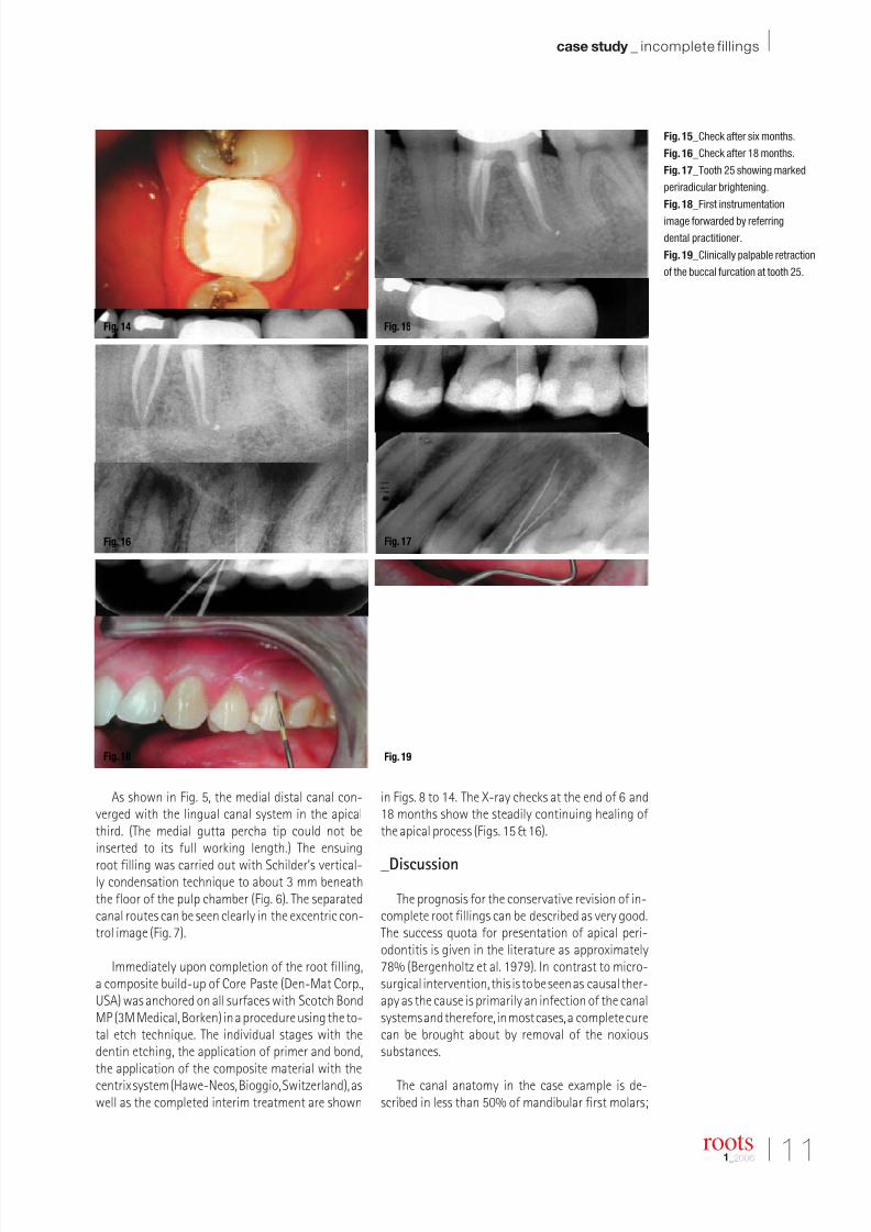
As shown in Fig. 5, the medial distal canal con-verged with the lingual canal system in the apicalthird. (The medial gutta percha tip could not beinserted to its full working length.) The ensuing
root filling was carried out with Schilder’s vertical-ly condensation technique to about 3 mm beneaththe floor of the pulp chamber (Fig. 6). The separatedcanal routes can be seen clearly in the excentric con-trol image (Fig. 7).
Immediately upon completion of the root filling,a composite build-up of Core Paste (Den-Mat Corp.,USA) was anchored on all surfaces with Scotch BondMP (3M Medical, Borken) in a procedure using the to-tal etch technique. The individual stages with thedentin etching, the application of primer and bond,the application of the composite material with thecentrix system (Hawe-Neos, Bioggio, Switzerland), aswell as the completed interim treatment are shown
in Figs. 8 to 14. The X-ray checks at the end of 6 and18 months show the steadily continuing healing of the apical process (Figs. 15 & 16).
_Discussion
The prognosis for the conservative revision of in-complete root fillings can be described as very good.The success quota for presentation of apical peri-odontitis is given in the literature as approximately78% (Bergenholtz et al. 1979). In contrast to micro-surgical intervention, this is to be seen as causal ther-apy as the cause is primarily an infection of the canalsystems and therefore, in most cases, a complete curecan be brought about by removal of the noxioussubstances.
The canal anatomy in the case example is de-scribed in less than 50% of mandibular first molars;
Fig. 14 Fig. 15
Fig. 16 Fig. 17
Fig. 18 Fig. 19
Fig. 15_Check after six months.
Fig. 16_Check after 18 months.
Fig. 17_Tooth 25 showing marked
periradicular brightening.
Fig. 18_First instrumentation
image forwarded by referring
dental practitioner.
Fig. 19_Clinically palpable retraction
of the buccal furcation at tooth 25.
8/13/2019 Dente de Roots 1
http://slidepdf.com/reader/full/dente-de-roots-1 10/58
12 I
I case study _ incomplete fill ings
it is more often found that the two distal main canalsflow into a common foramen in the apical region. Thethird distal canal shown may also be the slit-shapedoff-shoot of a “main canal”. However, the mechanicalwidening of such structures is necessary for thedisinfection of the complete canal system with rins-ing solutions. The subsequently implemented dentinadhesive build-up is increasingly discussed as post-
endodontic therapy.
To prevent any reinfection of the canal systems,bacteria-proof protection from the oral cavity is re-quired (Ray & Trope 1955). This can be achieved witha dentin bonding placed under a rubber dam. Teeththat have been endodontically treated are moreprone to factures, not because of their altered phys-ical dentin properties, but rather because of thesignificant loss of substance (Howe & Kendry 1990,Sedgley & Messer 1992, Bargholz 1996). The aim of post-endodontic therapy must therefore be to ensureoptimal stabilization and, under no circumstances,to cause further weakening of the hard tooth sub-stance by pin drilling.
_Case II
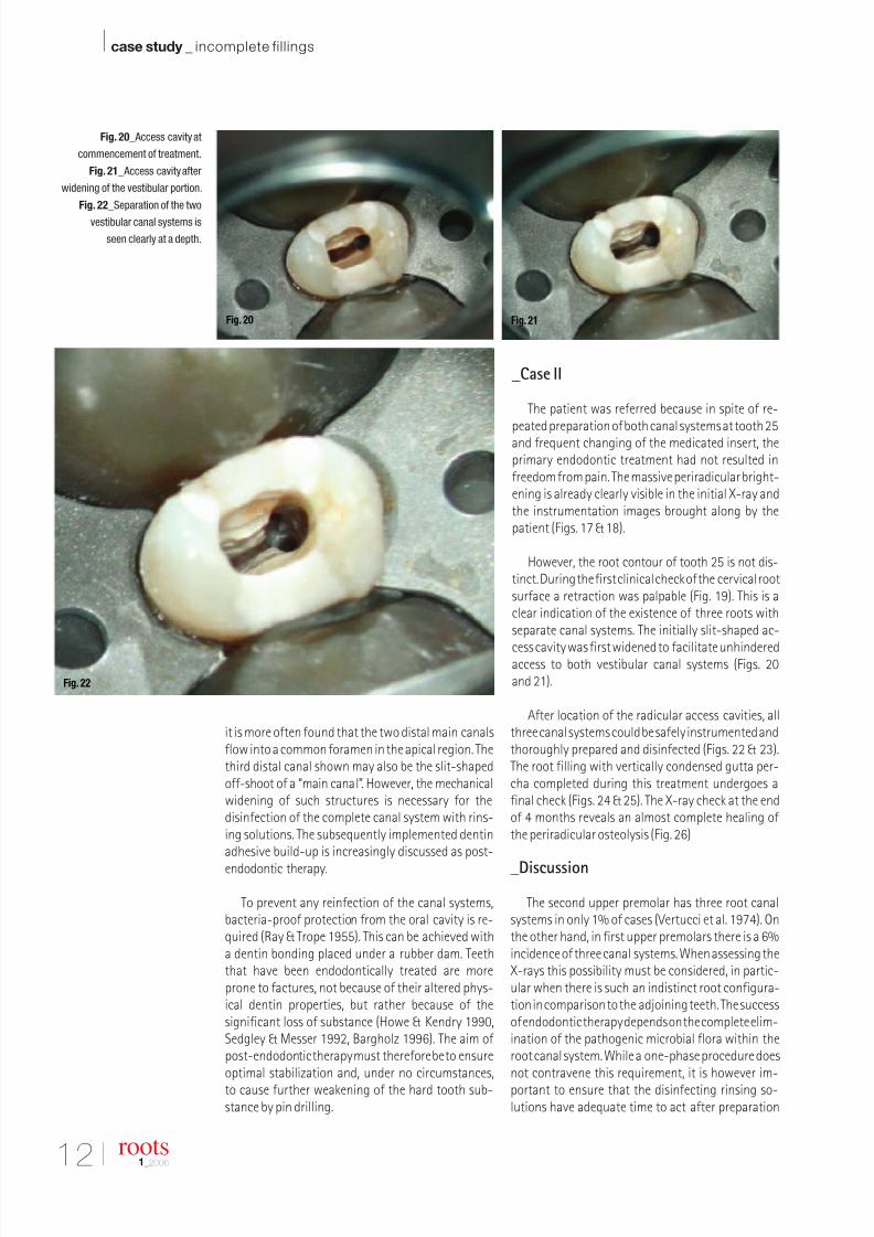
The patient was referred because in spite of re-peated preparation of both canal systems at tooth 25and frequent changing of the medicated insert, theprimary endodontic treatment had not resulted infreedom from pain. The massive periradicular bright-ening is already clearly visible in the initial X-ray andthe instrumentation images brought along by thepatient (Figs. 17 & 18).
However, the root contour of tooth 25 is not dis-tinct. During the first clinical check of the cervical rootsurface a retraction was palpable (Fig. 19). This is aclear indication of the existence of three roots withseparate canal systems. The initially slit-shaped ac-
cess cavity was first widened to facilitate unhinderedaccess to both vestibular canal systems (Figs. 20and 21).
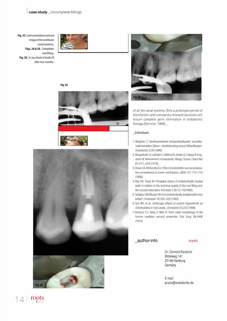
After location of the radicular access cavities, allthree canal systems could be safely instrumented andthoroughly prepared and disinfected (Figs. 22 & 23).The root filling with vertically condensed gutta per-cha completed during this treatment undergoes afinal check (Figs. 24 & 25). The X-ray check at the endof 4 months reveals an almost complete healing ofthe periradicular osteolysis (Fig. 26)
_Discussion
The second upper premolar has three root canalsystems in only 1% of cases (Vertucci et al. 1974). Onthe other hand, in first upper premolars there is a 6%incidence of three canal systems. When assessing theX-rays this possibility must be considered, in partic-ular when there is such an indistinct root configura-tion in comparison to the adjoining teeth. The successof endodontic therapy depends on the complete elim-ination of the pathogenic microbial flora within theroot canal system. While a one-phase procedure doesnot contravene this requirement, it is however im-portant to ensure that the disinfecting rinsing so-lutions have adequate time to act after preparation
Fig. 20_ Access cavity at
commencement of treatment.
Fig. 21_ Access cavity after
widening of the vestibular portion.
Fig. 22_Separation of the two
vestibular canal systems is
seen clearly at a depth.
roots1_2006
Fig. 20 Fig. 21
Fig. 22
8/13/2019 Dente de Roots 1
http://slidepdf.com/reader/full/dente-de-roots-1 11/58
8/13/2019 Dente de Roots 1
http://slidepdf.com/reader/full/dente-de-roots-1 12/58
I 15
scientific paper _ infection control I
roots1_2006
_In pulpitis, caused by a deep caries lesion, the inflam-matory reactions in the pulp start a long time beforebacteria are found in the pulp tissue. The initial inflam-matory reactions are initiated by bacterial antigens in-teracting with the local immune system (Bergenholtz1990, Pashley 1996, Jontell et al. 1998). As long as thebody of the carious lesion has not entered the pulp, theinflammatory process in the pulp is supposed to be re-versible and no endodontic therapy is usually required.With progressing caries, bacterial cells enter the super-ficial layers of the pulp. As long as there is vital pulp tis-sue, the pulp, even though heavily inflamed, is consid-ered to be relatively bacteria-free.
Apical periodontitis is an inflammatory process inthe periradicular tissue caused by microorganisms inthe necrotic root canal (Kakehashi et al. 1965). Somestudies have indicated that the prognosis of the treat-ment of apical periodontitis is lower if there are livingbacteria in the root canal at the time of filling (Engströmet al. 1964, Sjögren et al. 1997, Katebzadeh et al. 2000).It is generally accepted that successful treatment of pri-mary apical periodontitis rests on effective eliminationof the causative agents in the root canal system (Chu-gal et al. 2001). However, other studies have not beenable to show a difference in healing between teeth filledafter positive or negative cultures from the root canal
or between one and two-appointment treatments(Weiger et al. 2000, Peters & Wesselink 2002).
Elimination of endodontic infection is different fromelimination and control of most other infections in thehuman body. Because of the special anatomic environ-ment in the root canal and tooth, host measures thatin other sites are sufficient to eliminate the infectious or-ganisms are alone not enough for complete recovery inendodontic infections. Therefore, control of an endo-dontic infection is a concerted effort by several host andtreatment factors. Success in all aspects of this cooper-ation will eventually result in elimination of the infectivemicroorganisms and healing of the periapical lesion.
The necessary components in the elimination ofendodontic infection are: i) host defense system, ii) in
some cases systemic antibiotic therapy, iii) chemome-chanical preparation and irrigation, iv) local root canaldisinfecting medicaments, v) permanent root filling,vi) permanent coronal restoration. While the mainfocus of this article will be on chemomechanical prepa-ration and local disinfecting agents (factors iii and iv),the role of other contributory factors will also be brieflysummarized. Periapical actinomycosis and other ex-traradicular infections are left outside this review.
_Host defense
The host’s defense system is a key factor in prevent-
ing the spreading of the infection from the root canal tothe periapical tissues and bone. However, lack of circu-
Control and Elimination
of Endodontic Infection Author_ Dr. Markus Haapasalo, Canada & Dr. Unni Endal, Norway
The main goal in endodontics is the prevention and treatment of diseases of the dental pulp and periapical tissues. These
objectives can be best achieved if preventive measures and treatment procedures are based on a thorough and detailed
understanding of the etiology and pathogenesis of endodontic diseases.
Control of an endodontic infection is a con certedeffort by several host and treatment factors. Successin all aspects of this cooperation wil l eventually r esult
in elimination of the infective microorganisms andhealing of the periapical lesion. The necessary com-
ponents in the elim ination of endodontic infection arehost d efense system, chemom echanical preparation,
canal disi nfection, permanent root fill ing and coronalrestoration. The main focus in this article wil l be onchemomechanical preparation and lo cal disinfecting
agents, but the role of other contributor y factors willalso be briefly sum marized. At present it seems cor-
rect to conclude that no locally used root canal dis-infectant can predictably produce sterile canals in
the treatment of apical periodonti tis. Ho wever, theiruse further reduces the number of infecting micro-organisms after chemom echanical pr eparation to the
level of total elim ination. Future studies wil l probablyverify the new observation that root fillin g with gutta-
percha and sealer can have an actively d ualistic role
as a canal di sinfectant and as a permanent root fill ing.
_abstract roots
8/13/2019 Dente de Roots 1
http://slidepdf.com/reader/full/dente-de-roots-1 13/58
16 I
I scientific paper _ infection control
lation in the necrotic root canal makes it impossible forthe phagocytes and the rest of the immune system topenetrate into the root canal space for more than a fewhundred micrometers. Therefore, although of crucialimportance in maintaining general health, the defense
system is limited to achieving a balance between themicrobial intruders and the body, but it cannot elimi-nate the source of the infection in the root canal.
In chronic apical periodontitis the main mechanismresponsible for destruction of normal bone structureis activation of bone osteoclasts and inhibition of os-teoblast activity (Stashenko et al. 1992, 1998). The se-quence of events resulting in osteoclast stimulation isa network of immunological chain reactions where in-flammatory cytokines play a major role. Although al-ternative theories about the major route in osteoclastactivation have been presented, the key fact remainsthat it is the host’s own cells, osteoclasts, that removethe bone around the root tip. Nowadays, removal of bone is understood as an important and necessary de-fense strategy: bone has a poor capability to defend it-self against bacterial intruders, and osteomyelitis mightbe a result of the spreading of the intracanal infection.That is why bone is removed by the defense systembefore the infection reaches the periapical tissues. Inapical periodontitis the lesion is filled with phagocytesand other defense cells which effectively prevent fur-ther spreading of the microbial infection.
_Systemic antibiotics
Use of systemic antibiotics is not a routine part ofendodontic treatment of apical periodontitis. On thecontrary, antibiotics are only rarely used in endodontics.Minimizing the risk of post-treatment symptoms hasbeen one argument often used when prescribing an-tibiotics to endodontic patients. However, according toseveral studies, use of systemic antibiotics has not beenhelpful in reducing the number of patients with flare-ups or other acute problems after the start of the treat-ment (for review see: Fouad 2002). Neither is therescientific evidence that systemic antibiotic therapy hasa beneficial effect on the long-term prognosis of the
treatment of apical periodontitis. There is presently aconsensus in endodontics that systemic antibioticsshould be used only when general indications for theiruse are present (Fouad 2002).
Administration of systemic antibiotics should beconsidered with a spreading infection indicating failureof local host responses, or in cases of known reducedhost defense mechanisms that might expose the pa-tient to increased systemic risks (Fouad 2002, SiqueiraJF Jr. 2002).
Also, when the patient has a fever, antibiotics shouldbe given. The effectiveness of antibiotic therapy is neverfully predictable because of a variety of parameters af-fecting the outcome. Therefore, the focus must alwaysbe on local antimicrobial measures (chemomechanical
preparation and disinfection). Whenever there are gen-eral symptoms or spreading infection, the patient mustbe carefully monitored, and referral to hospital mustbe considered.
_Chemomechanical preparation& irrigation
Manual instrumentation
There is no disagreement on the fact that mechani-cal cleaning and high quality shaping of the root canalis the most important single factor for successful en-dodontic treatment. Together with the use of local irri-gating solutions with antibacterial activity, the majorityof, if not all bacteria in the root canal system will beeliminated. Mechanical instrumentation is a primarymeans of bacterial reduction in endodontic treatment.Byström & Sundqvist (1981) measured the reduction inbacterial counts cultured from the infected root canalwhen instrumented with manual steel instrumentsand saline irrigation. Fifteen root canals with necroticpulps and periapical lesions were instrumented at fivesequential appointments.
Mechanical instrumentation greatly reduced thenumber of cfu (colony forming units), usually 100–1,000-fold, but the number of bacteria-free root canalsincreased slowly. Even after five appointments withmechanical preparation and saline irrigation, severalcanals still showed growth. Corresponding observa-tions were reported also by Ørstavik et al. (1991). Since
it has become obvious that mechanical preparationwith manual instruments and irrigation with saline(which has practically no antibacterial activity) is unableto predictably produce sterile root canals, focus hasbeen put on the combined effect of instrumentationand strong antibacterial irrigating solutions.
Canal irrigation
Use of irrigating solutions is an important part ofeffective chemomechanical preparation. It facilitatesremoval of necrotic tissue and dentine chips from theroot canal and thus prevents packing of infected tissueapically in the root canal and into the periapical area.
In addition, many irrigating solutions have other bene-ficial effects. EDTA (ethylene-diamine-tetra-acetic acid,17% disodium salt, pH 7) is a chelating agent widelyused in endodontic preparation. It has low or no anti-bacterial activity, but it effectively removes the smearlayer by affecting the inorganic component of thedentine. Therefore, by facilitating cleaning and removalof infected tissue, EDTA contributes to the eliminationof bacteria in the root canal. It has also been shown thatremoval of the smear layer by EDTA (or citric acid) im-proves the antibacterial effect of locally used disinfect-ing agents in deeper layers of dentine (Haapasalo &Ørstavik 1987, Ørstavik & Haapasalo 1990).
Sodium hypochlorite (NaOCl), used in concentra-tions varying from 0.5% to 5.25%, is a strong antimi-
roots1_2006
Fig. 1
8/13/2019 Dente de Roots 1
http://slidepdf.com/reader/full/dente-de-roots-1 14/58
I 17
scientific paper _ infection control I
roots1_2006
crobial agent that is supposed to play an importantrole in dissolving the organic part of pulpal remnantsand dentine. Most importantly, it kills bacteria very rap-idly even at relatively low concentrations. Pashley et al.(1984) demonstrated greater cytotoxicity and caustic
effects on healthy tissue with 5.25% NaOCl than with0.5% and 1% solutions. No studies have clearly shownthat the stronger solutions have a better antibacterialeffect in vivo in the root canal. However, careless use of both NaOCl (in high and low concentrations) as well asEDTA will result in severe pain if they are introduced tothe periapical area. Niu et al. (2002) observed the ultra-structure on canal walls after EDTA and EDTA + NaOClirrigation by scanning electron microscopy. They re-ported that more debris was removed by irrigation withEDTA followed by NaOCl than with EDTA alone. Byström& Sundqvist (1983, 1985) showed that although 0.5%NaOCl, with or without EDTA, improved the efficiencyof preparation, all canals could not be made bacteria-free even after repeated appointments.
Sodium hypochlorite effectively kills bacteria, but iscaustic if pressed to the periapical area. In addition, ac-tive chlorine may cause damage to the patient’s clothesthrough its strong bleaching effect. Therefore, therehas been interest for alternative irrigating solutions thatcould replace NaOCl. Chlorhexidine gluconate (CHX)has long been used in dentistry because of its anti-microbial properties and its relatively low toxicity. It isalso increasingly used in endodontics. Although studiescomparing the antibacterial effect of NaOCl and CHX
have given somewhat conflicting results, it seems thatwhen used in identical concentrations, their antibacter-ial effect in the root canal and in infected dentine is rel-atively similar (Buck et al. 2001, Heling & Chandler 1998, Vahdaty et al. 1993, Ørstavik & Haapasalo 1990). How-ever, CHX lacks the tissue dissolving ability, which is oneof the obvious benefits of NaOCl. Waltimo et al. (1999)studied the antifungal effect of combinations of en-dodontic irrigants and found that the combinations of disinfectants were equally or less effective than themore effective component.
However, it has been shown that in certain concen-trations chlorhexidine and hydrogen peroxide have a
strong synergistic effect against Enterococcus faecalis,Streptococcus sobrinus and Staphylococcus aureus
(Heling & Chandler 1998, Steinberg et al. 1999).MTAD is a new irrigating solution with promising an-
tibacterial activity. However, more data will be neededbefore final evaluation of its antimicrobi-al effect can bedone (Shabahang & Torabinejad, 2003; Portenier et al.2006).
Rotary instrumentation
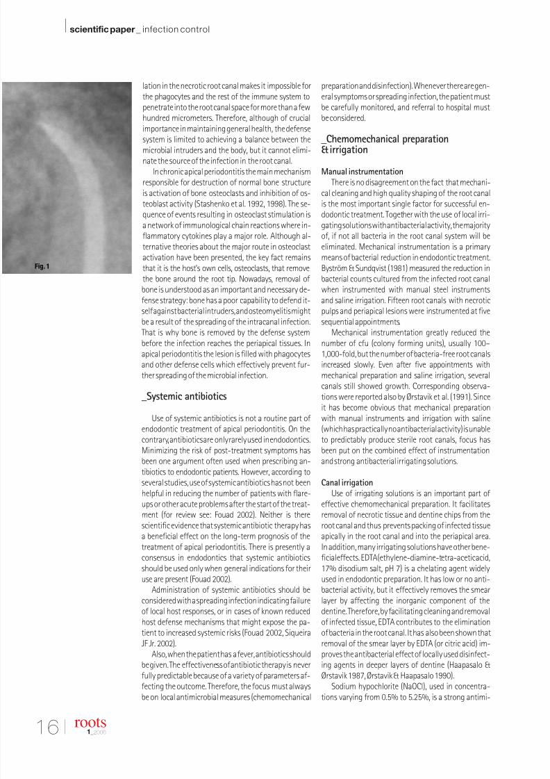
Use of rotary preparation with nickel-titanium in-struments undoubtedly offers several potential advan-tages. The most obvious of these are probably qualityof the apical preparation and efficiency (Fig. 1). How-ever, comparative studies have not always been in favor
of rotary instruments when the various aspects of preparation have been analyzed (Deplazes et al. 2001).Ahlqvist et al. (2001) showed that manual instrumen-tation produced cleaner canals than preparation withrotary instruments. Similar results have been reported
by Schäfer et al. (2002). However, rotary nickel-titaniuminstruments seem to maintain the original canal curva-ture better, particularly in the apical part of the rootcanal (Schäfer et al. 2002).
Dalton et al. (1998) compared steel K files and NiTirotary instruments in removing bacteria from infectedroot canals with saline as an irrigant. Only about a thirdof the teeth were made bacteria-free while no significantdifference could be detected between the two groups.However, larger preparation diameter of the apical ca-nal produced significant reduction in bacterial counts.Coldero et al. (2002) studied the effect of apical prepara-tion on the number of residual bacteria in the root canal.They concluded that additional apical enlargement to#35 did not further reduce the number of surviving bac-teria. However, the size of the original preparation in thisstudy is not given, and it is possible that even #35 is toosmall of a preparation size to show differences in bacte-rial elimination. In fact, Rollison et al. (2002) showed thatapical enlargement to #50 instead of #35 resulted in amore effective elimination of bacteria in the root canal,although absolute sterility was not obtained.
In a recent study Card et al. (2002) reported sterilityin a majority of root canals instrumented by rotary in-struments using large apical sizes and irrigation with
1% NaOCl. The instrumentation and bacterial samplingwas done in two phases: The first instrumentationutilized 1% NaOCl and 0.04 taper ProFile rotary files.The cuspid and bicuspid canals were instrumented to a#8 size and the molar canals to a #7 size. The second in-strumentation utilized LightSpeed files and 1% NaOClirrigation for further enlargement of the apical third.Typically, molars were instrumented to size #60 andcuspid/bicuspid canals to size #80. All of the cuspid/bicuspid canals and 81.5% of the molar canals werebacteria-free after the first instrumentation as shownby negative cultures from samples obtained from theroot canals. The molar results improved to 89% after
the second instrumentation. When the molar canalswere divided into two groups, one with no visible anas-tomoses between root canals and one with a complexroot canal anatomy, the proportion of sterile canalsin the first group was 93% already after the firstinstrumentation. The results of Card et al. (2002) are in-directly supported by earlier observations by Peters etal. (2001), who studied rotary preparation of root canalsof maxillary first molars. They compared the effects of four preparation techniques on canal volume and sur-face area using three-dimensionally reconstructedroot canals in extracted teeth. Micro CT data was usedto describe morphometric parameters related to thefour preparation techniques. Specimens were scannedbefore and after canals were prepared using Ni-Ti-K-
Fig. 2a
8/13/2019 Dente de Roots 1
http://slidepdf.com/reader/full/dente-de-roots-1 15/58
18 I
I scientific paper _ infection control
Files, Lightspeed instruments, ProFile. 04 and GT rotaryinstruments. Differences in dentine volume removed,canal straightening, the proportion of unchanged areaand canal transportation were calculated in this study(Peters et al. 2001).
The results showed that instrumentation of canalsincreased volume and surface area. Prepared canalswere significantly more rounded, had greater diametersand were straighter than unprepared canals. However,all instrumentation techniques left 35% or more ofthe canals’ surface area unchanged. Whilst there weresignificant differences between the three canal typesinvestigated, very few differences were found with re-spect to instrument types. The relatively large propor-tion of untouched canal walls in molar root canals of-fers a possible explanation why in the study of Card et al.(2002) it was difficult to totally eliminate bacteria fromsuch canals as compared to canines and premolars.
Apical preparation size
The main goals of mechanical preparation are thefollowing: i) to remove infected tissue from the rootcanal, ii) to facilitate the use and effectiveness ofirrigating solutions, iii) to create sufficient space foreffective delivery of intracanal medicaments betweenappointments, iv) to create sufficient space in the rootcanal to allow placement of permanent root filling of high quality. Despite these clearly defined and widelyaccepted general goals for preparation, there is no con-sensus about the recommended size for the apical
preparation in various teeth. Theoretically, optimal api-cal preparation would require an instrument size equalto or bigger than the largest diameter of the apical canal.This would guarantee that all walls in this criticallyimportant part of the canal would be engaged by theinstruments. Studies by Kerekes and Tronstad (1977a,1977b, 1977c) suggested that the final preparationsize should be quite high as compared to the sizesoften used in practice: #50 to #90 in incisors, caninesand premolars, and even in molar curved canals sizes#50 to #60.
These studies also demonstrated that in some roots,such as in maxillary first premolars, it was often im-
possible to obtain a round apical preparation withoutperforation of the root as the smaller external diameterof the root was in several cases smaller than the largerinternal diameter of the root canal. The same was con-cluded in another study of maxillary first molars by Ganiand Visvisian (1999).
In clinical practice, there are no methods availablethat would reliably measure the size of the apical rootcanal. Morfis et al. (1994) studied the size of apical fora-men in various tooth groups and found that the largestforamen was in the distal root of lower molars, the aver-age diameter being almost 0.4 mm (#40). Wu et al. (2002)studied if the first file to bind apically would correspond
to the diameter of the canal in the apical region. Thecanals were prepared three sizes larger than the first
binding file, and the quality of the final preparation wasthen analyzed. The result of this study showed that therewas no correlation between the first binding file andlarger diameter of the apical canal. At present, the typicalsize of the apical preparation in curved molar canals
varies from #20 to #60 in different parts of the world. Itis possible that in the treatment of vital pulp (pulpectomy)the size of the apical preparation is not of crucial impor-tance because of lack of microorganisms in the apical ca-nal. However, in the treatment of apical periodontitis api-cal enlargement may be more important in favor of largerpreparation size (Rollison et al. 2002, Card et al. 2002).
However, final evidence of the importance of thisto long-term prognosis is still lacking. It is obvious,however, that with size #25 as the final preparation in-strument in the apical canal, it leaves the walls in manycanals relatively untouched.
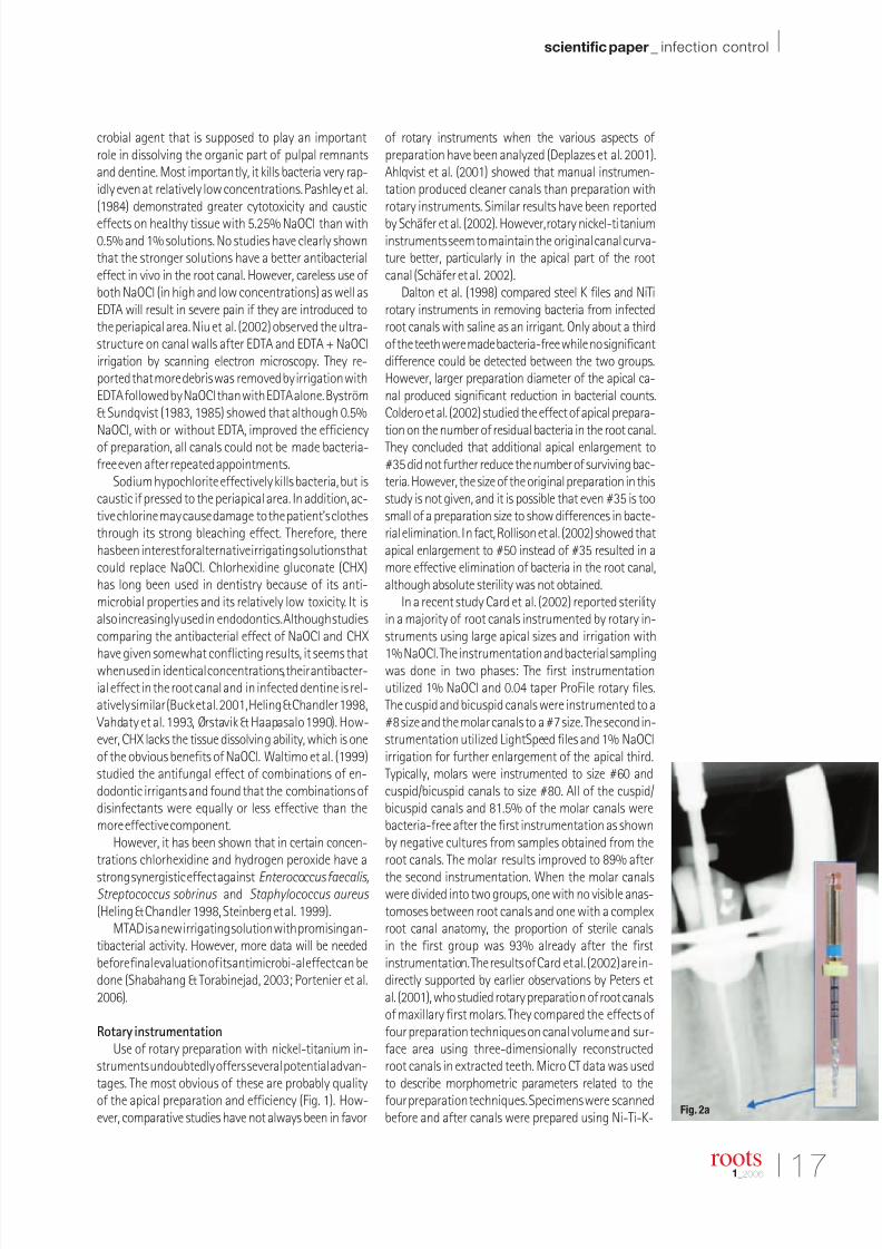
The quality of apical shaping and cleaning is af-fected, not only by the diameter of the last instrument,but also by the taper. For example, in manual prepara-tion, the typical 2% taper in the preparation of #30 givescanal diameters of #32, #34 and #36, 1, 2, and 3 mmfrom the working length. However, with a size #30 in-strument with 9% taper, the corresponding diametersare #39, #48 and #57 (Fig. 2). It has been speculated thatthe greater taper may facilitate the effect of antibac-terial irrigants in the apical canal (Coldero et al. 2002).However, at present there are no studies to show thepossible importance of the differences of apical taper.
Working length vs. apical foramenAnatomic studies about the location of the foramen
have demonstrated that it may often exist at a distanceof 0–3 mm from the anatomic apex (Burch & Hulen1972). Using the radiographic apex of the tooth as theonly guideline for working length determination wouldtherefore result in over instrumentation in a largenumber of cases. It is recommended that the workinglength is determined by use of radiographs and elec-tronic apex locators (Consensus report of the ESE onquality guidelines for endodontic treatment, 1994). Inpulpitis treatment, the recommended working length is1–2 mm short of the radiographic apex. In apical peri-
odontitis, elimination of root canal infection, not theleast in the apical canal, is the key to successful treat-ment. Therefore, in an optimal situation the root canalshould be instrumented, disinfected and filled to thelevel of the coronal side of the apical foramen (Fig. 3) toavoid the possibility of residual microbes surviving inthe uninstrumented apical canal (Trope and Bergen-holtz 2002).
Over instrumentation, with the possible exceptionof the smallest hand files of size #06 to #10 for certainsituations, should be avoided at all costs because ofthe following reasons: i) direct physical trauma to peri-apical tissue, ii) introduction of necrotic canal contentsand dead and living microorganisms into the periapicalarea persisting infection, periapical actinomycosis,
roots1_2006
Fig. 2b
8/13/2019 Dente de Roots 1
http://slidepdf.com/reader/full/dente-de-roots-1 16/58
I 19
scientific paper _ infection control I
roots1_2006
iii) bleeding into the root canalnutrients to intracanalbacteria, iv) growth of the foramen sizebetter possi-bilities for bacteria to get nutrients from the periapicalarea (inflammatory exudate), v) increased risk for extru-sion of irrigating solutions as well as overfilling with
sealer/gutta-percha, vi) in curved canals (most canals)creation of an oval foramen instead of a round onepoorer apical seal with a round gutta-percha masterpoint (complete compensation with a sealer is theo-retical)hide-out for residual microbes (Fig. 4).
_Disinfection of the root canalby intracanal medication
In pulpectomy, intracanal medication is not an inte-gral part of the treatment because the pulp is bacteriafree or only superficially infected. Only when a timelimitation has not allowed complete treatment in oneappointment, the canal space has been filled, e.g., withcalcium hydroxide, to prevent contamination of thecanal during appointments. In anatomically demandingteeth, inter-appointment calcium hydroxide may havebeen selected to facilitate removal of residual pulp tis-sue or to help to control bleeding.
In the treatment of apical periodontitis, intracanalmedication has been recommended to eradicate themicrobes that survive instrumentation and irrigation.A variety of different medications have been used forthis purpose. These include calcium hydroxide, phenolcompounds eugenol and camphorated parachloro-
phenol (CMCP), iodine potassium iodide (IPI), glutar-aldehyde, formocreosol, and pastes containing a mix-ture of antibiotics with or without corticoids. Byströmet al. (1985) showed that calcium hydroxide was moreeffective as an intracanal medication than CMCP orcamphorated phenol and made 34 out of 35 canalsbacteria free after four weeks. The effectiveness ofinter-appointment calcium hydroxide was also dem-onstrated by Sjögren et al. (1991) who showed that the7-day dressing with calcium hydroxide eliminated bac-teria that survived instrumentation and irrigation of thecanal, while the 10-minute application was ineffective.
However, the outstanding results in canal disinfec-
tion by calcium hydroxide have been to some extentchallenged by other studies that reported a residualflora in 7–35% of cases after the use of calcium hy-droxide (Ørstavik et al. 1991, Reit et al. 1999, Schupinget al. 2000). Peters et al. (2002) reported that thenumber of positive canals had increased in the periodbetween visits when calcium hydroxide was used asan intracanal dressing. However, the number of micro-organisms had only increased to about one percent of the original number. The different results may be partlyexplained by differences in the clinical cases studied(e.g., intact teeth vs. carious teeth), and in techniquesemployed in sampling and culturing the microbes.
Retreatment of root filled teeth with apical periodon-titis has a lower prognosis than treatment of primary api-
cal periodontitis (for review see Friedman 1998). This maybe due to several reasons such as technical complica-tions, difficult anatomy, unlocated root canals, etc. Onepossible explanation for poorer prognosis is the presenceof microflora that is more resistant to normal treatment
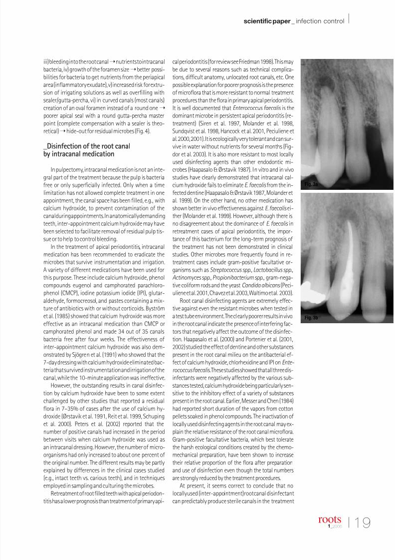
procedures than the flora in primary apical periodontitis.It is well documented that Enterococcus faecalis is thedominant microbe in persistent apical periodontitis (re-treatment) (Siren et al. 1997, Molander et al. 1998,Sundqvist et al. 1998, Hancock et al. 2001, Peciuliene etal. 2000, 2001). It is ecologically very tolerant and can sur-vive in water without nutrients for several months (Fig-dor et al. 2003). It is also more resistant to most locallyused disinfecting agents than other endodontic mi-crobes (Haapasalo & Ørstavik 1987). In vitro and in vivostudies have clearly demonstrated that intracanal cal-cium hydroxide fails to eliminate E. faecalis from the in-fected dentine (Haapasalo & Ørstavik 1987, Molander etal. 1999). On the other hand, no other medication hasshown better in vivo effectiveness against E. faecalis ei-ther (Molander et al. 1999). However, although there isno disagreement about the dominance of E. faecalis inretreatment cases of apical periodontitis, the impor-tance of this bacterium for the long-term prognosis of the treatment has not been demonstrated in clinicalstudies. Other microbes more frequently found in re-treatment cases include gram-positive facultative or-ganisms such as Streptococcus spp., Lactobacillus spp.,Actinomyces spp., Propionibacterium spp., gram-nega-tive coliform rods and the yeast Candida albicans (Peci-
uliene et al. 2001, Chavez et al. 2003, Waltimo et al. 2003).Root canal disinfecting agents are extremely effec-
tive against even the resistant microbes when tested ina test tube environment. The clearly poorer results in vivoin the root canal indicate the presence of interfering fac-tors that negatively affect the outcome of the disinfec-tion. Haapasalo et al. (2000) and Portenier et al. (2001,2002) studied the effect of dentine and other substancespresent in the root canal milieu on the antibacterial ef-fect of calcium hydroxide, chlorhexidine and IPI on Ente-
rococcus faecalis.These studies showed that all three dis-infectants were negatively affected by the various sub-stances tested, calcium hydroxide being particularly sen-
sitive to the inhibitory effect of a variety of substancespresent in the root canal. Earlier, Messer and Chen (1984)had reported short duration of the vapors from cottonpellets soaked in phenol compounds. The inactivation of locally used disinfecting agents in the root canal may ex-plain the relative resistance of the root canal microflora.Gram-positive facultative bacteria, which best toleratethe harsh ecological conditions created by the chemo-mechanical preparation, have been shown to increasetheir relative proportion of the flora after preparationand use of disinfection even though the total numbersare strongly reduced by the treatment procedures.
At present, it seems correct to conclude that nolocally used (inter-appointment) root canal disinfectantcan predictably produce sterile canals in the treatment
Fig. 3b
Fig. 3a
8/13/2019 Dente de Roots 1
http://slidepdf.com/reader/full/dente-de-roots-1 17/58
20 I
I scientific paper _ infection control
of apical periodontitis. However, it is clear from moststudies that their use further reduces the numberof infecting microorganisms after chemomechanicalpreparation. Furthermore, calcium hydroxide may helpto better remove the residual necrotic pulp tissue at
the second appointment as well as neutralize bacterialantigens remaining in the root canal system (Safavi &Nichols 1993, 1994).
Root filling & permanent restoration
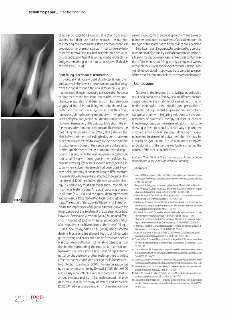
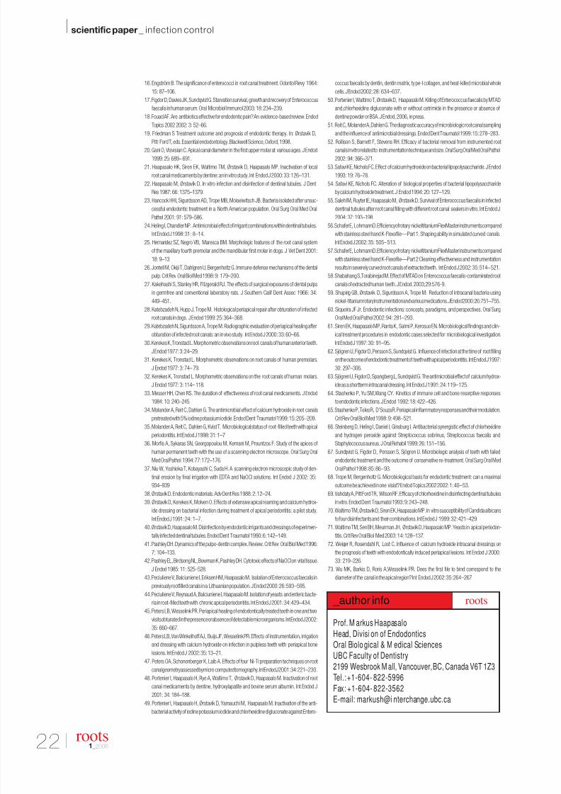
Eventually, all locally used disinfectants lose theirantibacterial effect over time and/or are washed awayfrom the canal through the apical foramen, e.g., per-manent root filling is necessary to secure that bacteriacannot reenter the root canal space after chemome-chanical preparation and disinfection. It has also beensuggested that the root filling entombs the residualbacteria in the root canal system so that they don’thave a possibility of coming in contact with living tissuein the periapical area, which could compromise healing.However, there is very little data available about the ef-fectiveness of entombment of root canal bacteria by theroot filling. Katebzadeh et al. (1999, 2000) studied theeffect of entombment on healing in dog teeth that wereexperimentally infected, followed by the developmentof apical lesions. Some of the canals were obturated atthe first appointment after instrumentation and irriga-tion with saline, while the rest were disinfected beforeroot canal filling with inter-appointment calcium hy-droxide dressing. The results showed better healing in
cases where calcium hydroxide had been used. How-ever, apical anatomy of dog teeth is quite different fromhuman teeth, which may have affected the results. Her-nandez et al. (2001) evaluated the root canal morphol-ogy in 72 maxillary fourth premolar and 59 mandibularfirst molar teeth in dogs. An apical delta was presentin all roots (n = 334), and the apical delta representedapproximately 12 to 18% of the total root length for allroots. The results of the study by Sjögren et al. (1997) in-dicate the importance of negative bacterial growth forthe prognosis of the treatment of apical periodontitis.However, Peters and Wesselink (2002) found no differ-ence in healing of teeth with apical periodontitis filled
after negative or positive culture at the time of filling.In a new study, Saleh et al. (2004) using infected
dentine blocks in vitro showed that root filling withgutta-percha and sealer AH plus or Grossman’s sealerwas clearly more effective in eliminatingE. faecalis fromthe dentine surrounding the root canal than calciumhydroxide one week after filling. Root fillings made of gutta-percha and several other sealers proved to be lesseffective than calcium hydroxide againstE. faecalis den-tine infection (Saleh et al. 2004). The result is supportedby an earlier observation by Ørstavik (1988) that AH 26was clearly more effective in killing bacteria in dentinearound the root canal than other sealers tested. It may be
of interest that in the study of Peters and Wesselink(2002), AH 26 was used as a sealer. In future studies com-
paring the outcome of single-appointment and two-ap-pointment endodontic treatment of apical periodontitis,the type of the sealer has to be taken into consideration.
Finally, all root fillings must be protected by a coronalrestoration of high quality. Lack of coronal restoration or
a leaking restoration may result in bacterial contamina-tion of the whole root filling in only a couple of weeks.Although the clinical relevance of coronal leakage is notyet fully understood, it is obvious that a considerable partof the need for retreatment is caused by coronal leakage.
_Conclusions
Success in the treatment of apical periodontitis is aresult of a combined effort by several different factorscontributing to the inhibition of spreading of the in-fection, elimination of the infection, and prevention of reinfection. A high level of asepsis and chemomechan-ical preparation with irrigating solutions are the cor-nerstones of successful therapy. In light of presentknowledge, interappointment use of calcium hydroxidedressing in the root canal is a secure way to guaranteeeffective antimicrobial strategy. However, one-ap-pointment treatment of apical periodontitis may bea reachable goal in the future with more completeunderstanding of the various key factors affecting thecontrol of the root canal infection._
Editorial Note: Parts of this article were published in Endo-dontic Topics, 2003,6:29-56 (Blackwell Publishing).
_Literature
1. Ahlquist M, Henningsson O, Hultenby K, Ohlin J. The effectiveness of manual and rotary
techniques in the cleaning of root canals: a scanning electron microscopy study. Int Endod
J 2001: 34: 533–537.
2. Bergenholtz G. Pathogenic mechanisms in pulpal disease. J Endod 1990: 16: 98–101.
3. Buck RA, Eleazer PD, Staat RH, Scheetz JP. Effectiveness of three endodontic irrigants
at various tubular depths in human dentin. J Endod 2001: 27: 206–208.
4. Burch JG, Hulen S. The relationship of the apical foramen to the anatomic apex of the
tooth root. Oral Surg Oral Med Oral Pathol 1972: 34: 262– 268.
5. Byström A, Claesson R, Sundqvist G. The antibacterial effect of camphorated paramo-
nochlorophenol, camphorated phenol and calcium hydroxide in the treatment of infected
root canals. Endod Dent Traumatol 1985: 1: 170–175.
6. Byström A, Sundqvist G. Bacteriologic evaluation of the efficacy of mechanical root canal
instrumentation in endodontic therapy. Scand J Dent Res 1981: 89: 321– 328.
7. Byström A, Sundqvist G. Bacteriologic evaluation of the effect of 0.5 percent sodium
hypochlorite in endodontic therapy. Oral Surg Oral Med Oral Pathol 1983: 55: 307–312.
8. Byström A, Sundqvist G. The antibacterial action of sodium hypochlorite and EDTA in
60 cases of endodontic therapy. Int Endod J 1985: 18: 35–40.
9. Card SJ, Sigurdsson A, Ørstavik D, Trope M. The effectiveness of increased apical en-
largement in reducing intracanal bacteria. J Endod 2002: 28: 779–783.
10. Chavez De Paz LE, Dahlen G, Molander A, Moller A, Bergenholtz G. Bacteria recovered from
teeth with apical periodontitis after antimicrobial endodontic treatment. Int Endod J 2003:
36: 500–508.
11. Chugal NM, Clive JM, Spangberg LS. A prognostic model for assessment of the outcome
of endodontic treatment: effect of biologic and diagnostic variables. Oral Surg Oral Med Oral
Pathol 2001: 91: 342–352.
12.Coldero LG, McHugh S, Mackenzie D, Saunders WP. Reduction in intracanal bacteria during
root canal preparation with and without apical enlargement. Int Endod J 2002: 35: 437–446.
13. Consensus report of the European Society of Endodontology on quality guidelines for
endodontic treatment. Int Endod J 1994: 27: 115–124.
14. Dalton BC, Ørstavik D, Phillips C, Pettiette M, Trope M. Bacterial reduction with nickel-
titanium rotary instrumentation. J Endod 1998: 24: 763–767.
15. Deplazes P, Peters O, Barbakow F. Comparing apical preparations of root canals shapedby nickel-titanium rotary instruments and nickel-titanium hand instruments. J Endod 2001:
27: 196–202.
roots1_2006
Fig. 4a
Fig. 4b
8/13/2019 Dente de Roots 1
http://slidepdf.com/reader/full/dente-de-roots-1 18/58
22 I
I scientific paper _ infection control
16. Engström B. The significance of enterococci in root canal treatment. Odontol Revy 1964:
15: 87–106.
17. Figdor D, Davies JK, Sundqvist G. Starvation survival, growth and recovery of Enterococcus
faecalis in human serum. Oral Microbiol Immunol 2003: 18: 234–239.
18. Fouad AF. Are antibiotics effective for endodontic pain? An evidence-based review. Endod
Topics 2002 2002: 3: 52–66.
19. Friedman S Treatment outcome and prognosis of endodontic therapy. In: Ørstavik D,
Pitt-Ford T, eds. Essential endodontology. Blackwell Science, Oxford, 1998.
20. Gani O, Visvisian C. Apical canal diameter in the first upper molar at various ages. J Endod
1999: 25: 689– 691.
21. Haapasalo HK, Siren EK, Waltimo TM, Ørstavik D, Haapasalo MP. Inactivation of local
root canal medicaments by dentine: an in vitro study. Int Endod J 2000: 33: 126–131.
22. Haapasalo M, Ørstavik D. In vitro infection and disinfection of dentinal tubules. J Dent
Res 1987: 66: 1375–1379.
23. Hancock HHI, Sigurdsson AD, Trope MB, Moiseiwitsch JB. Bacteria isolated after unsuc-
cessful endodontic treatment in a North American population. Oral Surg Oral Med Oral
Pathol 2001: 91: 579–586.
24. Heling I, Chandler NP. Antimicrobial effect of irrigant combinations within dentinal tubules.
Int Endod J 1998: 31: 8–14.
25. Hernandez SZ, Negro VB, Maresca BM. Morphologic features of the root canal system
of the maxillary fourth premolar and the mandibular first molar in dogs. J Vet Dent 2001:
18: 9–13.
26. Jontell M, Okiji T, Dahlgren U, Bergenholtz G. Immune defense mechanisms of the dental
pulp. Crit Rev Oral Biol Med 1998: 9: 179–200.27. Kakehashi S, Stanley HR, Fitzgerald RJ. The effects of surgical exposures of dental pulps
in germfree and conventional laboratory rats. J Southern Calif Dent Assoc 1966: 34:
449–451.
28. Katebzadeh N, Hupp J, Trope M. Histological periapical repair after obturation of infected
root canals in dogs. J Endod 1999: 25: 364–368.
29. Katebzadeh N, Sigurdsson A, Trope M. Radiographic evaluation of periapical healing after
obturation of infected root canals: an in vivo study. Int Endod J 2000: 33: 60–66.
30. Kerekes K, Tronstad L. Morphometric observations on root canals of human anterior teeth.
J Endod 1977: 3: 24–29.
31. Kerekes K, Tronstad L. Morphometric observations on root canals of human premolars.
J Endod 1977: 3: 74– 79.
32. Kerekes K, Tronstad L. Morphometric observations on the root canals of human molars.
J Endod 1977: 3: 114– 118.
33. Messer HH, Chen RS. The duration of effectiveness of root canal medicaments. J Endod
1984: 10: 240–245.
34. Molander A, Reit C, Dahlen G. The antimicrobial effect of calcium hydroxide in root canals
pretreated with 5% iodine potassium iodide. Endod Dent Traumatol 1999: 15: 205–209.35. Molander A, Reit C, Dahlen G, Kvist T. Microbiological status of root-filled teeth with apical
periodontitis. Int Endod J 1998: 31: 1–7
36. Morfis A, Sykaras SN, Georgopoulou M, Kernani M, Prountzos F. Study of the apices of
human permanent teeth with the use of a scanning electron microscope. Oral Surg Oral
Med Oral Pathol 1994: 77: 172–176.
37. Niu W, Yoshioka T, Kobayashi C, Suda H. A scanning electron microscopic study of den-
tinal erosion by final irrigation with EDTA and NaOCl solutions. Int Endod J 2002: 35:
934–939.
38. Ørstavik D. Endodontic materials. Adv Dent Res 1988: 2: 12–24.
39. Ørstavik D, Kerekes K, Molven O. Effects of extensive apical reaming and calcium hydrox-
ide dressing on bacterial infection during treatment of apical periodontitis: a pilot study.
Int Endod J 1991: 24: 1–7.
40. Ørstavik D, Haapasalo M. Disinfection by endodontic irrigants and dressings of experimen-
tally infected dentinal tubules. Endod Dent Traumatol 1990: 6: 142–149.
41. Pashley DH. Dynamics of the pulpo-dentin complex. Review. Crit Rev Oral Biol Med 1996:
7: 104–133.
42. Pashley EL, Birdsong NL, Bowman K, Pashley DH. Cytotoxic effects of NaOCl on vital tissue.
J Endod 1985: 11: 525–528.
43. Peciuliene V, Balciuniene I, Eriksen HM, Haapasalo M. Isolation of Enterococcus faecalis in
previously rootfilled canals in a Lithuanian population. J Endod 2000: 26: 593–595.
44. Peciuliene V, Reynaud A, Balciuniene I, Haapasalo M. Isolation of yeasts and enteric bacte-
ria in root-filled teeth with chronic apical periodontitis. Int Endod J 2001: 34: 429–434.
45. Peters LB, Wesselink PR. Periapical healing of endodontically treated teeth in one and two
visits obturated in the presence or absence of detectable microorganisms. Int Endod J 2002:
35: 660–667.
46. Peters LB, Van Winkelhoff AJ, Buijs JF, Wesselink PR. Effects of instrumentation, irrigation
and dressing with calcium hydroxide on infection in pulpless teeth with periapical bone
lesions. Int Endod J 2002: 35: 13–21.
47. Peters OA, Schonenberger K, Laib A. Effects of four Ni-Ti preparation techniques on root
canal geometry assessed by micro computed tomography. Int Endod J 2001: 34: 221–230.
48. Portenier I, Haapasalo H, Rye A, Waltimo T, Ørstavik D, Haapasalo M. Inactivation of root
canal medicaments by dentine, hydroxylapatite and bovine serum albumin. Int Endod J
2001: 34: 184–188.49. Portenier I, Haapasalo H, Ørstavik D, Yamauchi M, Haapasalo M. Inactivation of the anti-
bacterial activity of iodine potassium iodide and chlorhexidine digluconate against Entero-
coccus faecalis by dentin, dentin matrix, type-I collagen, and heat-killed microbial whole
cells. J Endod 2002: 28: 634–637.
50. Portenier I, Waltimo T, Ørstavik D, Haapasalo M. Killing of Enterococcus faecalis by MTAD
and,chlorhexidine digluconate with or without cetrimide in the presence or absence of
dentine powder or BSA. J Endod, 2006, in press.
51. Reit C, Molander A, Dahlen G. The diagnostic accuracy of microbiologic root canal sampling
and the influence of antimicrobial dressings. Endod Dent Traumatol 1999: 15: 278–283.
52. Rollison S, Barnett F, Stevens RH. Efficacy of bacterial removal from instrumented root
canals in vitro related to instrumentation technique and size. Oral Surg Oral Med Oral Pathol
2002: 94: 366–371.
53. Safavi KE, Nichols FC. Effect of calcium hydroxide on bacterial lipopolysaccharide. J Endod
1993: 19: 76–78.
54. Safavi KE, Nichols FC. Alteration of biological properties of bacterial lipopolysaccharide
by calcium hydroxide treatment. J Endod 1994: 20: 127–129.
55. Saleh IM, Ruyter IE, Haapasalo M, Ørstavik D. Survival of Enterococcus faecalis in infected
dentinal tubules after root canal filling with different root canal sealers in vitro. Int Endod J
2004: 37: 193–198.
56. Schafer E, Lohmann D. Efficiency of rotary nickeltitanium FlexMaster instruments compared
with stainless steel hand K-Flexofile—Part 1. Shaping ability in simulated curved canals.
Int Endod J 2002: 35: 505– 513.
57. Schafer E, Lohmann D. Efficiency of rotary nickeltitanium FlexMaster instruments compared
with stainless steel hand K-Flexofile—Part 2 Cleaning effectiveness and instrumentation
results in severely curved root canals of extracted teeth. Int Endod J 2002: 35: 514– 521.
58. Shabahang S,Torabinejad M. Effect of MTAD on Enterococcus faecalis-contaminated rootcanals of extracted human teeth. J Endod. 2003;29:576-9.
59. Shuping GB, Ørstavik D, Sigurdsson A, Trope M. Reduction of intracanal bacteria using
nickel-titanium rotary instrumentation and various medications. J Endod 2000: 26: 751–755.
60. Siqueira JF Jr. Endodontic infections: concepts, paradigms, and perspectives. Oral Surg
Oral Med Oral Pathol 2002: 94: 281–293.
61. Siren EK, Haapasalo MP, Ranta K, Salmi P, Kerosuo EN. Microbiological findings and clin-
ical treatment procedures in endodontic cases selected for microbiological investigation.
Int Endod J 1997: 30: 91–95.
62. Sjögren U, Figdor D, Persson S, Sundqvist G. Influence of infection at the time of root filling
on the outcome of endodontic treatment of teeth with apical periodontitis. Int Endod J 1997:
30: 297–306.
63. Sjögren U, Figdor D, Spangberg L, Sundqvist G. The antimicrobial effect of calcium hydrox-
ide as a shortterm intracanal dressing. Int Endod J 1991: 24: 119– 125.
64. Stashenko P, Yu SM,Wang CY. Kinetics of immune cell and bone resorptive responses
to endodontic infections. J Endod 1992: 18: 422–426.
65. Stashenko P, Teles R, D’Souza R. Periapical inflammatory responses and their modulation.
Crit Rev Oral Biol Med 1998: 9: 498–521.66. Steinberg D, Heling I, Daniel I, Ginsburg I. Antibacterial synergistic effect of chlorhexidine
and hydrogen peroxide against Streptococcus sobrinus, Streptococcus faecalis and
Staphylococcus aureus. J Oral Rehabil 1999: 26: 151–156.
67. Sundqvist G, Figdor D, Persson S, Sjögren U. Microbiologic analysis of teeth with failed
endodontic treatment and the outcome of conservative re-treatment. Oral Surg Oral Med
Oral Pathol 1998: 85: 86– 93.
68. Trope M, Bergenholtz G. Microbiological basis for endodontic treatment: can a maximal
outcome be achieved in one visist? Endod Topics 2002 2002: 1: 40– 53.
69. Vahdaty A, Pitt Ford TR, Wilson RF. Efficacy of chlorhexidine in disinfecting dentinal tubules
in vitro. Endod Dent Traumatol 1993: 9: 243–248.
70. Waltimo TM, Ørstavik D, Siren EK, Haapasalo MP. In vitro susceptibility of Candida albicans
to four disinfectants and their combinations. Int Endod J 1999: 32: 421–429.
71. Waltimo TM, Sen BH, Meurman JH, Ørstavik D, Haapasalo MP. Yeasts in apical periodon-
titis. Crit Rev Oral Biol Med 2003: 14: 128–137.
72. Weiger R, Rosendahl R, Lost C. Influence of calcium hydroxide intracanal dressings on
the prognosis of teeth with endodontically induced periapical lesions. Int Endod J 2000:
33: 219–226.
73. Wu MK, Barkis D, Roris A,Wesselink PR. Does the first file to bind correspond to the
diameter of the canal in the apical region? Int Endod J 2002: 35: 264–267.
roots1_2006
Prof. M arkus Haapasalo
Head, Divisi on of EndodonticsOral Biolo gical & M edical SciencesUBC Faculty of Dentistry
2199 Wesbrook M all, Vancouver, BC, Canada V6T 1Z3Tel.: +1-604- 822-5996
Fax: +1-604- 822-3562
E-mail: markush@i nterchange.ubc.ca
_author info roots
8/13/2019 Dente de Roots 1
http://slidepdf.com/reader/full/dente-de-roots-1 19/58
8/13/2019 Dente de Roots 1
http://slidepdf.com/reader/full/dente-de-roots-1 20/58
24 I
I scientific paper _ apex locator
In 1942, Suzuki discovered that the electricalresistance (single current source) between an in-strument inserted into a root canal and an elec-trode attached to the oral mucosa registered aconsistent value. In 1962 Sunada, using a directcurrent device with a simple circuit, demonstratedthat the consistent electrical resistance betweenthe periodontium and the mucous membrane was6.5 kΩ (DC Resistance). Through the 1970s, fre-
quency measurements were measured throughthe feedback of an oscillator loop by calibrationat the periodontal pocket of each tooth. This cul-minated with the efforts of Hasedgawa in 1979with the use of high frequency waves and a spe-
cially coated file that could record in conductivefluids.
In 1983, Ushiyama introduced the voltagegradient method where a concentric bipolarelectrode measured the current density evoked ina limited area of the canal. Maximum potentialwas reached when the electrode was at the apicalconstriction. The mid ’80s saw the developmentof a relative value of frequency response methodwhere the apical constriction was picked by filter-ing the difference between two direct potentialsafter a 1 kHz rectilinear wave was applied to thecanal space.
A third generation electronic foramenal loca-tor (EFL) developed in the late ’80s by Kobayashiused multi-channel impedance/ratio based tech-nology to simultaneously measure the impedanceof two different frequencies, calculate the quo-tient of the impedance and express it in termsof the position of the electrode (file) in thecanal. This formed the basis of the technologyused in the Root ZX® (J. Morita USA Inc., Irvine,CA) where no calibration was required and a
microprocessor calculated the impedance quo-tient.
Fourth generation EFLs (Elements Diagnostic,SybronEndo, Orange, CA) measure resistance andcapacitance separately rather than the resultantimpedance value—impedance being a function of resistance and capacitance (Fig. 4a).
There can be different combinations of valuesof capacitance and resistance that provide thesame impedance, and thus the same foramenal
roots1_2006
Fig. 2b
Fig. 2a
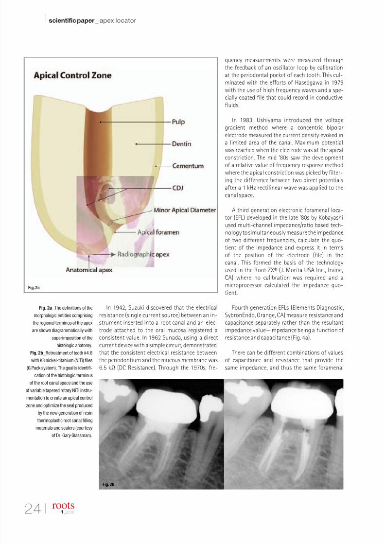
Fig. 2a_The definitions of the
morphologic entities comprising
the regional terminus of the apex
are shown diagrammatically with
superimposition of the
histologic anatomy.
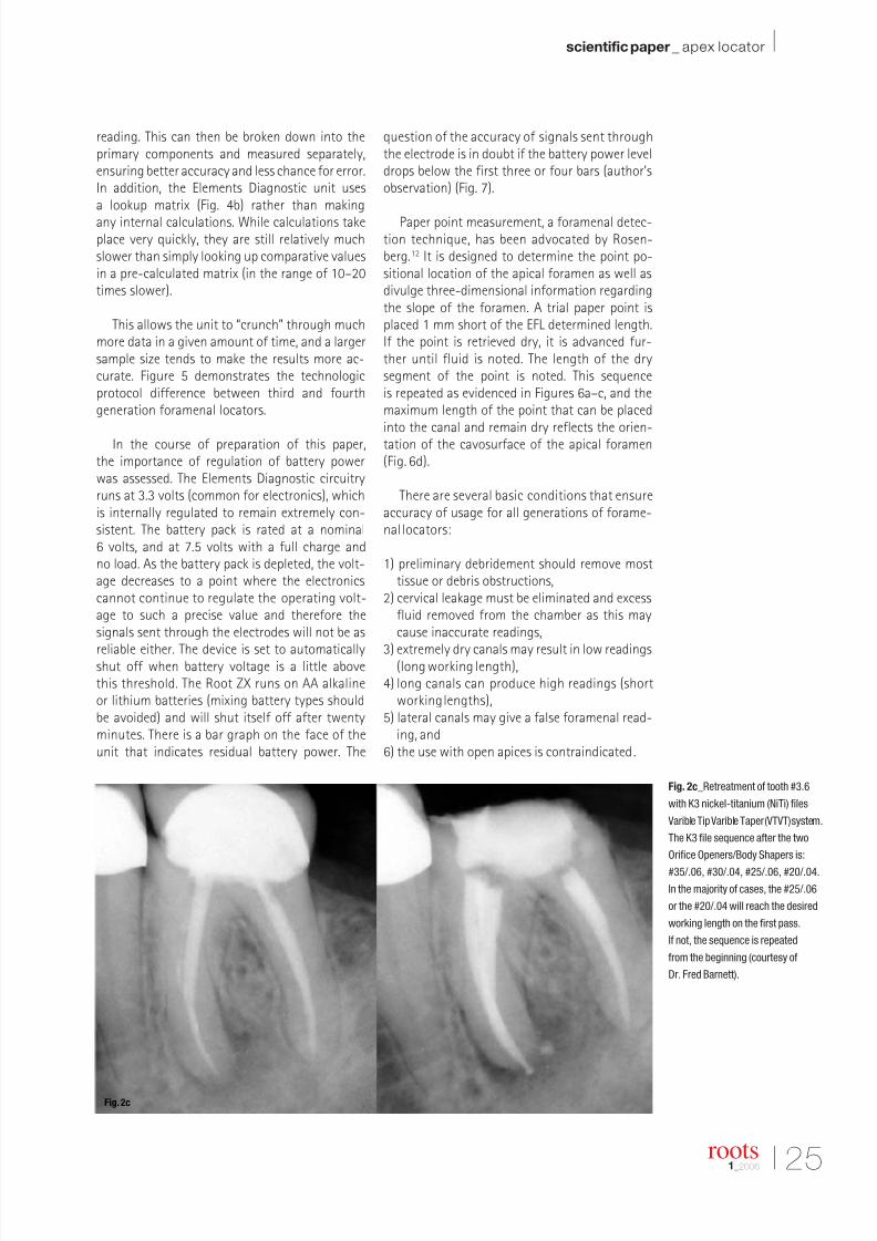
Fig. 2b_Retreatment of tooth #4.6
with K3 nickel-titanium (NiTi) files
(G Pack system). The goal is identifi-
cation of the histologic terminus
of the root canal space and the use
of variable tapered rotary NiTi instru-
mentation to create an apical control
zone and optimize the seal produced
by the new generation of resin
thermoplastic root canal filling
materials and sealers (courtesy
of Dr. Gary Glassman).
8/13/2019 Dente de Roots 1
http://slidepdf.com/reader/full/dente-de-roots-1 21/58
I 25
scientific paper _ apex locator I
roots1_2006
reading. This can then be broken down into theprimary components and measured separately,ensuring better accuracy and less chance for error.In addition, the Elements Diagnostic unit usesa lookup matrix (Fig. 4b) rather than making
any internal calculations. While calculations takeplace very quickly, they are still relatively muchslower than simply looking up comparative valuesin a pre-calculated matrix (in the range of 10–20times slower).
This allows the unit to “crunch” through muchmore data in a given amount of time, and a largersample size tends to make the results more ac-curate. Figure 5 demonstrates the technologicprotocol difference between third and fourthgeneration foramenal locators.
In the course of preparation of this paper,the importance of regulation of battery powerwas assessed. The Elements Diagnostic circuitryruns at 3.3 volts (common for electronics), whichis internally regulated to remain extremely con-sistent. The battery pack is rated at a nominal6 volts, and at 7.5 volts with a full charge andno load. As the battery pack is depleted, the volt-age decreases to a point where the electronicscannot continue to regulate the operating volt-age to such a precise value and therefore thesignals sent through the electrodes will not be as
reliable either. The device is set to automaticallyshut off when battery voltage is a little abovethis threshold. The Root ZX runs on AA alkalineor lithium batteries (mixing battery types shouldbe avoided) and will shut itself off after twentyminutes. There is a bar graph on the face of theunit that indicates residual battery power. The
question of the accuracy of signals sent throughthe electrode is in doubt if the battery power leveldrops below the first three or four bars (author’sobservation) (Fig. 7).
Paper point measurement, a foramenal detec-tion technique, has been advocated by Rosen-berg.12 It is designed to determine the point po-sitional location of the apical foramen as well asdivulge three-dimensional information regardingthe slope of the foramen. A trial paper point isplaced 1 mm short of the EFL determined length.If the point is retrieved dry, it is advanced fur-ther until fluid is noted. The length of the drysegment of the point is noted. This sequenceis repeated as evidenced in Figures 6a–c, and themaximum length of the point that can be placedinto the canal and remain dry reflects the orien-tation of the cavosurface of the apical foramen(Fig. 6d).
There are several basic conditions that ensureaccuracy of usage for all generations of forame-nal locators:
1) preliminary debridement should remove mosttissue or debris obstructions,
2) cervical leakage must be eliminated and excessfluid removed from the chamber as this maycause inaccurate readings,
3) extremely dry canals may result in low readings(long working length),
4) long canals can produce high readings (shortworking lengths),
5) lateral canals may give a false foramenal read-ing, and
6) the use with open apices is contraindicated.
Fig. 2c_Retreatment of tooth #3.6
with K3 nickel-titanium (NiTi) files
Varible Tip Varible Taper (VTVT) system.
The K3 file sequence after the two
Orifice Openers/Body Shapers is:
#35/.06, #30/.04, #25/.06, #20/.04.
In the majority of cases, the #25/.06
or the #20/.04 will reach the desired
working length on the first pass.
If not, the sequence is repeated
from the beginning (courtesy of
Dr. Fred Barnett).
Fig. 2c
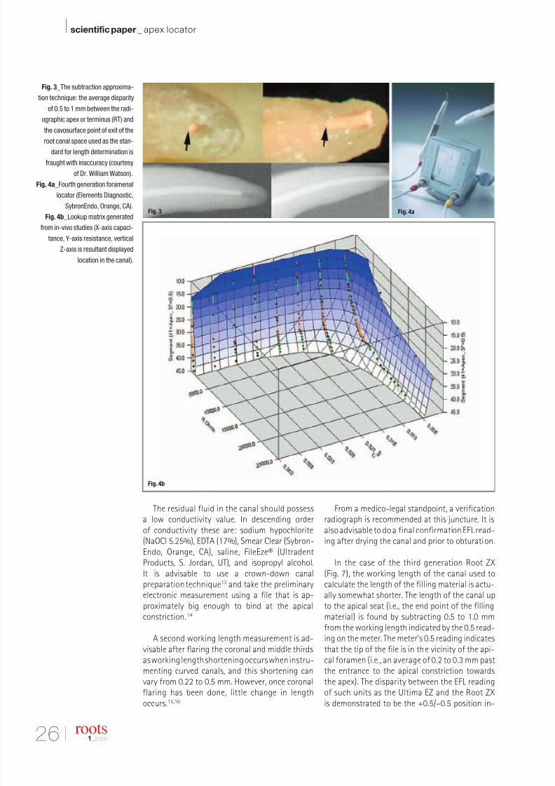
8/13/2019 Dente de Roots 1
http://slidepdf.com/reader/full/dente-de-roots-1 22/58
26 I
I scientific paper _ apex locator
The residual fluid in the canal should possessa low conductivity value. In descending orderof conductivity these are: sodium hypochlorite(NaOCl 5.25%), EDTA (17%), Smear Clear (Sybron-
Endo, Orange, CA), saline, FileEze® (UltradentProducts, S. Jordan, UT), and isopropyl alcohol.It is advisable to use a crown-down canalpreparation technique13 and take the preliminaryelectronic measurement using a file that is ap-proximately big enough to bind at the apicalconstriction.14
A second working length measurement is ad-visable after flaring the coronal and middle thirdsas working length shortening occurs when instru-menting curved canals, and this shortening canvary from 0.22 to 0.5 mm. However, once coronalflaring has been done, little change in lengthoccurs.15,16
From a medico-legal standpoint, a verificationradiograph is recommended at this juncture. It isalso advisable to do a final confirmation EFL read-ing after drying the canal and prior to obturation.
In the case of the third generation Root ZX(Fig. 7), the working length of the canal used tocalculate the length of the filling material is actu-ally somewhat shorter. The length of the canal upto the apical seat (i.e., the end point of the fillingmaterial) is found by subtracting 0.5 to 1.0 mmfrom the working length indicated by the 0.5 read-ing on the meter. The meter’s 0.5 reading indicatesthat the tip of the file is in the vicinity of the api-cal foramen (i.e., an average of 0.2 to 0.3 mm pastthe entrance to the apical constriction towardsthe apex). The disparity between the EFL readingof such units as the Ultima EZ and the Root ZXis demonstrated to be the +0.5/–0.5 position in-
Fig. 3_The subtraction approxima-
tion technique: the average disparity
of 0.5 to 1 mm between the radi-
ographic apex or terminus (RT) and
the cavosurface point of exit of the
root canal space used as the stan-
dard for length determination is
fraught with inaccuracy (courtesy
of Dr. William Watson).
Fig. 4a_Fourth generation foramenal
locator (Elements Diagnostic,
SybronEndo, Orange, CA).
Fig. 4b_Lookup matrix generated
from in-vivo studies (X-axis capaci-
tance, Y-axis resistance, vertical
Z-axis is resultant displayed
location in the canal).
roots1_2006
Fig. 4b
Fig. 3 Fig. 4a
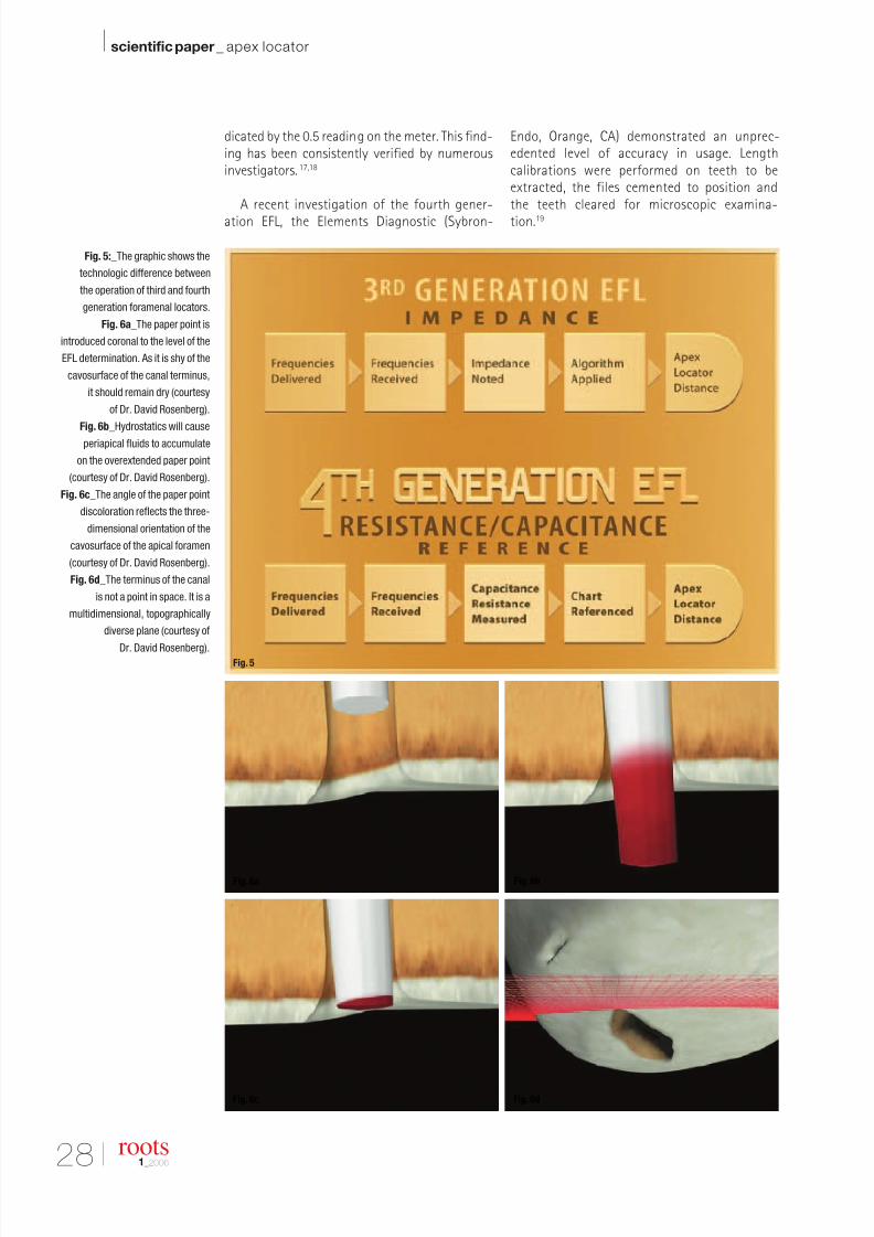
8/13/2019 Dente de Roots 1
http://slidepdf.com/reader/full/dente-de-roots-1 23/58
28 I
I scientific paper _ apex locator
roots1_2006
dicated by the 0.5 reading on the meter. This find-ing has been consistently verified by numerousinvestigators.17,18
A recent investigation of the fourth gener-
ation EFL, the Elements Diagnostic (Sybron-
Endo, Orange, CA) demonstrated an unprec-edented level of accuracy in usage. Lengthcalibrations were performed on teeth to beextracted, the files cemented to position andthe teeth cleared for microscopic examina-
tion.19
Fig. 5:_The graphic shows the
technologic difference between
the operation of third and fourth
generation foramenal locators.
Fig. 6a_The paper point is
introduced coronal to the level of the
EFL determination. As it is shy of the
cavosurface of the canal terminus,
it should remain dry (courtesy
of Dr. David Rosenberg).
Fig. 6b_Hydrostatics will cause
periapical fluids to accumulate
on the overextended paper point
(courtesy of Dr. David Rosenberg).
Fig. 6c_The angle of the paper point
discoloration reflects the three-
dimensional orientation of the
cavosurface of the apical foramen
(courtesy of Dr. David Rosenberg).
Fig. 6d_The terminus of the canal
is not a point in space. It is a
multidimensional, topographically
diverse plane (courtesy of
Dr. David Rosenberg).
Fig. 6a Fig. 6b
Fig. 6c Fig. 6d
Fig. 5
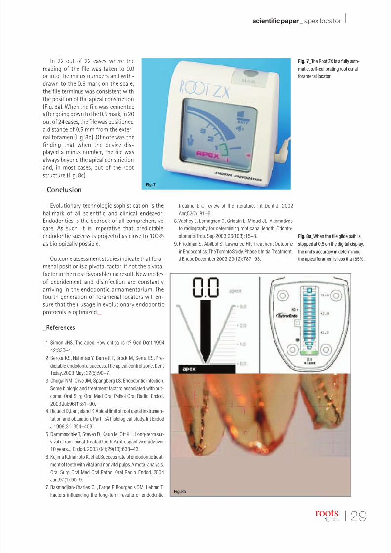
8/13/2019 Dente de Roots 1
http://slidepdf.com/reader/full/dente-de-roots-1 24/58
I 29
scientific paper _ apex locator I
roots1_2006
In 22 out of 22 cases where thereading of the file was taken to 0.0or into the minus numbers and with-drawn to the 0.5 mark on the scale,the file terminus was consistent with
the position of the apical constriction(Fig. 8a). When the file was cementedafter going down to the 0.5 mark, in 20out of 24 cases, the file was positioneda distance of 0.5 mm from the exter-nal foramen (Fig. 8b). Of note was thefinding that when the device dis-played a minus number, the file wasalways beyond the apical constrictionand, in most cases, out of the rootstructure (Fig. 8c).
_ConclusionEvolutionary technologic sophistication is the
hallmark of all scientific and clinical endeavor.Endodontics is the bedrock of all comprehensivecare. As such, it is imperative that predictableendodontic success is projected as close to 100%as biologically possible.
Outcome assessment studies indicate that fora-menal position is a pivotal factor, if not the pivotalfactor in the most favorable end result. New modesof debridement and disinfection are constantly
arriving in the endodontic armamentarium. Thefourth generation of foramenal locators will en-sure that their usage in evolutionary endodonticprotocols is optimized._
_References
1. Simon JHS. The apex: How critical is it? Gen Dent 1994
42:330–4.
2. Serota KS, Nahmias Y, Barnett F, Brock M, Senia ES. Pre-
dictable endodontic success.The apical control zone. Dent
Today.2003 May; 22(5):90–7.
3. Chugal NM, Clive JM, Spangberg LS. Endodontic infection:
Some biologic and treatment factors associated with out-come. Oral Surg Oral Med Oral Pathol Oral Radiol Endod.
2003 Jul;96(1):81–90.
4. Ricucci D,Langeland K.Apical limit of root canal instrumen-
tation and obturation, Part II:A histological study. Int Endod
J 1998;31: 394–409.
5. Dammaschke T, Steven D, Kaup M, Ott KH. Long-term sur-
vival of root-canal-treated teeth:A retrospective study over
10 years.J Endod. 2003 Oct;29(10):638–43.
6. Kojima K,Inamoto K,et al.Success rate of endodontic treat-
ment of teeth with vital and nonvital pulps.A meta-analysis.
Oral Surg Oral Med Oral Pathol Oral Radiol Endod. 2004
Jan;97(1):95–9.
7. Basmadjian-Charles CL, Farge P, Bourgeois DM, Lebrun T.
Factors influencing the long-term results of endodontic
treatment: a review of the literature. Int Dent J. 2002
Apr;52(2) : 81–6.
8.Vachey E, Lemagnen G, Grislain L, Miquel JL. Alternatives
to radiography for determining root canal length. Odonto-
stomatol Trop. Sep 2003;26(103):15–8.
9. Friedman S, Abitbol S, Lawrence HP. Treatment Outcome
in Endodontics:The Toronto Study.Phase I: Initial Treatment.
J Endod December 2003;29(12):787–93.
Fig. 7
Fig. 8a
Fig. 7_The Root ZX is a fully auto-
matic, self-calibrating root canal
foramenal locator.
Fig. 8a_When the file glide path is
stopped at 0.5 on the digital display,
the unit’s accuracy in determining
the apical foramen is less than 85%.
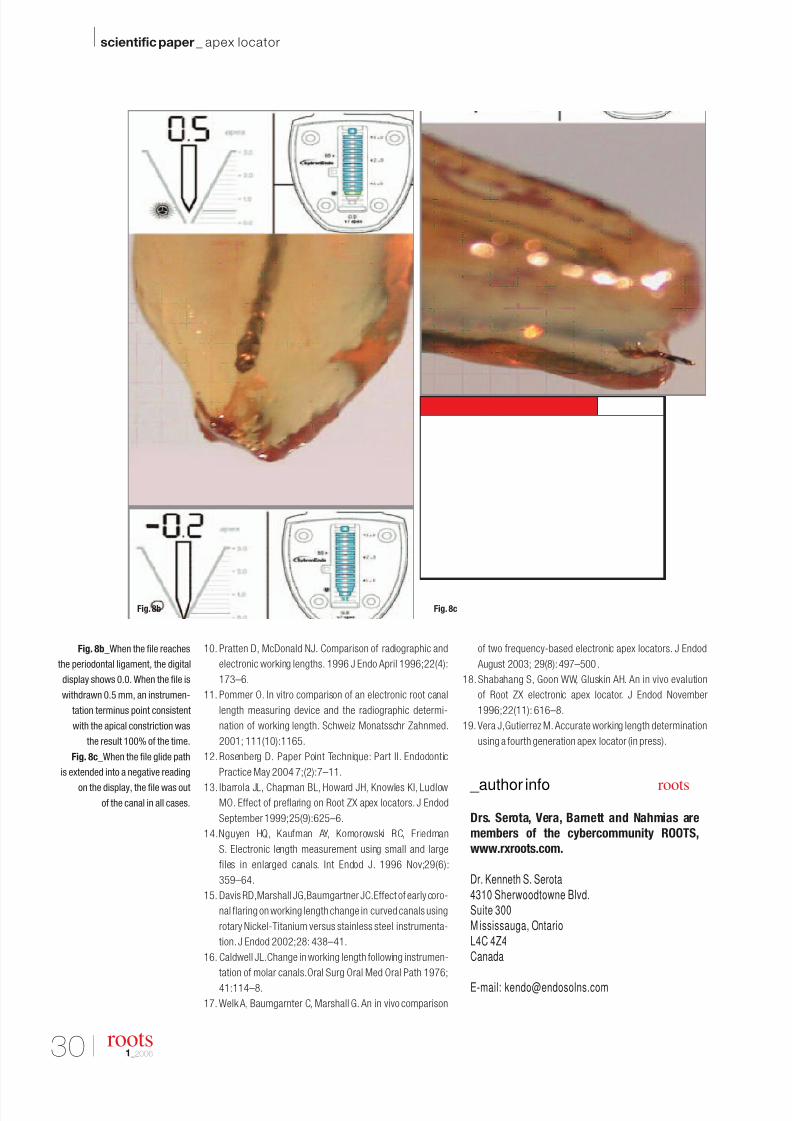
8/13/2019 Dente de Roots 1
http://slidepdf.com/reader/full/dente-de-roots-1 25/58
30 I
I scientific paper _ apex locator
10. Pratten D, McDonald NJ. Comparison of radiographic and
electronic working lengths. 1996 J Endo April 1996;22(4):
173–6.
11. Pommer O. In vitro comparison of an electronic root canal
length measuring device and the radiographic determi-
nation of working length. Schweiz Monatsschr Zahnmed.
2001; 111(10):1165.
12. Rosenberg D. Paper Point Technique: Part II. Endodontic
Practice May 2004 7;(2):7–11.
13. Ibarrola JL, Chapman BL, Howard JH, Knowles KI, LudlowMO. Effect of preflaring on Root ZX apex locators. J Endod
September 1999;25(9):625–6.
14.Nguyen HQ, Kaufman AY, Komorowski RC, Friedman
S. Electronic length measurement using small and large
files in enlarged canals. Int Endod J. 1996 Nov;29(6):
359–64.
15. Davis RD,Marshall JG,Baumgartner JC.Effect of early coro-
nal flaring on working length change in curved canals using
rotary Nickel-Titanium versus stainless steel instrumenta-
tion. J Endod 2002;28: 438–41.
16. Caldwell JL.Change in working length following instrumen-
tation of molar canals.Oral Surg Oral Med Oral Path 1976;
41:114–8.
17. Welk A, Baumgarnter C, Marshall G. An in vivo comparison
of two frequency-based electronic apex locators. J Endod
August 2003; 29(8):497–500.
18. Shabahang S, Goon WW, Gluskin AH. An in vivo evalution
of Root ZX electronic apex locator. J Endod November
1996;22(11): 616–8.
19. Vera J,Gutierrez M. Accurate working length determination
using a fourth generation apex locator (in press).
Fig. 8b_When the file reaches
the periodontal ligament, the digital
display shows 0.0. When the file is
withdrawn 0.5 mm, an instrumen-
tation terminus point consistent
with the apical constriction was
the result 100% of the time.
Fig. 8c_When the file glide path
is extended into a negative reading
on the display, the file was out
of the canal in all cases.
roots1_2006
Drs. Serota, Vera, Barnett and Nahmias are
members of the cybercommunity ROOTS,
www.rxroots.com.
Dr. Kenneth S. Serota
4310 Sherwoodtowne Blvd.Suite 300M ississauga, Ontario
L4C 4Z4Canada
E-mail: [email protected]
_author info roots
Fig. 8cFig. 8b
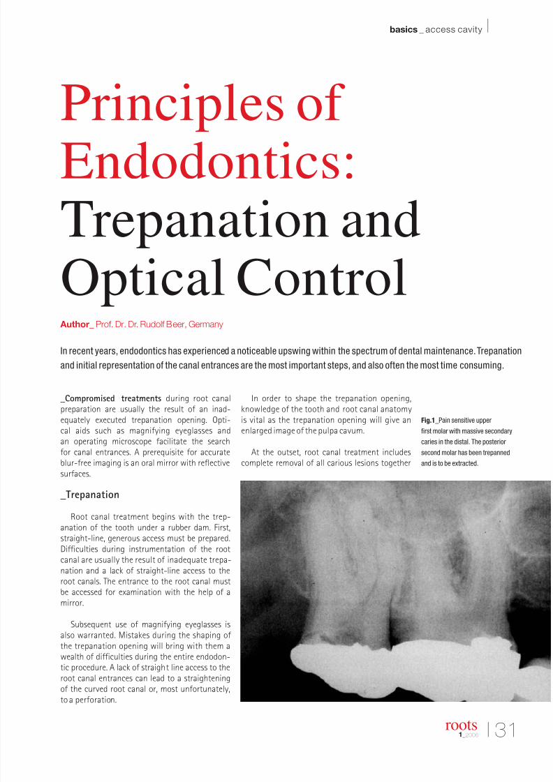
8/13/2019 Dente de Roots 1
http://slidepdf.com/reader/full/dente-de-roots-1 26/58
I 31
basics _ access cavity I
roots1_2006
_Compromised treatments during root canal
preparation are usually the result of an inad-equately executed trepanation opening. Opti-cal aids such as magnifying eyeglasses andan operating microscope facilitate the searchfor canal entrances. A prerequisite for accurateblur-free imaging is an oral mirror with reflectivesurfaces.
_Trepanation
Root canal treatment begins with the trep-anation of the tooth under a rubber dam. First,straight-line, generous access must be prepared.
Difficulties during instrumentation of the rootcanal are usually the result of inadequate trepa-nation and a lack of straight-line access to theroot canals. The entrance to the root canal mustbe accessed for examination with the help of amirror.
Subsequent use of magnifying eyeglasses isalso warranted. Mistakes during the shaping of the trepanation opening will bring with them awealth of difficulties during the entire endodon-tic procedure. A lack of straight line access to theroot canal entrances can lead to a straighteningof the curved root canal or, most unfortunately,to a perforation.
In order to shape the trepanation opening,
knowledge of the tooth and root canal anatomyis vital as the trepanation opening will give anenlarged image of the pulpa cavum.
At the outset, root canal treatment includescomplete removal of all carious lesions together
Fig.1_Pain sensitive upper
first molar with massive secondary
caries in the distal. The posterior
second molar has been trepanned
and is to be extracted.
Principles ofEndodontics:Trepanation and
Optical Control Author_ Prof. Dr. Dr. Rudolf Beer, Germany
In recent years, endodontics has experienced a noticeable upswing within the spectrum of dental maintenance.Trepanation
and initial representation of the canal entrances are the most important steps, and also often the most time consuming.
Fig. 1
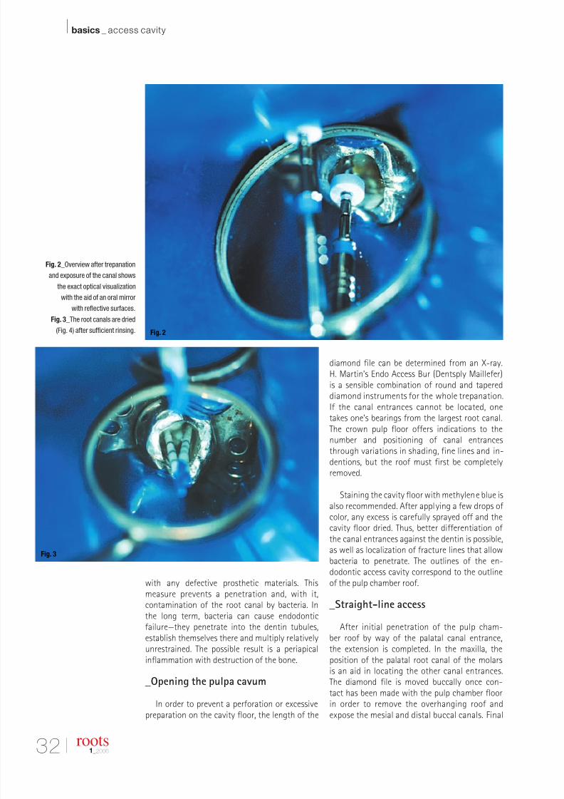
8/13/2019 Dente de Roots 1
http://slidepdf.com/reader/full/dente-de-roots-1 27/58
Fig. 2_Overview after trepanation
and exposure of the canal shows
the exact optical visualization
with the aid of an oral mirror
with reflective surfaces.
Fig. 3_The root canals are dried
(Fig. 4) after sufficient rinsing.
32 I
I basics _ access cavity
with any defective prosthetic materials. Thismeasure prevents a penetration and, with it,contamination of the root canal by bacteria. Inthe long term, bacteria can cause endodonticfailure—they penetrate into the dentin tubules,establish themselves there and multiply relativelyunrestrained. The possible result is a periapicalinflammation with destruction of the bone.
_Opening the pulpa cavum
In order to prevent a perforation or excessivepreparation on the cavity floor, the length of the
diamond file can be determined from an X-ray.H. Martin’s Endo Access Bur (Dentsply Maillefer)is a sensible combination of round and tapereddiamond instruments for the whole trepanation.If the canal entrances cannot be located, one
takes one’s bearings from the largest root canal.The crown pulp floor offers indications to thenumber and positioning of canal entrancesthrough variations in shading, fine lines and in-dentions, but the roof must first be completelyremoved.
Staining the cavity floor with methylene blue isalso recommended. After applying a few drops of color, any excess is carefully sprayed off and thecavity floor dried. Thus, better differentiation of the canal entrances against the dentin is possible,as well as localization of fracture lines that allow
bacteria to penetrate. The outlines of the en-dodontic access cavity correspond to the outlineof the pulp chamber roof.
_Straight-line access
After initial penetration of the pulp cham-ber roof by way of the palatal canal entrance,the extension is completed. In the maxilla, theposition of the palatal root canal of the molarsis an aid in locating the other canal entrances.The diamond file is moved buccally once con-tact has been made with the pulp chamber floorin order to remove the overhanging roof andexpose the mesial and distal buccal canals. Final
roots1_2006
Fig. 2
Fig. 3
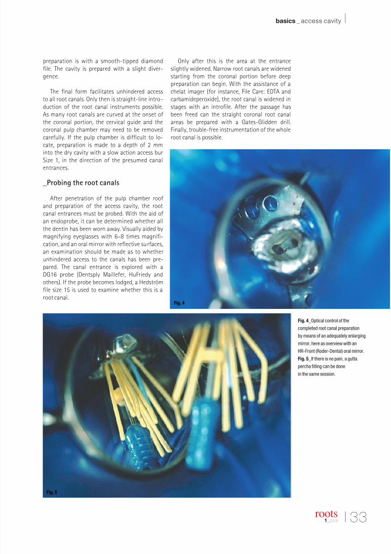
8/13/2019 Dente de Roots 1
http://slidepdf.com/reader/full/dente-de-roots-1 28/58
I 33
basics _ access cavity I
roots1_2006
preparation is with a smooth-tipped diamondfile. The cavity is prepared with a slight diver-gence.
The final form facilitates unhindered access
to all root canals. Only then is straight-line intro-duction of the root canal instruments possible.As many root canals are curved at the onset ofthe coronal portion, the cervical guide and thecoronal pulp chamber may need to be removedcarefully. If the pulp chamber is difficult to lo-cate, preparation is made to a depth of 2 mminto the dry cavity with a slow action access burSize 1, in the direction of the presumed canalentrances.
_Probing the root canals
After penetration of the pulp chamber roofand preparation of the access cavity, the rootcanal entrances must be probed. With the aid ofan endoprobe, it can be determined whether allthe dentin has been worn away. Visually aided bymagnifying eyeglasses with 6–8 times magnifi-cation, and an oral mirror with reflective surfaces,an examination should be made as to whetherunhindered access to the canals has been pre-pared. The canal entrance is explored with aDG16 probe (Dentsply Maillefer, HuFriedy andothers). If the probe becomes lodged, a Hedström
file size 15 is used to examine whether this is aroot canal.
Only after this is the area at the entranceslightly widened. Narrow root canals are widenedstarting from the coronal portion before deeppreparation can begin. With the assistance of achelat imager (for instance, File Care: EDTA and
carbamideperoxide), the root canal is widened instages with an introfile. After the passage hasbeen freed can the straight coronal root canalareas be prepared with a Gates-Glidden drill.Finally, trouble-free instrumentation of the wholeroot canal is possible.
Fig. 4_Optical control of the
completed root canal preparation
by means of an adequately enlarging
mirror, here as overview with an
HR-Front (Roder-Dental) oral mirror.
Fig. 5_If there is no pain, a gutta
percha filling can be done
in the same session.
Fig. 4
Fig. 5
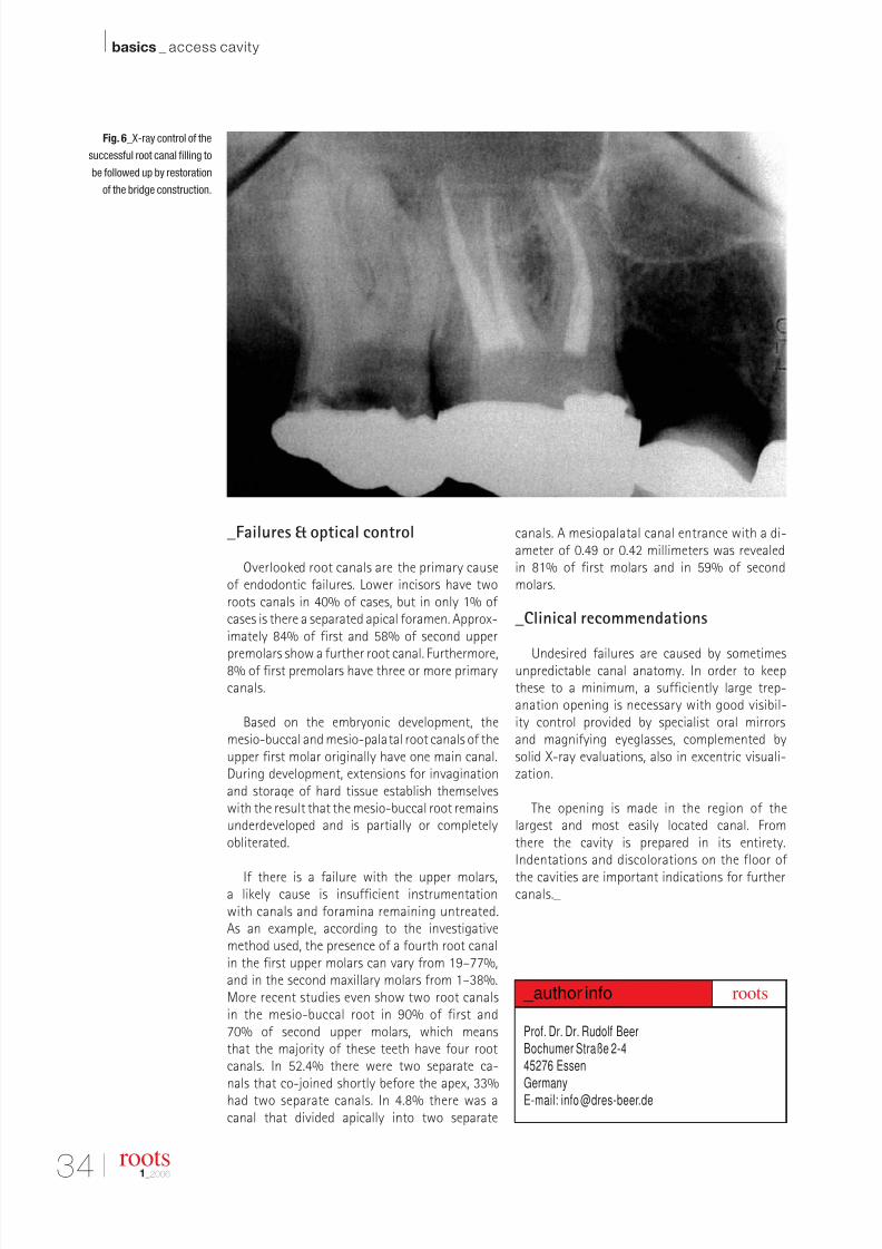
8/13/2019 Dente de Roots 1
http://slidepdf.com/reader/full/dente-de-roots-1 29/58
Fig. 6_X-ray control of the
successful root canal filling to
be followed up by restoration
of the bridge construction.
34 I
I basics _ access cavity
_Failures & optical control
Overlooked root canals are the primary causeof endodontic failures. Lower incisors have tworoots canals in 40% of cases, but in only 1% of cases is there a separated apical foramen. Approx-imately 84% of first and 58% of second upper
premolars show a further root canal. Furthermore,8% of first premolars have three or more primarycanals.
Based on the embryonic development, themesio-buccal and mesio-palatal root canals of theupper first molar originally have one main canal.During development, extensions for invaginationand storage of hard tissue establish themselveswith the result that the mesio-buccal root remainsunderdeveloped and is partially or completelyobliterated.
If there is a failure with the upper molars,a likely cause is insufficient instrumentationwith canals and foramina remaining untreated.As an example, according to the investigativemethod used, the presence of a fourth root canalin the first upper molars can vary from 19–77%,and in the second maxillary molars from 1–38%.More recent studies even show two root canalsin the mesio-buccal root in 90% of first and70% of second upper molars, which meansthat the majority of these teeth have four rootcanals. In 52.4% there were two separate ca-nals that co-joined shortly before the apex, 33%had two separate canals. In 4.8% there was acanal that divided apically into two separate
canals. A mesiopalatal canal entrance with a di-ameter of 0.49 or 0.42 millimeters was revealedin 81% of first molars and in 59% of secondmolars.
_Clinical recommendations
Undesired failures are caused by sometimesunpredictable canal anatomy. In order to keepthese to a minimum, a sufficiently large trep-anation opening is necessary with good visibil-ity control provided by specialist oral mirrorsand magnifying eyeglasses, complemented bysolid X-ray evaluations, also in excentric visuali-zation.
The opening is made in the region of thelargest and most easily located canal. Fromthere the cavity is prepared in its entirety.Indentations and discolorations on the floor of
the cavities are important indications for furthercanals._
roots1_2006
Prof. Dr. Dr. Rudolf BeerBochumer Straße 2-445276 Essen
Germany
E-mail: info @dres-beer.de
_author info roots
Fig. 6
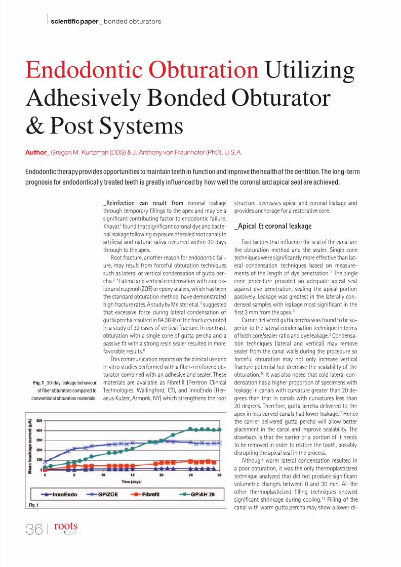
8/13/2019 Dente de Roots 1
http://slidepdf.com/reader/full/dente-de-roots-1 30/58
36 I
I scientific paper _ bonded obturators
_Reinfection can result from coronal leakagethrough temporary fillings to the apex and may be asignificant contributing factor to endodontic failure.Khayat1 found that significant coronal dye and bacte-rial leakage following exposure of sealed root canals toartificial and natural saliva occurred within 30 daysthrough to the apex.
Root fracture, another reason for endodontic fail-ure, may result from forceful obturation techniquessuch as lateral or vertical condensation of gutta per-cha.2–4 Lateral and vertical condensation with zinc ox-ide and eugenol (ZOE) or epoxy sealers, which has been
the standard obturation method, have demonstratedhigh fracture rates. A study by Meister et al.5 suggestedthat excessive force during lateral condensation of gutta percha resulted in 84.38 % of the fractures notedin a study of 32 cases of vertical fracture. In contrast,obturation with a single cone of gutta percha and apassive fit with a strong resin sealer resulted in morefavorable results.6
This communication reports on the clinical use andin vitro studies performed with a fiber-reinforced ob-turator combined with an adhesive and sealer. Thesematerials are available as Fibrefill (Pentron ClinicalTechnologies, Wallingford, CT), and InnoEndo (Her-
aeus Kulzer, Armonk, NY) which strengthens the root
structure, decreases apical and coronal leakage andprovides anchorage for a restorative core.
_Apical & coronal leakage
Two factors that influence the seal of the canal arethe obturation method and the sealer. Single conetechniques were significantly more effective than lat-eral condensation techniques based on measure-ments of the length of dye penetration.7 The singlecone procedure provided an adequate apical sealagainst dye penetration, sealing the apical portion
passively. Leakage was greatest in the laterally con-densed samples with leakage most significant in thefirst 3 mm from the apex.8
Carrier delivered gutta percha was found to be su-perior to the lateral condensation technique in termsof both core/sealer ratio and dye leakage.9 Condensa-tion techniques (lateral and vertical) may removesealer from the canal walls during the procedure soforceful obturation may not only increase verticalfracture potential but decrease the sealability of theobturation.10 It was also noted that cold lateral con-densation has a higher proportion of specimens withleakage in canals with curvature greater than 20 de-
grees than that in canals with curvatures less than20 degrees. Therefore, gutta percha delivered to theapex in less curved canals had lower leakage.11 Hencethe carrier-delivered gutta percha will allow betterplacement in the canal and improve sealability. Thedrawback is that the carrier or a portion of it needsto be removed in order to restore the tooth, possiblydisrupting the apical seal in the process.
Although warm lateral condensation resulted ina poor obturation, it was the only thermoplasticizedtechnique analyzed that did not produce significantvolumetric changes between 0 and 30 min. All theother thermoplasticized filling techniques showedsignificant shrinkage during cooling.12 Filling of thecanal with warm gutta percha may show a lower di-
Fig. 1_30-day leakage behaviour
of fiber obturators compared to
conventional obturation materials.
roots1_2006
Endodontic Obturation Utilizing
Adhesively Bonded Obturator& Post Systems
Author_ Gregori M. Kurtzman (DDS) & J. Anthony von Fraunhofer (PhD), U.S.A.
Endodontic therapy provides opportunities to maintain teeth in function and improve the health of the dentition.The long-term
prognosis for endodontically treated teeth is greatly influenced by how well the coronal and apical seal are achieved.
Fig. 1
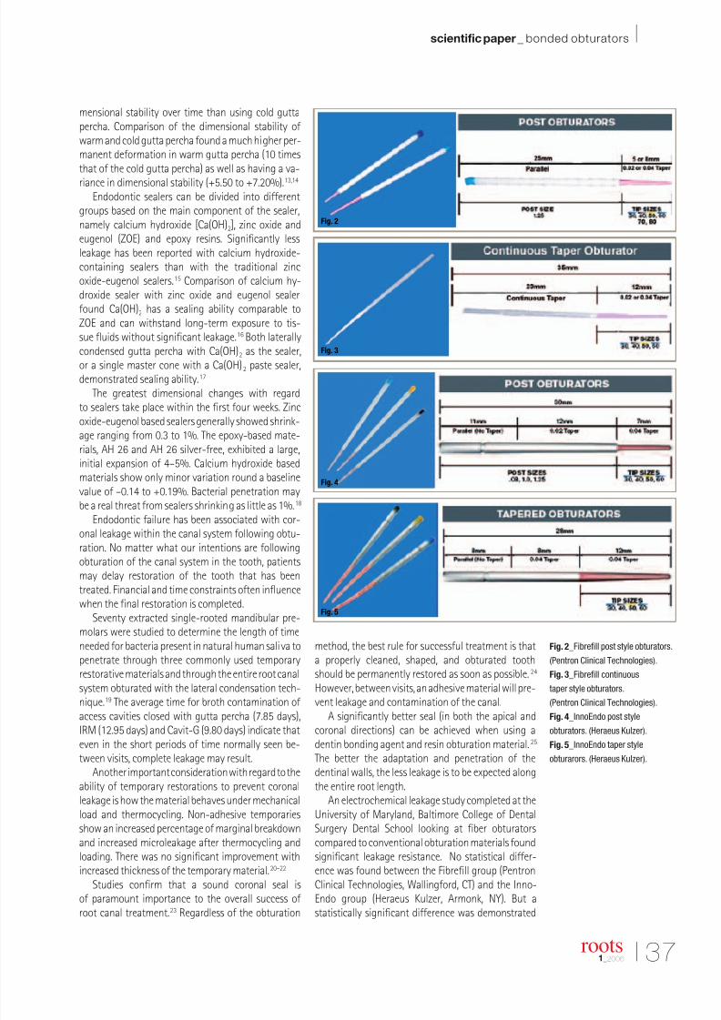
8/13/2019 Dente de Roots 1
http://slidepdf.com/reader/full/dente-de-roots-1 31/58
I 37
scientific paper _ bonded obturators I
roots1_2006
mensional stability over time than using cold guttapercha. Comparison of the dimensional stability of warm and cold gutta percha found a much higher per-manent deformation in warm gutta percha (10 timesthat of the cold gutta percha) as well as having a va-
riance in dimensional stability (+5.50 to +7.20%).13,14
Endodontic sealers can be divided into differentgroups based on the main component of the sealer,namely calcium hydroxide [Ca(OH)2], zinc oxide andeugenol (ZOE) and epoxy resins. Significantly lessleakage has been reported with calcium hydroxide-containing sealers than with the traditional zincoxide-eugenol sealers.15 Comparison of calcium hy-droxide sealer with zinc oxide and eugenol sealerfound Ca(OH)2 has a sealing ability comparable toZOE and can withstand long-term exposure to tis-sue fluids without significant leakage.16 Both laterallycondensed gutta percha with Ca(OH)
2as the sealer,
or a single master cone with a Ca(OH)2 paste sealer,demonstrated sealing ability.17
The greatest dimensional changes with regardto sealers take place within the first four weeks. Zincoxide-eugenol based sealers generally showed shrink-age ranging from 0.3 to 1%. The epoxy-based mate-rials, AH 26 and AH 26 silver-free, exhibited a large,initial expansion of 4–5%. Calcium hydroxide basedmaterials show only minor variation round a baselinevalue of –0.14 to +0.19%. Bacterial penetration maybe a real threat from sealers shrinking as little as 1%.18
Endodontic failure has been associated with cor-
onal leakage within the canal system following obtu-ration. No matter what our intentions are followingobturation of the canal system in the tooth, patientsmay delay restoration of the tooth that has beentreated. Financial and time constraints often influencewhen the final restoration is completed.
Seventy extracted single-rooted mandibular pre-molars were studied to determine the length of timeneeded for bacteria present in natural human saliva topenetrate through three commonly used temporaryrestorative materials and through the entire root canalsystem obturated with the lateral condensation tech-nique.19 The average time for broth contamination of
access cavities closed with gutta percha (7.85 days),IRM (12.95 days) and Cavit-G (9.80 days) indicate thateven in the short periods of time normally seen be-tween visits, complete leakage may result.
Another important consideration with regard to theability of temporary restorations to prevent coronalleakage is how the material behaves under mechanicalload and thermocycling. Non-adhesive temporariesshow an increased percentage of marginal breakdownand increased microleakage after thermocycling andloading. There was no significant improvement withincreased thickness of the temporary material.20–22
Studies confirm that a sound coronal seal isof paramount importance to the overall success of root canal treatment.23 Regardless of the obturation
method, the best rule for successful treatment is thata properly cleaned, shaped, and obturated toothshould be permanently restored as soon as possible.24
However, between visits, an adhesive material will pre-vent leakage and contamination of the canal.
A significantly better seal (in both the apical andcoronal directions) can be achieved when using adentin bonding agent and resin obturation material.25
The better the adaptation and penetration of thedentinal walls, the less leakage is to be expected alongthe entire root length.
An electrochemical leakage study completed at theUniversity of Maryland, Baltimore College of DentalSurgery Dental School looking at fiber obturatorscompared to conventional obturation materials foundsignificant leakage resistance. No statistical differ-ence was found between the Fibrefill group (PentronClinical Technologies, Wallingford, CT) and the Inno-Endo group (Heraeus Kulzer, Armonk, NY). But astatistically significant difference was demonstrated
Fig. 2_Fibrefill post style obturators.
(Pentron Clinical Technologies).
Fig. 3_Fibrefill continuous
taper style obturators.
(Pentron Clinical Technologies).
Fig. 4_InnoEndo post style
obturators. (Heraeus Kulzer).
Fig. 5_InnoEndo taper style
obturarors. (Heraeus Kulzer).
Fig. 2
Fig. 3
Fig. 4
Fig. 5
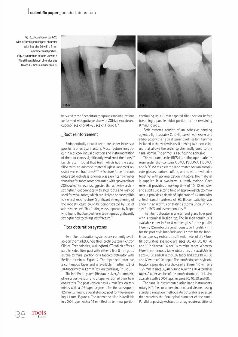
8/13/2019 Dente de Roots 1
http://slidepdf.com/reader/full/dente-de-roots-1 32/58
38 I
I scientific paper _ bonded obturators
between these fiber obturator groups and obturationsperformed with gutta percha with ZOE (zinc oxide andeugenol) sealer or AH-26 sealer, Figure 1.26
_Root reinforcement
Endodontically treated teeth are under increasedpossibility of vertical fracture. Most fracture lines oc-cur in a bucco-lingual direction and instrumentationof the root canals significantly weakened the roots.27
Lertchirakarn found that teeth which had the canalfilled with an adhesive material (glass ionomer) re-sisted vertical fractures.28 The fracture force for roots
obturated with glass ionomer was significantly higherthan that for tooth roots obturated with epoxy resin orZOE sealer. The results suggested that adhesive sealersstrengthen endodontically treated roots and may beused for weak roots, which are likely to be susceptibleto vertical root fracture. Significant strengthening of the root structure could be demonstrated by use ofadhesive sealers. This finding was supported by Trope,who found that bonded resin techniques significantlystrengthened teeth against fracture.29
_Fiber obturation systems
Two fiber obturation systems are currently avail-able on the market. One is the Fibrefill System (PentronClinical Technologies, Wallingford, CT) which offers aparallel sided fiber post with either a 5 or 8 mm guttapercha terminal portion or a tapered obturator withResilon terminus, Figure 2. The taper obturator hasa continuous taper and is available in either .02 or.04 tapers with a 12 mm Resilon terminus, Figure 3.
The InnoEndo system (Hearaus Kulzer, Armonk, NY)offers a post version and a taper version of their fiberobturators. The post version has a 7 mm Resilon ter-minus with a .02 taper segment for the subsequent12 mm turning to a parallel-sided post for the remain-ing 11 mm, Figure 4. The tapered version is availablein a 0.04 taper with a 12 mm Resilon terminal portion
continuing as a 8 mm tapered fiber portion beforebecoming a parallel-sided portion for the remaining8 mm, Figure 5.
Both systems consist of an adhesive bondingagent, a light-curable Ca(OH)2 based resin sealer anda fiber post with an apical terminus of Resilon. A primerincluded in the system is a self etching two-bottle liq-uid that allows the sealer to chemically bond to thecanal dentin. The primer is a self curing adhesive.
The root canal sealer (RCS) is a radiopaque dual cureresin sealer that contains UDMA, PEGDMA, HDDMA,and BISGMA resins with silane treated barium borosil-cate glasses, barium sulfate, and calcium hydroxide
together with polymerization initiators. The materialis supplied in a two-barrel automix syringe. Oncemixed, it provides a working time of 10–12 minutesand a self cure setting time of approximately 25 min-utes. It provides a depth of light cure of 1.7 mm witha final Barcol hardness of 80. Biocompatibility wasshown in agar diffusion testing at Loma Linda Univer-sity for RCS and its components.30
The fiber obturator is a resin and glass fiber postwith a terminal Resilon tip. The Resilon terminus isavailable either in 5 or 8 mm lengths for the parallelFibrefill, 12 mm for the continuous taper Fibrefill, 7 mmfor the post style InnoEndo and 12 mm for the Inno-
Endo taper style obturators. The diameter of the Fibre-fill obturators available are sizes 30, 40, 50, 60, 70and 80 in either a 0.02 or 0.04 terminal taper. Whereas,Fibrefill continuous taper obturators are available insizes 40, 50 and 60 in the 0.02 taper and sizes 30. 40, 50and 60 with a 0.04 taper. The InnoEndo post style ob-turator is provided in a choice of a .8 mm, 1.0 mm or a1.25 mm in sizes 30, 40, 50 and 60 with a 0.04 terminaltaper. A taper version of the InnoEndo obturator is alsoavailable with a 0.04 taper in sizes 30, 40, 50 and 60.
The canal is instrumented using hand instruments,rotary NiTi files or a combination, and cleaned usingstandard irrigation methods. An obturator is selectedthat matches the final apical diameter of the canal.Parallel or post style obturators may require additional
Fig. 6_Obturation of tooth 20
with a Fibrefill parallel post obturator
with final size 30 with a 5 mm
apical terminal portion.
Fig. 7_Obturation of tooth 20 with a
Fibrefill parallel post obturator size
50 with a 5 mm Resilon terminus.
roots1_2006
Fig. 6 Fig. 7
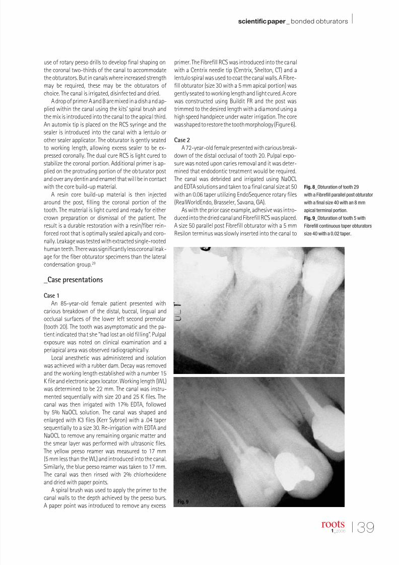
8/13/2019 Dente de Roots 1
http://slidepdf.com/reader/full/dente-de-roots-1 33/58
I 39
scientific paper _ bonded obturators I
roots1_2006
use of rotary peeso drills to develop final shaping onthe coronal two-thirds of the canal to accommodatethe obturators. But in canals where increased strengthmay be required, these may be the obturators of choice. The canal is irrigated, disinfected and dried.
A drop of primer A and B are mixed in a dish and ap-plied within the canal using the kits’ spiral brush andthe mix is introduced into the canal to the apical third.An automix tip is placed on the RCS syringe and thesealer is introduced into the canal with a lentulo orother sealer applicator. The obturator is gently seatedto working length, allowing excess sealer to be ex-pressed coronally. The dual cure RCS is light cured tostabilize the coronal portion. Additional primer is ap-plied on the protruding portion of the obturator postand over any dentin and enamel that will be in contactwith the core build-up material.
A resin core build-up material is then injectedaround the post, filling the coronal portion of thetooth. The material is light cured and ready for eithercrown preparation or dismissal of the patient. Theresult is a durable restoration with a resin/fiber rein-forced root that is optimally sealed apically and coro-nally. Leakage was tested with extracted single-rootedhuman teeth. There was significantly less coronal leak-age for the fiber obturator specimens than the lateralcondensation group.29
_Case presentations
Case 1An 85-year-old female patient presented with
carious breakdown of the distal, buccal, lingual andocclusal surfaces of the lower left second premolar(tooth 20). The tooth was asymptomatic and the pa-tient indicated that she “had lost an old filling”. Pulpalexposure was noted on clinical examination and aperiapical area was observed radiographically.
Local anesthetic was administered and isolationwas achieved with a rubber dam. Decay was removedand the working length established with a number 15K file and electronic apex locator. Working length (WL)was determined to be 22 mm. The canal was instru-
mented sequentially with size 20 and 25 K files. Thecanal was then irrigated with 17% EDTA, followedby 5% NaOCL solution. The canal was shaped andenlarged with K3 files (Kerr Sybron) with a .04 tapersequentially to a size 30. Re-irrigation with EDTA andNaOCL to remove any remaining organic matter andthe smear layer was performed with ultrasonic files.The yellow peeso reamer was measured to 17 mm(5 mm less than the WL) and introduced into the canal.Similarly, the blue peeso reamer was taken to 17 mm.The canal was then rinsed with 2% chlorhexideneand dried with paper points.
A spiral brush was used to apply the primer to thecanal walls to the depth achieved by the peeso burs.A paper point was introduced to remove any excess
primer. The Fibrefill RCS was introduced into the canalwith a Centrix needle tip (Centrix, Shelton, CT) and alentulo spiral was used to coat the canal walls. A Fibre-fill obturator (size 30 with a 5 mm apical portion) wasgently seated to working length and light cured. A core
was constructed using Buildit FR and the post wastrimmed to the desired length with a diamond using ahigh speed handpiece under water irrigation. The corewas shaped to restore the tooth morphology (Figure 6).
Case 2
A 72-year-old female presented with carious break-down of the distal occlusal of tooth 20. Pulpal expo-sure was noted upon caries removal and it was deter-mined that endodontic treatment would be required.The canal was debrided and irrigated using NaOCLand EDTA solutions and taken to a final canal size at 50with an 0.06 taper utilizing EndoSequence rotary files(RealWorldEndo, Brasseler, Savana, GA).
As with the prior case example, adhesive was intro-duced into the dried canal and Fibrefill RCS was placed.A size 50 parallel post Fibrefill obturator with a 5 mmResilon terminus was slowly inserted into the canal to
Fig. 8_Obturation of tooth 29
with a Fibrefill parallel post obturator
with a final size 40 with an 8 mm
apical terminal portion.
Fig. 9_Obturation of tooth 5 with
Fibrefill continuous taper obturators
size 40 with a 0.02 taper.
Fig. 8
Fig. 9
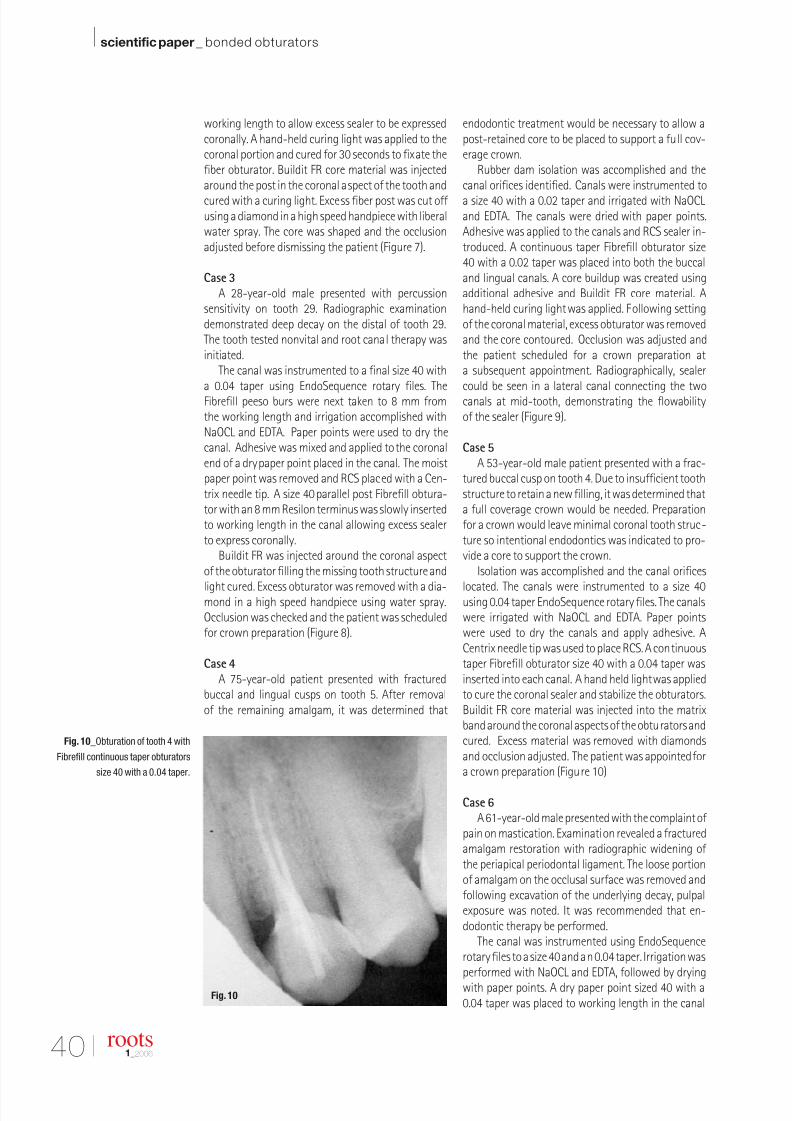
8/13/2019 Dente de Roots 1
http://slidepdf.com/reader/full/dente-de-roots-1 34/58
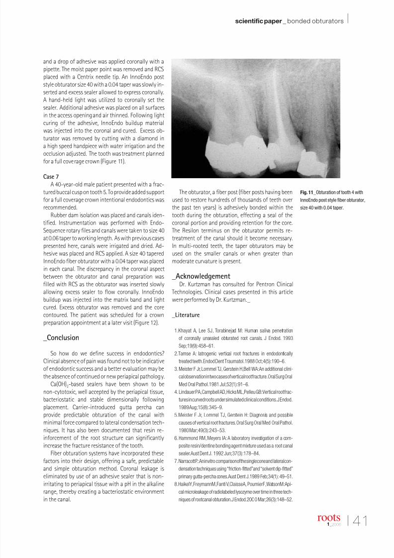
8/13/2019 Dente de Roots 1
http://slidepdf.com/reader/full/dente-de-roots-1 35/58
I 41
scientific paper _ bonded obturators I
roots1_2006
and a drop of adhesive was applied coronally with apipette. The moist paper point was removed and RCSplaced with a Centrix needle tip. An InnoEndo poststyle obturator size 40 with a 0.04 taper was slowly in-serted and excess sealer allowed to express coronally.
A hand-held light was utilized to coronally set thesealer. Additional adhesive was placed on all surfacesin the access opening and air thinned. Following lightcuring of the adhesive, InnoEndo buildup materialwas injected into the coronal and cured. Excess ob-turator was removed by cutting with a diamond ina high speed handpiece with water irrigation and theocclusion adjusted. The tooth was treatment plannedfor a full coverage crown (Figure 11).
Case 7
A 40-year-old male patient presented with a frac-tured buccal cusp on tooth 5. To provide added supportfor a full coverage crown intentional endodontics wasrecommended.
Rubber dam isolation was placed and canals iden-tified. Instrumentation was performed with Endo-Sequence rotary files and canals were taken to size 40at 0.06 taper to working length. As with previous casespresented here, canals were irrigated and dried. Ad-hesive was placed and RCS applied. A size 40 taperedInnoEndo fiber obturator with a 0.04 taper was placedin each canal. The discrepancy in the coronal aspectbetween the obturator and canal preparation wasfilled with RCS as the obturator was inserted slowly
allowing excess sealer to flow coronally. InnoEndobuildup was injected into the matrix band and lightcured. Excess obturator was removed and the corecontoured. The patient was scheduled for a crownpreparation appointment at a later visit (Figure 12).
_Conclusion
So how do we define success in endodontics?Clinical absence of pain was found not to be indicativeof endodontic success and a better evaluation may bethe absence of continued or new periapical pathology.
Ca(OH)2-based sealers have been shown to be
non-cytotoxic, well accepted by the periapical tissue,bacteriostatic and stable dimensionally followingplacement. Carrier-introduced gutta percha canprovide predictable obturation of the canal withminimal force compared to lateral condensation tech-niques. It has also been documented that resin re-inforcement of the root structure can significantlyincrease the fracture resistance of the tooth.
Fiber obturation systems have incorporated thesefactors into their design, offering a safe, predictableand simple obturation method. Coronal leakage iseliminated by use of an adhesive sealer that is non-irritating to periapical tissue with a pH in the alkalinerange, thereby creating a bacteriostatic environmentin the canal.
The obturator, a fiber post (fiber posts having beenused to restore hundreds of thousands of teeth overthe past ten years) is adhesively bonded within thetooth during the obturation, effecting a seal of thecoronal portion and providing retention for the core.The Resilon terminus on the obturator permits re-treatment of the canal should it become necessary.In multi-rooted teeth, the taper obturators may beused on the smaller canals or when greater thanmoderate curvature is present.
_AcknowledgementDr. Kurtzman has consulted for Pentron Clinical
Technologies. Clinical cases presented in this articlewere performed by Dr. Kurtzman._
_Literature
1.Khayat A, Lee SJ, Torabinejad M: Human saliva penetration
of coronally unsealed obturated root canals. J Endod. 1993
Sep;19(9):458–61.
2.Tamse A: Iatrogenic vertical root fractures in endodontically
treated teeth.Endod Dent Traumatol.1988 Oct;4(5):190–6.
3.Meister F Jr, Lommel TJ, Gerstein H,Bell WA:An additional clini-
cal observation in two cases of vertical root fracture.Oral Surg Oral
Med Oral Pathol.1981 Jul;52(1):91–6.
4.Lindauer PA,Campbell AD,Hicks ML,Pelleu GB:Vertical root frac-tures in curved roots under simulated clinical conditions.J Endod.
1989 Aug;15(8):345–9.
5.Meister F Jr, Lommel TJ, Gerstein H: Diagnosis and possible
causes of vertical root fractures.Oral Surg Oral Med Oral Pathol.
1980 Mar;49(3):243–53.
6.Hammond RM, Meyers IA:A laboratory investigation of a com-
posite resin/dentine bonding agent mixture used as a root canal
sealer.Aust Dent J. 1992 Jun;37(3):178–84.
7.Narracott P.:An in vitro comparison of the single cone and lateral con-
densation techniques using “friction-fitted”and “solvent dip-fitted”
primary gutta-percha cones.Aust Dent J.1989 Feb;34(1): 49–51.
8.Haikel Y,Freymann M,Fanti V,Claisse A, Poumier F,Watson M:Api-
cal microleakage of radiolabeled lysozyme over time in three tech-
niques of root canal obturation.J Endod.2000 Mar;26(3):148–52.
Fig. 11_Obturation of tooth 4 with
InnoEndo post style fiber obturator,
size 40 with 0.04 taper.
Fig. 11
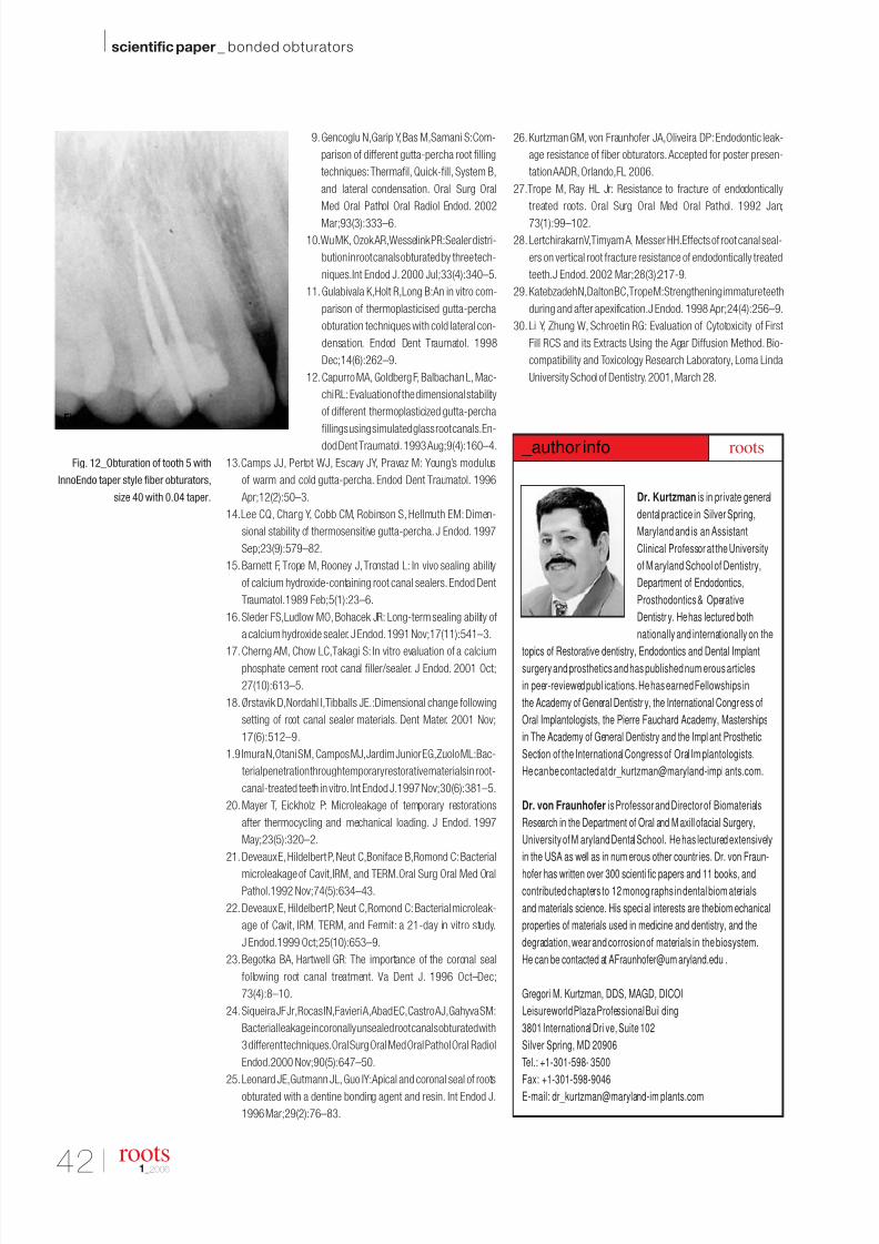
8/13/2019 Dente de Roots 1
http://slidepdf.com/reader/full/dente-de-roots-1 36/58
42 I
I scientific paper _ bonded obturators
9.Gencoglu N,Garip Y,Bas M,Samani S:Com-
parison of different gutta-percha root filling
techniques:Thermafil, Quick-fill, System B,
and lateral condensation. Oral Surg Oral
Med Oral Pathol Oral Radiol Endod. 2002
Mar;93(3):333–6.
10.Wu MK, Ozok AR,Wesselink PR:Sealer distri-
bution in root canals obturated by three tech-
niques.Int Endod J.2000 Jul;33(4):340–5.
11.Gulabivala K,Holt R,Long B:An in vitro com-
parison of thermoplasticised gutta-percha
obturation techniques with cold lateral con-
densation. Endod Dent Traumatol. 1998
Dec;14(6):262–9.
12.Capurro MA, Goldberg F, Balbachan L, Mac-
chi RL:Evaluation of the dimensional stability
of different thermoplasticized gutta-percha
fillings using simulated glass root canals.En-
dod Dent Traumatol. 1993 Aug;9(4):160–4.
13.Camps JJ, Pertot WJ, Escavy JY, Pravaz M: Young’s modulus
of warm and cold gutta-percha. Endod Dent Traumatol. 1996
Apr;12(2):50–3.
14.Lee CQ, Chang Y, Cobb CM, Robinson S, Hellmuth EM: Dimen-
sional stability of thermosensitive gutta-percha.J Endod. 1997
Sep;23(9):579–82.
15. Barnett F, Trope M, Rooney J,Tronstad L: In vivo sealing ability
of calcium hydroxide-containing root canal sealers. Endod Dent
Traumatol.1989 Feb;5(1):23–6.
16. Sleder FS,Ludlow MO, Bohacek JR: Long-term sealing ability of
a calcium hydroxide sealer. J Endod.1991 Nov;17(11):541–3.
17. Cherng AM, Chow LC,Takagi S: In vitro evaluation of a calciumphosphate cement root canal filler/sealer. J Endod. 2001 Oct;
27(10):613–5.
18. Ørstavik D,Nordahl I,Tibballs JE.:Dimensional change following
setting of root canal sealer materials. Dent Mater. 2001 Nov;
17(6):512–9.
1.9 Imura N,Otani SM,Campos MJ,Jardim Junior EG,Zuolo ML:Bac-
terial penetration through temporary restorative materials in root-
canal-treated teeth in vitro. Int Endod J.1997 Nov;30(6):381–5.
20. Mayer T, Eickholz P: Microleakage of temporary restorations
after thermocycling and mechanical loading. J Endod. 1997
May;23(5):320–2.
21. Deveaux E, Hildelbert P, Neut C,Boniface B,Romond C:Bacterial
microleakage of Cavit,IRM, and TERM.Oral Surg Oral Med OralPathol.1992 Nov;74(5):634–43.
22. Deveaux E, Hildelbert P, Neut C,Romond C: Bacterial microleak-
age of Cavit, IRM, TERM, and Fermit: a 21-day in vitro study.
J Endod.1999 Oct;25(10):653–9.
23. Begotka BA, Hartwell GR: The importance of the coronal seal
following root canal treatment. Va Dent J. 1996 Oct–Dec;
73(4):8–10.
24. Siqueira JF Jr,Rocas IN,Favieri A,Abad EC,Castro AJ,Gahyva SM:
Bacterial leakage in coronally unsealed root canals obturated with
3 different techniques.Oral Surg Oral Med Oral Pathol Oral Radiol
Endod.2000 Nov;90(5):647–50.
25. Leonard JE,Gutmann JL,Guo IY:Apical and coronal seal of roots
obturated with a dentine bonding agent and resin. Int Endod J.
1996 Mar;29(2):76–83.
26. Kurtzman GM, von Fraunhofer JA,Oliveira DP:Endodontic leak-
age resistance of fiber obturators.Accepted for poster presen-
tation AADR,Orlando,FL 2006.
27.Trope M, Ray HL Jr: Resistance to fracture of endodontically
treated roots. Oral Surg Oral Med Oral Pathol. 1992 Jan;
73(1):99–102.
28. Lertchirakarn V,Timyam A,Messer HH.Effects of root canal seal-
ers on vertical root fracture resistance of endodontically treated
teeth.J Endod. 2002 Mar;28(3):217-9.
29. Katebzadeh N,Dalton BC,Trope M:Strengthening immature teeth
during and after apexification.J Endod. 1998 Apr;24(4):256–9.
30. Li Y, Zhung W, Schroetin RG: Evaluation of Cytotoxicity of First
Fill RCS and its Extracts Using the Agar Diffusion Method. Bio-
compatibility and Toxicology Research Laboratory, Loma Linda
University School of Dentistry. 2001,March 28.
Fig. 12_Obturation of tooth 5 with
InnoEndo taper style fiber obturators,
size 40 with 0.04 taper.
roots1_2006
Dr. Kurtzman is in pr ivate general
dental practice in Silver Spring,
Maryland and is an Assistant
Clinical Professor at the University
of M aryland School of Dentistry,
Department of Endodontics,
Prosthodontics & Operative
Dentistr y. He has lectured both
nationally and internationally on the
topics of Restorative dentistry, Endodontics and Dental Implantsurgery and prosthetics and has published num erous articles
in peer-reviewed publ ications. He has earned Fellowships in
the Academy of General Dentistr y, the International Congr ess of
Oral Implantologists, the Pierre Fauchard Academy, Masterships
in The Academy of General Dentistry and the Impl ant Prosthetic
Section of the International Congress of Oral Im plantologists.
He can be contacted at dr_kurtzman@maryland-impl ants.com.
Dr. von Fraunhofer is Professor and Director of Biomaterials
Research in the Department of Oral and M axill ofacial Surgery,
University of M aryland Dental School. He has lectured extensively
in the USA as well as in num erous other countr ies. Dr. von Fraun-
hofer has written over 300 scienti fic papers and 11 books, andcontributed chapters to 12 monog raphs in dental biom aterials
and materials science. His speci al interests are the biom echanical
properties of materials used in medicine and dentistry, and the
degradation, wear and corrosion of materials in the biosystem.
He can be contacted at AFraunhofer@um aryland.edu .
Gregori M. Kurtzman, DDS, MAGD, DICOI
Leisureworld Plaza Professional Buil ding
3801 International Dri ve, Suite 102
Silver Spring, MD 20906
Tel.: +1-301-598- 3500
Fax: +1-301-598-9046
E-mail: dr_kurtzman@maryland-im plants.com
_author info roots
Fig. 12
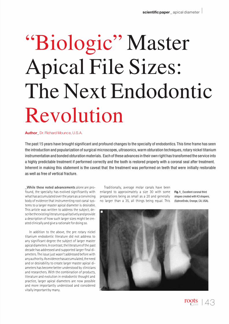
8/13/2019 Dente de Roots 1
http://slidepdf.com/reader/full/dente-de-roots-1 37/58
I 43
scientific paper _ apical diameter I
roots1_2006
_While these noted advancements alone are pro-found, the specialty has evolved significantly withwhat has accumulated over the years as a convincingbody of evidence that instrumenting root canal sys-tems to a larger master apical diameter is desirable.This article was written to address the subject, de-scribe the existing literature qualitatively and providea description of how such larger sizes might be cre-ated clinically and give a rationale for doing so.
In addition to the above, the pre rotary nickeltitanium endodontic literature did not address toany significant degree the subject of larger masterapical diameters. In contrast, the literature of the pastdecade has addressed and supported larger final di-ameters. The issue just wasn’t addressed before withany authority. As evidence has accumulated, the needand or desirability to create larger master apical di-ameters has become better understood by cliniciansand researchers. With the combination of products,literature and evolution in endodontic thought andpractice, larger apical diameters are now possibleand more importantly understood and consideredvitally important by many.
Traditionally, average molar canals have beenenlarged to approximately a size 30 with somepreparations being as small as a 20 and generallyno larger than a 35, all things being equal. This
Fig. 1_ Excellent coronal third
shapes created with K3 shapers,
(SybronEndo, Orange, CA, USA).
“Biologic” MasterApical File Sizes:The Next Endodontic
Revolution Author_ Dr. Richard Mounce, U.S.A.
The past 15 years have brought significant and profound changes to the specialty of endodontics. This time frame has seen
the introduction and popularization of surgical microscopes, ultrasonics,warm obturation techniques, rotary nickel titanium
instrumentation and bonded obturation materials. Each of these advances in their own right has transformed the service into
a highly predictable treatment if performed correctly and the tooth is restored properly with a coronal seal after treatment.
Inherent in making this statement is the caveat that the treatment was performed on teeth that were initially restorable
as well as free of vertical fracture.
Fig. 1
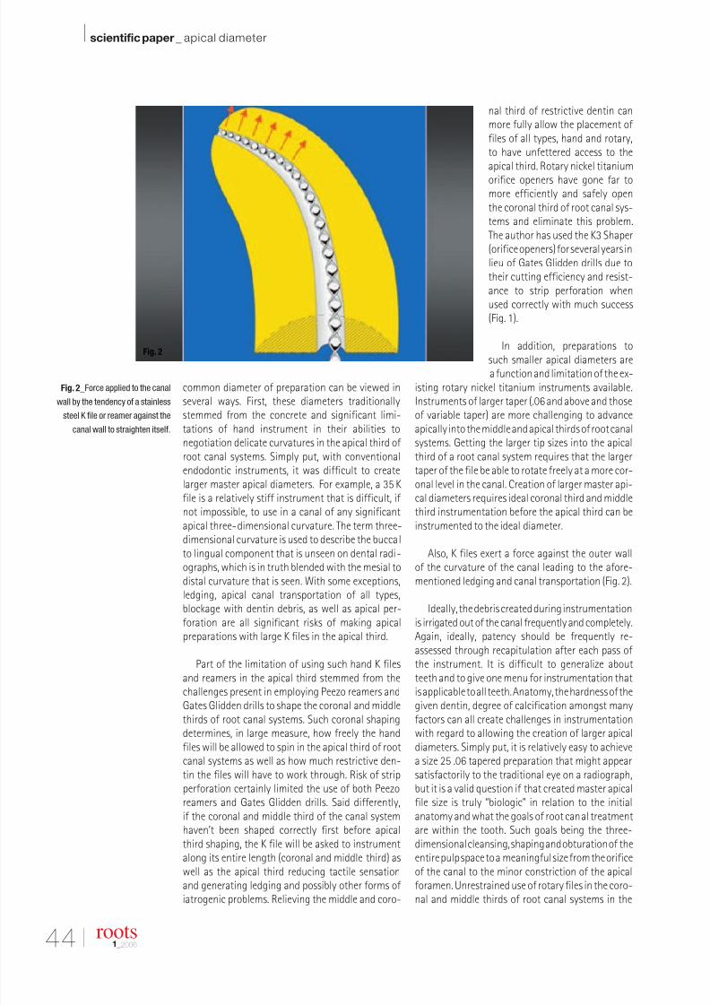
8/13/2019 Dente de Roots 1
http://slidepdf.com/reader/full/dente-de-roots-1 38/58
44 I
I scientific paper _ apical diameter
roots1_2006
common diameter of preparation can be viewed inseveral ways. First, these diameters traditionallystemmed from the concrete and significant limi-tations of hand instrument in their abilities tonegotiation delicate curvatures in the apical third of root canal systems. Simply put, with conventionalendodontic instruments, it was difficult to createlarger master apical diameters. For example, a 35 Kfile is a relatively stiff instrument that is difficult, if not impossible, to use in a canal of any significant
apical three-dimensional curvature. The term three-dimensional curvature is used to describe the buccalto lingual component that is unseen on dental radi-ographs, which is in truth blended with the mesial todistal curvature that is seen. With some exceptions,ledging, apical canal transportation of all types,blockage with dentin debris, as well as apical per-foration are all significant risks of making apicalpreparations with large K files in the apical third.
Part of the limitation of using such hand K filesand reamers in the apical third stemmed from thechallenges present in employing Peezo reamers and
Gates Glidden drills to shape the coronal and middlethirds of root canal systems. Such coronal shapingdetermines, in large measure, how freely the handfiles will be allowed to spin in the apical third of rootcanal systems as well as how much restrictive den-tin the files will have to work through. Risk of stripperforation certainly limited the use of both Peezoreamers and Gates Glidden drills. Said differently,if the coronal and middle third of the canal systemhaven’t been shaped correctly first before apicalthird shaping, the K file will be asked to instrumentalong its entire length (coronal and middle third) aswell as the apical third reducing tactile sensationand generating ledging and possibly other forms of iatrogenic problems. Relieving the middle and coro-
nal third of restrictive dentin canmore fully allow the placement of files of all types, hand and rotary,to have unfettered access to theapical third. Rotary nickel titanium
orifice openers have gone far tomore efficiently and safely openthe coronal third of root canal sys-tems and eliminate this problem.The author has used the K3 Shaper(orifice openers) for several years inlieu of Gates Glidden drills due totheir cutting efficiency and resist-ance to strip perforation whenused correctly with much success(Fig. 1).
In addition, preparations tosuch smaller apical diameters area function and limitation of the ex-
isting rotary nickel titanium instruments available.Instruments of larger taper (.06 and above and thoseof variable taper) are more challenging to advanceapically into the middle and apical thirds of root canalsystems. Getting the larger tip sizes into the apicalthird of a root canal system requires that the largertaper of the file be able to rotate freely at a more cor-onal level in the canal. Creation of larger master api-cal diameters requires ideal coronal third and middlethird instrumentation before the apical third can be
instrumented to the ideal diameter.
Also, K files exert a force against the outer wallof the curvature of the canal leading to the afore-mentioned ledging and canal transportation (Fig. 2).
Ideally, the debris created during instrumentationis irrigated out of the canal frequently and completely.Again, ideally, patency should be frequently re-assessed through recapitulation after each pass of the instrument. It is difficult to generalize aboutteeth and to give one menu for instrumentation thatis applicable to all teeth. Anatomy, the hardness of the
given dentin, degree of calcification amongst manyfactors can all create challenges in instrumentationwith regard to allowing the creation of larger apicaldiameters. Simply put, it is relatively easy to achievea size 25 .06 tapered preparation that might appearsatisfactorily to the traditional eye on a radiograph,but it is a valid question if that created master apicalfile size is truly “biologic” in relation to the initialanatomy and what the goals of root canal treatmentare within the tooth. Such goals being the three-dimensional cleansing, shaping and obturation of theentire pulp space to a meaningful size from the orificeof the canal to the minor constriction of the apicalforamen. Unrestrained use of rotary files in the coro-nal and middle thirds of root canal systems in the
Fig. 2_Force applied to the canal
wall by the tendency of a stainless
steel K file or reamer against the
canal wall to straighten itself.
Fig. 2
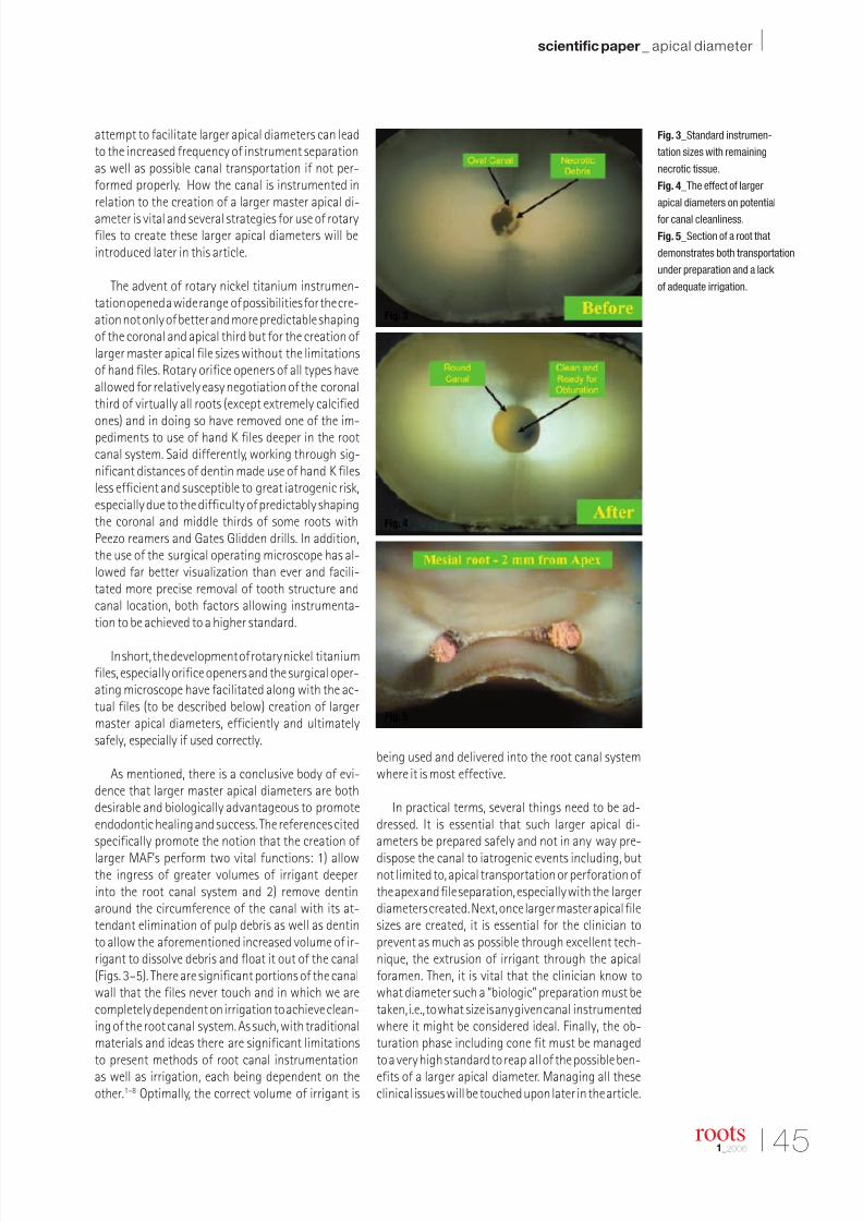
8/13/2019 Dente de Roots 1
http://slidepdf.com/reader/full/dente-de-roots-1 39/58
I 45
scientific paper _ apical diameter I
roots1_2006
attempt to facilitate larger apical diameters can leadto the increased frequency of instrument separationas well as possible canal transportation if not per-formed properly. How the canal is instrumented inrelation to the creation of a larger master apical di-
ameter is vital and several strategies for use of rotaryfiles to create these larger apical diameters will beintroduced later in this article.
The advent of rotary nickel titanium instrumen-tation opened a wide range of possibilities for the cre-ation not only of better and more predictable shapingof the coronal and apical third but for the creation of larger master apical file sizes without the limitationsof hand files. Rotary orifice openers of all types haveallowed for relatively easy negotiation of the coronalthird of virtually all roots (except extremely calcifiedones) and in doing so have removed one of the im-pediments to use of hand K files deeper in the rootcanal system. Said differently, working through sig-nificant distances of dentin made use of hand K filesless efficient and susceptible to great iatrogenic risk,especially due to the difficulty of predictably shapingthe coronal and middle thirds of some roots withPeezo reamers and Gates Glidden drills. In addition,the use of the surgical operating microscope has al-lowed far better visualization than ever and facili-tated more precise removal of tooth structure andcanal location, both factors allowing instrumenta-tion to be achieved to a higher standard.
In short, the development of rotary nickel titaniumfiles, especially orifice openers and the surgical oper-ating microscope have facilitated along with the ac-tual files (to be described below) creation of largermaster apical diameters, efficiently and ultimatelysafely, especially if used correctly.
As mentioned, there is a conclusive body of evi-dence that larger master apical diameters are bothdesirable and biologically advantageous to promoteendodontic healing and success. The references citedspecifically promote the notion that the creation of
larger MAF’s perform two vital functions: 1) allowthe ingress of greater volumes of irrigant deeperinto the root canal system and 2) remove dentinaround the circumference of the canal with its at-tendant elimination of pulp debris as well as dentinto allow the aforementioned increased volume of ir-rigant to dissolve debris and float it out of the canal(Figs. 3–5). There are significant portions of the canalwall that the files never touch and in which we arecompletely dependent on irrigation to achieve clean-ing of the root canal system. As such, with traditionalmaterials and ideas there are significant limitationsto present methods of root canal instrumentationas well as irrigation, each being dependent on theother.1–8 Optimally, the correct volume of irrigant is
being used and delivered into the root canal systemwhere it is most effective.
In practical terms, several things need to be ad-dressed. It is essential that such larger apical di-ameters be prepared safely and not in any way pre-
dispose the canal to iatrogenic events including, butnot limited to, apical transportation or perforation of the apex and file separation, especially with the largerdiameters created. Next, once larger master apical filesizes are created, it is essential for the clinician toprevent as much as possible through excellent tech-nique, the extrusion of irrigant through the apicalforamen. Then, it is vital that the clinician know towhat diameter such a “biologic” preparation must betaken, i.e., to what size is any given canal instrumentedwhere it might be considered ideal. Finally, the ob-turation phase including cone fit must be managedto a very high standard to reap all of the possible ben-efits of a larger apical diameter. Managing all theseclinical issues will be touched upon later in the article.
Fig. 3_Standard instrumen-
tation sizes with remaining
necrotic tissue.
Fig. 4_The effect of larger
apical diameters on potential
for canal cleanliness.
Fig. 5_Section of a root that
demonstrates both transportation
under preparation and a lack
of adequate irrigation.
Fig. 3
Fig. 4
Fig. 5
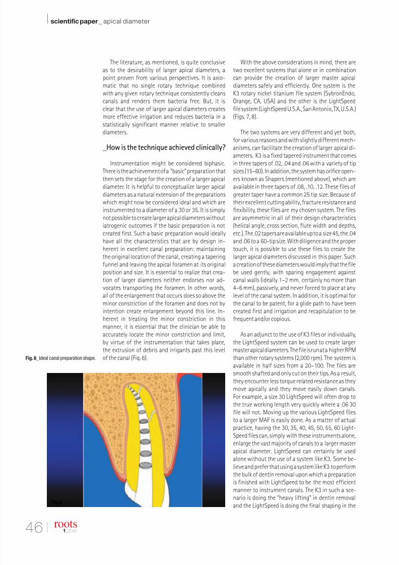
8/13/2019 Dente de Roots 1
http://slidepdf.com/reader/full/dente-de-roots-1 40/58
46 I
I scientific paper _ apical diameter
The literature, as mentioned, is quite conclusiveas to the desirability of larger apical diameters, apoint proven from various perspectives. It is axio-matic that no single rotary technique combinedwith any given rotary technique consistently cleans
canals and renders them bacteria free. But, it isclear that the use of larger apical diameters createsmore effective irrigation and reduces bacteria in astatistically significant manner relative to smallerdiameters.
_How is the technique achieved clinically?
Instrumentation might be considered biphasic.There is the achievement of a “basic” preparation thatthen sets the stage for the creation of a larger apicaldiameter. It is helpful to conceptualize larger apicaldiameters as a natural extension of the preparationswhich might now be considered ideal and which areinstrumented to a diameter of a 30 or 35. It is simplynot possible to create larger apical diameters withoutiatrogenic outcomes if the basic preparation is notcreated first. Such a basic preparation would ideallyhave all the characteristics that are by design in-herent in excellent canal preparation: maintainingthe original location of the canal, creating a taperingfunnel and leaving the apical foramen at its originalposition and size. It is essential to realize that crea-tion of larger diameters neither endorses nor ad-vocates transporting the foramen. In other words,
all of the enlargement that occurs does so above theminor constriction of the foramen and does not byintention create enlargement beyond this line. In-herent in treating the minor constriction in thismanner, it is essential that the clinician be able toaccurately locate the minor constriction and limit,by virtue of the instrumentation that takes place,the extrusion of debris and irrigants past this levelof the canal (Fig. 6).
With the above considerations in mind, there aretwo excellent systems that alone or in combinationcan provide the creation of larger master apicaldiameters safely and efficiently. One system is theK3 rotary nickel titanium file system (SybronEndo,
Orange, CA, USA) and the other is the LightSpeedfile system (LightSpeed U.S.A., San Antonio, TX, U.S.A.)(Figs. 7, 8).
The two systems are very different and yet both,for various reasons and with slightly different mech-anisms, can facilitate the creation of larger apical di-ameters. K3 is a fixed tapered instrument that comesin three tapers of .02, .04 and .06 with a variety of tipsizes (15–60). In addition, the system has orifice open-ers known as Shapers (mentioned above), which areavailable in three tapers of .08, .10, .12. These files of greater taper have a common 25 tip size. Because of their excellent cutting ability, fracture resistance andflexibility, these files are my chosen system. The filesare asymmetric in all of their design characteristics(helical angle, cross section, flute width and depths,etc.). The .02 tapers are available up to a size 45, the .04and .06 to a 60-tip size. With diligence and the propertouch, it is possible to use these files to create thelarger apical diameters discussed in this paper. Sucha creation of these diameters would imply that the filebe used gently, with sparing engagement againstcanal walls (ideally 1–2 mm, certainly no more than4–6 mm), passively, and never forced to place at any
level of the canal system. In addition, it is optimal forthe canal to be patent, for a glide path to have beencreated first and irrigation and recapitulation to befrequent and/or copious.
As an adjunct to the use of K3 files or individually,the LightSpeed system can be used to create largermaster apical diameters. The file is run at a higher RPMthan other rotary systems (2,000 rpm). The system isavailable in half sizes from a 20–100. The files aresmooth shafted and only cut on their tips. As a result,they encounter less torque related resistance as theymove apically and they move easily down canals.
For example, a size 30 LightSpeed will often drop tothe true working length very quickly where a .06 30file will not. Moving up the various LightSpeed filesto a larger MAF is easily done. As a matter of actualpractice, having the 30, 35, 40, 45, 50, 55, 60 Light-Speed files can, simply with these instruments alone,enlarge the vast majority of canals to a larger masterapical diameter. LightSpeed can certainly be usedalone without the use of a system like K3. Some be-lieve and prefer that using a system like K3 to performthe bulk of dentin removal upon which a preparationis finished with LightSpeed to be the most efficientmanner to instrument canals. The K3 in such a sce-nario is doing the “heavy lifting” in dentin removaland the LightSpeed is doing the final shaping in the
Fig. 6_Ideal canal preparation shape.
roots1_2006
Fig. 6
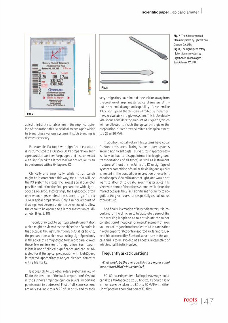
8/13/2019 Dente de Roots 1
http://slidepdf.com/reader/full/dente-de-roots-1 41/58
I 47
scientific paper _ apical diameter I
roots1_2006
apical third of the canal system. In the empirical opin-ion of the author, this is the ideal means upon whichto blend these various systems if such blending isdeemed necessary.
For example, if a tooth with significant curvatureis instrumented to a .06 25 or 30 K3 preparation, sucha preparation can then be gauged and instrumentedwith LightSpeed to a larger MAF (as desired) or it canbe performed with a .04 tapered K3.
Clinically and empirically, while not all canalsmight be instrumented this way, the author will usethe K3 system to create the largest apical diameterpossible and refine the final preparation with Light-Speed as desired. Interestingly, the LightSpeed oftenonly encounters minimal resistance to go from a30–60 apical preparation. Only a minor amount of shaping need be done or dentin be removed to allowthe canal to be opened to a larger master apical di-ameter (Figs. 9, 10).
The only drawback to LightSpeed instrumentation
which might be viewed as the objection of a purist isthat because the instrument only cuts at its tip end,the preparations which result using LightSpeed onlyin the apical third might tend to be more parallel overthose few millimeters of preparation. Such paral-lelism is not of clinical significance and can be ad- justed for if the apical preparation with LightSpeedis tapered appropriately and/or blended correctlywith a file like K3.
Is it possible to use other rotary systems in lieu of K3 for the creation of the basic preparation? Yes, butin the author’s empirical opinion several importantpoints must be addressed. First of all, some systemsare only available to a MAF of 30 or 35 and by their
very design they have limited the clinician away fromthe creation of larger master apical diameters. With-out the extended range and capability of a system likeK3 or LightSpeed, the clinician is limited by the largestfile size available in a given system. This is absolutelyvital if one considers the amount of irrigation, whichwill be allowed to reach the apical third given thepreparation in its entirety, is limited at its apical extentto a 25 or 30 MAF.
In addition, not all rotary file systems have equalfracture resistance. Taking some rotary systemsaround significant pigtail curvatures inappropriatelyis likely to lead to disappointment in ledging (andtransportations of all types) as well as instrumentfracture. Without the flexibility of a K3 or LightSpeed
system or something of similar flexibility, one quicklyis limited in the possibilities in creation of excellentcanal shapes. Viewed in another light, one would notwant to attempt to create larger master apical filesizes with some of the other systems available on themarket because they lack significant flexibility to ne-gotiate the given curvature, especially a small radiusof curvature.
And finally, in creation of larger diameters, it is im-portant for the clinician to be absolutely sure of thetrue working length so as to not violate the minorconstriction of the apical foramen. Placement of large
volumes of irrigant into the apical third in canals thathave been perforated or transported are far more sus-ceptible to morbidity. Such misadventure in the api-cal third is to be avoided at all costs, irrespective of which canal third is involved.
_Frequently asked questions
_What would be the average MAF for a molar canal
such as the MB of a lower molar?
50–60, case dependent. Taking the average molarcanal to a 06-tapered size 35 tip size, K3 could easilyin most cases be taken to a 50 or a 60 MAF with eitherLightSpeed or a combination of K3 files.
Fig. 7_The K3 rotary nickel
titanium system by SybronEndo,
Orange, CA, USA.
Fig. 8_The LightSpeed rotary
nickel titanium system by
LightSpeed Technologies,
San Antonio, TX, USA.
Fig. 7
Fig. 8
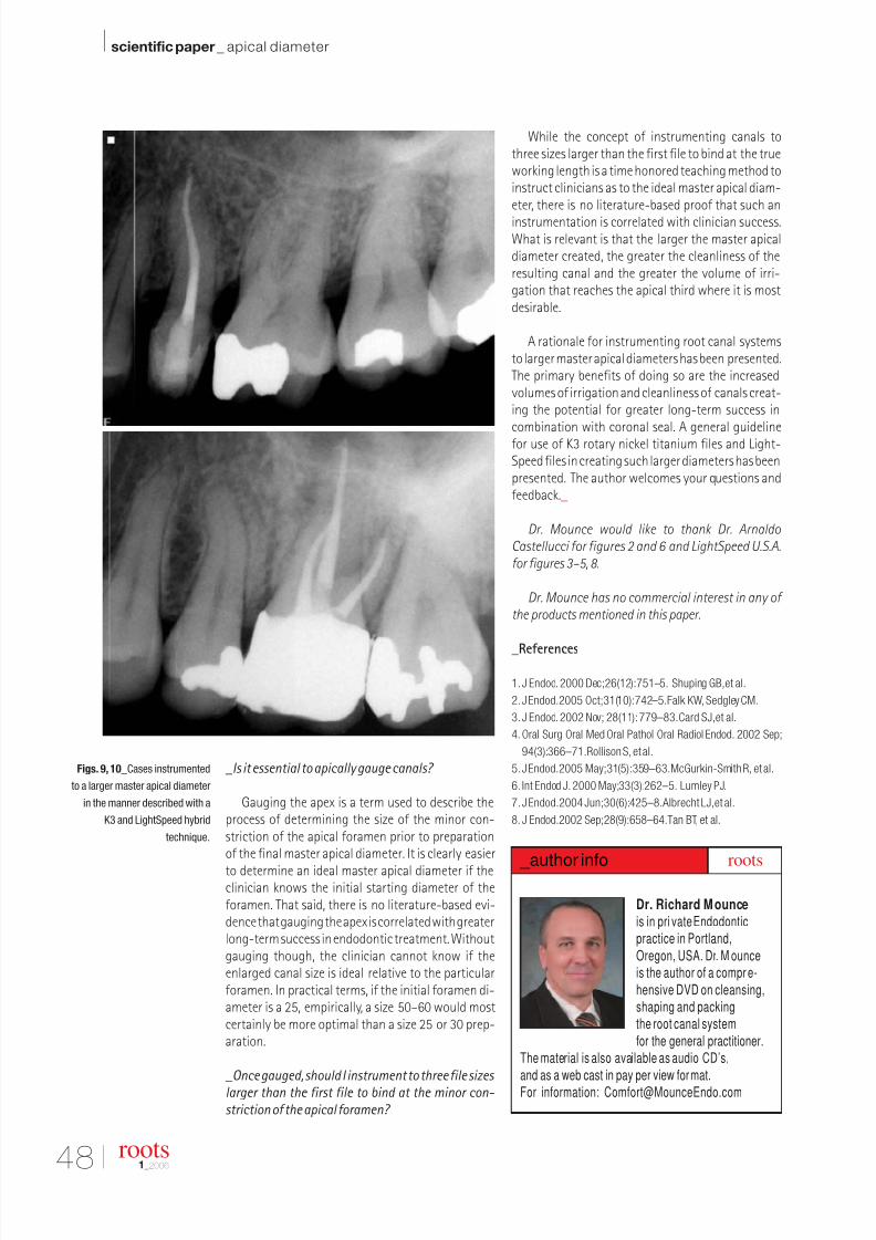
8/13/2019 Dente de Roots 1
http://slidepdf.com/reader/full/dente-de-roots-1 42/58
48 I
I scientific paper _ apical diameter
_Is it essential to apically gauge canals?
Gauging the apex is a term used to describe theprocess of determining the size of the minor con-striction of the apical foramen prior to preparationof the final master apical diameter. It is clearly easier
to determine an ideal master apical diameter if theclinician knows the initial starting diameter of theforamen. That said, there is no literature-based evi-dence that gauging the apex is correlated with greaterlong-term success in endodontic treatment. Withoutgauging though, the clinician cannot know if theenlarged canal size is ideal relative to the particularforamen. In practical terms, if the initial foramen di-ameter is a 25, empirically, a size 50–60 would mostcertainly be more optimal than a size 25 or 30 prep-aration.
_Once gauged, should I instrument to three file sizes
larger than the first file to bind at the minor con-
striction of the apical foramen?
While the concept of instrumenting canals tothree sizes larger than the first file to bind at the trueworking length is a time honored teaching method toinstruct clinicians as to the ideal master apical diam-eter, there is no literature-based proof that such an
instrumentation is correlated with clinician success.What is relevant is that the larger the master apicaldiameter created, the greater the cleanliness of theresulting canal and the greater the volume of irri-gation that reaches the apical third where it is mostdesirable.
A rationale for instrumenting root canal systemsto larger master apical diameters has been presented.The primary benefits of doing so are the increasedvolumes of irrigation and cleanliness of canals creat-ing the potential for greater long-term success incombination with coronal seal. A general guidelinefor use of K3 rotary nickel titanium files and Light-Speed files in creating such larger diameters has beenpresented. The author welcomes your questions andfeedback._
Dr. Mounce would like to thank Dr. Arnaldo
Castellucci for figures 2 and 6 and LightSpeed U.S.A.
for figures 3–5, 8.
Dr. Mounce has no commercial interest in any of
the products mentioned in this paper.
_References
1.J Endod. 2000 Dec;26(12):751–5. Shuping GB,et al.
2. J Endod.2005 Oct;31(10):742–5.Falk KW, Sedgley CM.
3.J Endod. 2002 Nov; 28(11):779–83.Card SJ,et al.
4. Oral Surg Oral Med Oral Pathol Oral Radiol Endod. 2002 Sep;
94(3):366–71.Rollison S,et al.
5. J Endod.2005 May;31(5):359–63.McGurkin-Smith R, et al.
6. Int Endod J. 2000 May;33(3):262–5. Lumley PJ.
7. J Endod.2004 Jun;30(6):425–8.Albrecht LJ,et al.
8. J Endod.2002 Sep;28(9):658–64.Tan BT, et al.
Figs. 9, 10_Cases instrumented
to a larger master apical diameter
in the manner described with a
K3 and LightSpeed hybrid
technique.
roots1_2006
Dr. Richard Mounce
is in pri vate Endodonticpractice in Portland,
Oregon, USA. Dr. M ounceis the author of a compr e-
hensive DVD on cleansing,shaping and packingthe root canal system
for the general practitioner.The material is also available as audio CD’s,
and as a web cast in pay per view format.
For information: [email protected]
_author info roots
Fig. 9
Fig. 10
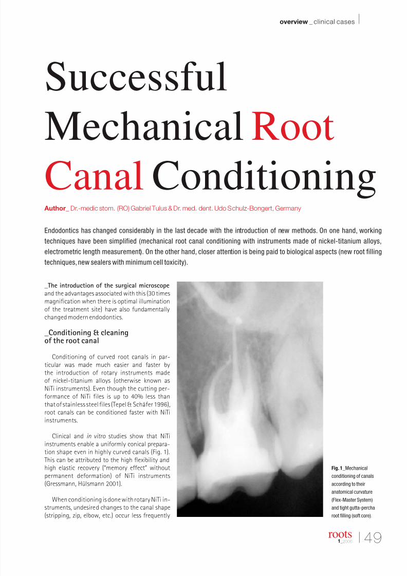
8/13/2019 Dente de Roots 1
http://slidepdf.com/reader/full/dente-de-roots-1 43/58
I 49
overview _ clinical cases I
roots1_2006
_The introduction of the surgical microscope
and the advantages associated with this (30 times
magnification when there is optimal illuminationof the treatment site) have also fundamentallychanged modern endodontics.
_Conditioning & cleaningof the root canal
Conditioning of curved root canals in par-ticular was made much easier and faster bythe introduction of rotary instruments madeof nickel-titanium alloys (otherwise known asNiTi instruments). Even though the cutting per-formance of NiTi files is up to 40% less than
that of stainless steel files (Tepel & Schäfer 1996),root canals can be conditioned faster with NiTiinstruments.
Clinical and in vitro studies show that NiTiinstruments enable a uniformly conical prepara-tion shape even in highly curved canals (Fig. 1).This can be attributed to the high flexibility andhigh elastic recovery (“memory effect” withoutpermanent deformation) of NiTi instruments(Gressmann, Hülsmann 2001).
When conditioning is done with rotary NiTi in-struments, undesired changes to the canal shape(stripping, zip, elbow, etc.) occur less frequently
Fig. 1_Mechanical
conditioning of canals
according to their
anatomical curvature
(Flex-Master System)
and tight gutta-percha
root filling (soft core).
Successful
Mechanical Root
Canal Conditioning Author_ Dr.-medic stom. (RO) Gabriel Tulus & Dr. med. dent. Udo Schulz-Bongert, Germany
Endodontics has changed considerably in the last decade with the introduction of new methods. On one hand, working
techniques have been simplified (mechanical root canal conditioning with instruments made of nickel-titanium alloys,
electrometric length measurement). On the other hand, closer attention is being paid to biological aspects (new root filling
techniques, new sealers with minimum cell toxicity).
Fig. 1
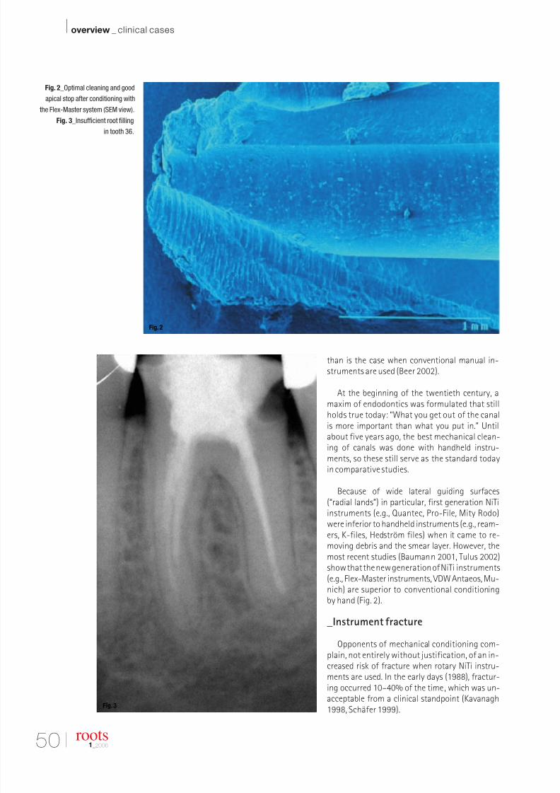
8/13/2019 Dente de Roots 1
http://slidepdf.com/reader/full/dente-de-roots-1 44/58
50 I
I overview _ clinical cases
roots1_2006
than is the case when conventional manual in-struments are used (Beer 2002).
At the beginning of the twentieth century, amaxim of endodontics was formulated that still
holds true today: “What you get out of the canalis more important than what you put in.” Untilabout five years ago, the best mechanical clean-ing of canals was done with handheld instru-ments, so these still serve as the standard todayin comparative studies.
Because of wide lateral guiding surfaces(“radial lands”) in particular, first generation NiTiinstruments (e.g., Quantec, Pro-File, Mity Rodo)were inferior to handheld instruments (e.g., ream-ers, K-files, Hedström files) when it came to re-moving debris and the smear layer. However, the
most recent studies (Baumann 2001, Tulus 2002)show that the new generation of NiTi instruments(e.g., Flex-Master instruments, VDW Antaeos, Mu-nich) are superior to conventional conditioningby hand (Fig. 2).
_Instrument fracture
Opponents of mechanical conditioning com-plain, not entirely without justification, of an in-creased risk of fracture when rotary NiTi instru-ments are used. In the early days (1988), fractur-ing occurred 10–40% of the time, which was un-acceptable from a clinical standpoint (Kavanagh1998, Schäfer 1999).
Fig. 2_Optimal cleaning and good
apical stop after conditioning with
the Flex-Master system (SEM view).
Fig. 3_Insufficient root filling
in tooth 36.
Fig. 2
Fig. 3
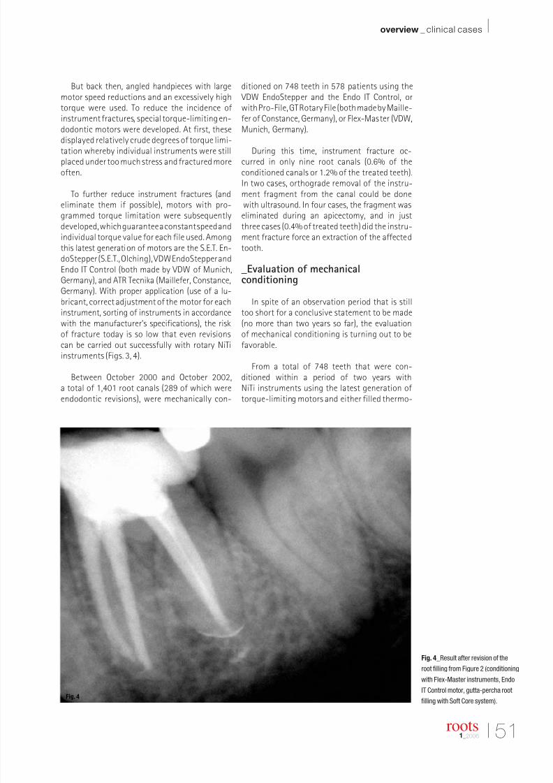
8/13/2019 Dente de Roots 1
http://slidepdf.com/reader/full/dente-de-roots-1 45/58
I 51
overview _ clinical cases I
roots1_2006
But back then, angled handpieces with largemotor speed reductions and an excessively hightorque were used. To reduce the incidence ofinstrument fractures, special torque-limiting en-dodontic motors were developed. At first, these
displayed relatively crude degrees of torque limi-tation whereby individual instruments were stillplaced under too much stress and fractured moreoften.
To further reduce instrument fractures (andeliminate them if possible), motors with pro-grammed torque limitation were subsequentlydeveloped, which guarantee a constant speed andindividual torque value for each file used. Amongthis latest generation of motors are the S.E.T. En-doStepper (S.E.T., Olching), VDW EndoStepper andEndo IT Control (both made by VDW of Munich,Germany), and ATR Tecnika (Maillefer, Constance,Germany). With proper application (use of a lu-bricant, correct adjustment of the motor for eachinstrument, sorting of instruments in accordancewith the manufacturer’s specifications), the riskof fracture today is so low that even revisionscan be carried out successfully with rotary NiTiinstruments (Figs. 3, 4).
Between October 2000 and October 2002,a total of 1,401 root canals (289 of which wereendodontic revisions), were mechanically con-
ditioned on 748 teeth in 578 patients using the VDW EndoStepper and the Endo IT Control, orwith Pro-File, GT Rotary File (both made by Maille-fer of Constance, Germany), or Flex-Master (VDW,Munich, Germany).
During this time, instrument fracture oc-curred in only nine root canals (0.6% of theconditioned canals or 1.2% of the treated teeth).In two cases, orthograde removal of the instru-ment fragment from the canal could be donewith ultrasound. In four cases, the fragment waseliminated during an apicectomy, and in justthree cases (0.4% of treated teeth) did the instru-ment fracture force an extraction of the affectedtooth.
_Evaluation of mechanicalconditioning
In spite of an observation period that is stilltoo short for a conclusive statement to be made(no more than two years so far), the evaluationof mechanical conditioning is turning out to befavorable.
From a total of 748 teeth that were con-ditioned within a period of two years withNiTi instruments using the latest generation of torque-limiting motors and either filled thermo-
Fig. 4_Result after revision of the
root filling from Figure 2 (conditioning
with Flex-Master instruments, Endo
IT Control motor, gutta-percha root
filling with Soft Core system).Fig. 4
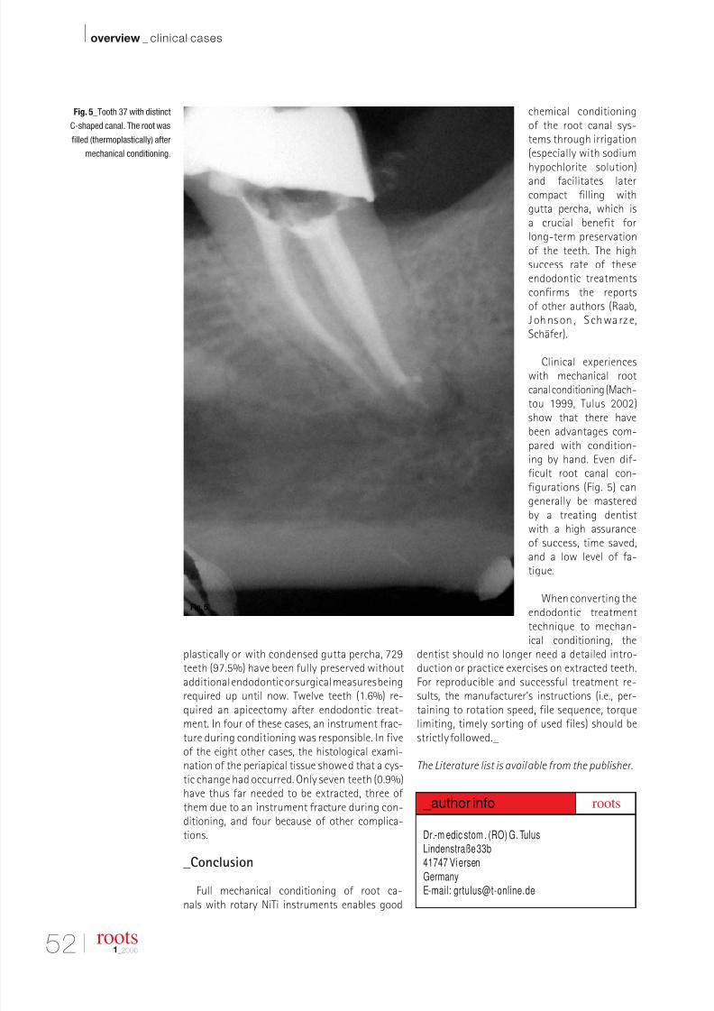
8/13/2019 Dente de Roots 1
http://slidepdf.com/reader/full/dente-de-roots-1 46/58
52 I
I overview _ clinical cases
roots1_2006
plastically or with condensed gutta percha, 729teeth (97.5%) have been fully preserved withoutadditional endodontic or surgical measures beingrequired up until now. Twelve teeth (1.6%) re-
quired an apicectomy after endodontic treat-ment. In four of these cases, an instrument frac-ture during conditioning was responsible. In fiveof the eight other cases, the histological exami-nation of the periapical tissue showed that a cys-tic change had occurred. Only seven teeth (0.9%)have thus far needed to be extracted, three of them due to an instrument fracture during con-ditioning, and four because of other complica-tions.
_Conclusion
Full mechanical conditioning of root ca-nals with rotary NiTi instruments enables good
chemical conditioningof the root canal sys-tems through irrigation(especially with sodiumhypochlorite solution)
and facilitates latercompact filling withgutta percha, which isa crucial benefit forlong-term preservationof the teeth. The highsuccess rate of theseendodontic treatmentsconfirms the reportsof other authors (Raab,J ohnson , S chwarze,Schäfer).
Clinical experienceswith mechanical rootcanal conditioning (Mach-tou 1999, Tulus 2002)show that there havebeen advantages com-pared with condition-ing by hand. Even dif-ficult root canal con-figurations (Fig. 5) cangenerally be masteredby a treating dentist
with a high assuranceof success, time saved,and a low level of fa-tigue.
When converting theendodontic treatmenttechnique to mechan-ical conditioning, the
dentist should no longer need a detailed intro-duction or practice exercises on extracted teeth.For reproducible and successful treatment re-sults, the manufacturer’s instructions (i.e., per-
taining to rotation speed, file sequence, torquelimiting, timely sorting of used files) should bestrictly followed._
The Literature list is available from the publisher.
Fig. 5_Tooth 37 with distinct
C-shaped canal. The root was
filled (thermoplastically) after
mechanical conditioning.
Dr.-m edic stom . (RO) G. TulusLindenstraße 33b41747 Vi ersen
Germany
E-mail: [email protected]
_author info roots
Fig. 5
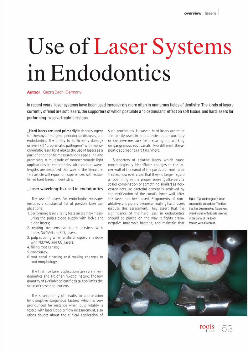
8/13/2019 Dente de Roots 1
http://slidepdf.com/reader/full/dente-de-roots-1 47/58
I 53
overview _ lasers I
roots1_2006
_Hard lasers are used primarily in dental surgery,for therapy of marginal periodontal diseases, andendodontics. The ability to sufficiently damageor even kill “problematic pathogens” with mono-chromatic laser light makes the use of lasers as apart of endodontic measures look appealing andpromising. A multitude of monochromatic lightapplications in endodontics with various wave-lengths are described this way in the literature.This article will report on experiences with estab-lished hard lasers in dentistry.
_Laser wavelengths used in endodontics
The use of lasers for endodontic measuresincludes a substantial list of possible laser ap-plications:1. performing laser vitality tests on teeth by meas-
uring the pulp’s blood supply with HeNe anddiode lasers;
2. treating oversensitive tooth cervices withdiode, Nd:YAG and CO2 lasers;
3. pulp capping when artificial exposure is donewith Nd:YAG and CO2 lasers;
4. filling root canals;5. endosurgy;6. root canal cleaning and making changes to
root morphology.
The first five laser applications are rare in en-dodontics and are of an “exotic” nature. The lowquantity of available scientific data also limits thevalue of these applications.
The susceptibility of results to adulterationby disruptive exogenous factors, which is verypronounced for instance when pulp vitality istested with laser Doppler flow measurement, alsoraises doubts about the clinical application of
such procedures. However, hard lasers are morefrequently used in endodontics as an auxiliaryor exclusive measure for preparing and workingon gangrenous root canals. Two different thera-peutic approaches are taken here.
Supporters of ablative lasers, which causemorphologically identifiable changes to the in-ner wall of the canal of the particular root to betreated, now even claim that they no longer regarda root filling in the proper sense (gutta-percha
sealer combination or something similar) as nec-essary because bacterial density is achieved bythe vitrification of the canal’s inner wall afterthe laser has been used. Proponents of non-ablative and purely decontaminating hard lasersdispute this assessment. They assert that thesignificance of the hard laser in endodonticsshould be placed on the way it fights gram-negative anaerobic bacteria, and maintain that
Fig. 1_Typical image of a laser
endodontic procedure. The fiber
that has been marked (to prevent
over-instrumentation) is inserted
in the canal of the tooth
treated with a trephine.
Use of Laser Systemsin Endodontics
Author_ Georg Bach, Germany
In recent years, laser systems have been used increasingly more often in numerous fields of dentistry. The kinds of lasers
currently offered are soft lasers, the supporters of which postulate a “biostimulant” effect on soft tissue,and hard lasers for
performing invasive treatment steps.
Fig. 1
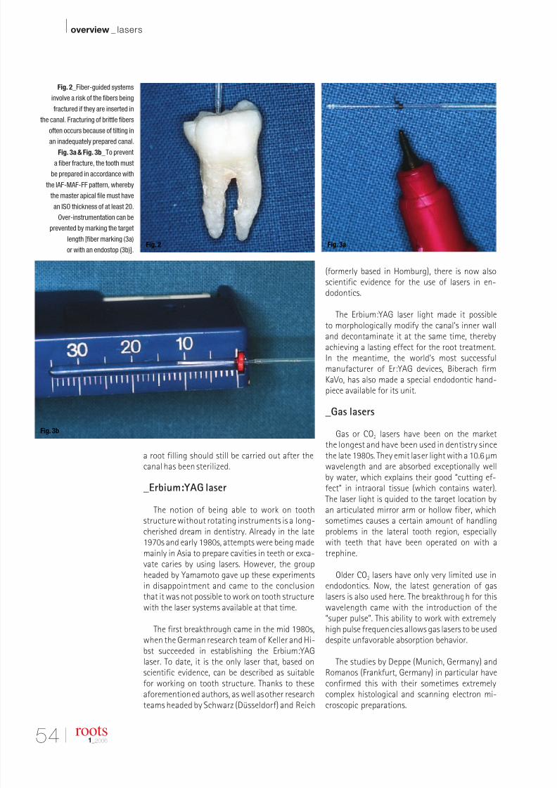
8/13/2019 Dente de Roots 1
http://slidepdf.com/reader/full/dente-de-roots-1 48/58
54 I
I overview _ lasers
roots1_2006
a root filling should still be carried out after thecanal has been sterilized.
_Erbium:YAG laser
The notion of being able to work on toothstructure without rotating instruments is a long-cherished dream in dentistry. Already in the late1970s and early 1980s, attempts were being made
mainly in Asia to prepare cavities in teeth or exca-vate caries by using lasers. However, the groupheaded by Yamamoto gave up these experimentsin disappointment and came to the conclusionthat it was not possible to work on tooth structurewith the laser systems available at that time.
The first breakthrough came in the mid 1980s,when the German research team of Keller and Hi-bst succeeded in establishing the Erbium:YAGlaser. To date, it is the only laser that, based onscientific evidence, can be described as suitablefor working on tooth structure. Thanks to theseaforementioned authors, as well as other researchteams headed by Schwarz (Düsseldorf) and Reich
(formerly based in Homburg), there is now alsoscientific evidence for the use of lasers in en-dodontics.
The Erbium:YAG laser light made it possibleto morphologically modify the canal’s inner walland decontaminate it at the same time, therebyachieving a lasting effect for the root treatment.In the meantime, the world’s most successfulmanufacturer of Er:YAG devices, Biberach firmKaVo, has also made a special endodontic hand-piece available for its unit.
_Gas lasers
Gas or CO2 lasers have been on the marketthe longest and have been used in dentistry sincethe late 1980s. They emit laser light with a 10.6 µmwavelength and are absorbed exceptionally wellby water, which explains their good “cutting ef-fect” in intraoral tissue (which contains water).The laser light is guided to the target location byan articulated mirror arm or hollow fiber, whichsometimes causes a certain amount of handlingproblems in the lateral tooth region, especiallywith teeth that have been operated on with a
trephine.
Older CO2 lasers have only very limited use inendodontics. Now, the latest generation of gaslasers is also used here. The breakthrough for thiswavelength came with the introduction of the“super pulse”. This ability to work with extremelyhigh pulse frequencies allows gas lasers to be useddespite unfavorable absorption behavior.
The studies by Deppe (Munich, Germany) andRomanos (Frankfurt, Germany) in particular haveconfirmed this with their sometimes extremelycomplex histological and scanning electron mi-croscopic preparations.
Fig. 2_Fiber-guided systems
involve a risk of the fibers being
fractured if they are inserted in
the canal. Fracturing of brittle fibers
often occurs because of tilting in
an inadequately prepared canal.
Fig. 3a & Fig. 3b_To prevent
a fiber fracture, the tooth must
be prepared in accordance with
the IAF-MAF-FF pattern, whereby
the master apical file must have
an ISO thickness of at least 20.
Over-instrumentation can be
prevented by marking the target
length [fiber marking (3a)
or with an endostop (3b)].Fig. 2 Fig. 3a
Fig. 3b
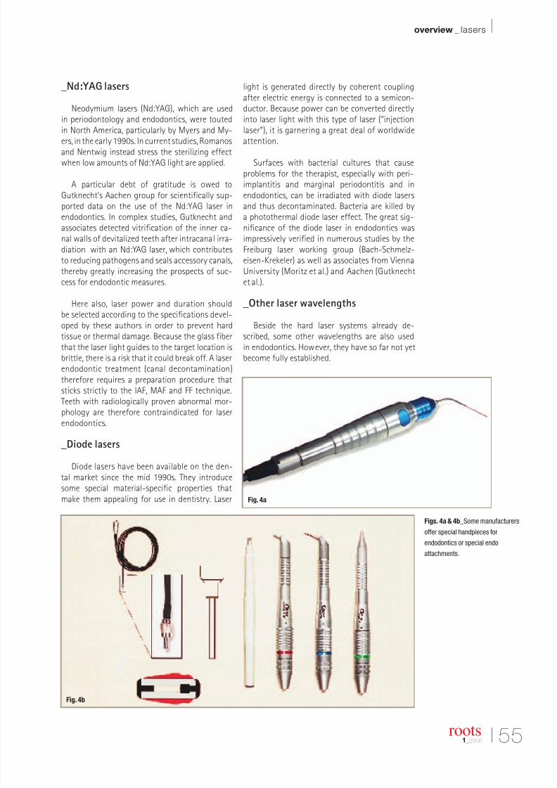
8/13/2019 Dente de Roots 1
http://slidepdf.com/reader/full/dente-de-roots-1 49/58
I 55
overview _ lasers I
roots1_2006
_Nd:YAG lasers
Neodymium lasers (Nd:YAG), which are usedin periodontology and endodontics, were toutedin North America, particularly by Myers and My-
ers, in the early 1990s. In current studies, Romanosand Nentwig instead stress the sterilizing effectwhen low amounts of Nd:YAG light are applied.
A particular debt of gratitude is owed toGutknecht’s Aachen group for scientifically sup-ported data on the use of the Nd:YAG laser inendodontics. In complex studies, Gutknecht andassociates detected vitrification of the inner ca-nal walls of devitalized teeth after intracanal irra-diation with an Nd:YAG laser, which contributesto reducing pathogens and seals accessory canals,thereby greatly increasing the prospects of suc-cess for endodontic measures.
Here also, laser power and duration shouldbe selected according to the specifications devel-oped by these authors in order to prevent hardtissue or thermal damage. Because the glass fiberthat the laser light guides to the target location isbrittle, there is a risk that it could break off. A laserendodontic treatment (canal decontamination)therefore requires a preparation procedure thatsticks strictly to the IAF, MAF and FF technique.Teeth with radiologically proven abnormal mor-
phology are therefore contraindicated for laserendodontics.
_Diode lasers
Diode lasers have been available on the den-tal market since the mid 1990s. They introducesome special material-specific properties thatmake them appealing for use in dentistry. Laser
light is generated directly by coherent couplingafter electric energy is connected to a semicon-ductor. Because power can be converted directlyinto laser light with this type of laser (“injectionlaser”), it is garnering a great deal of worldwide
attention.
Surfaces with bacterial cultures that causeproblems for the therapist, especially with peri-implantitis and marginal periodontitis and inendodontics, can be irradiated with diode lasersand thus decontaminated. Bacteria are killed bya photothermal diode laser effect. The great sig-nificance of the diode laser in endodontics wasimpressively verified in numerous studies by theFreiburg laser working group (Bach-Schmelz-eisen-Krekeler) as well as associates from ViennaUniversity (Moritz et al.) and Aachen (Gutknechtet al.).
_Other laser wavelengths
Beside the hard laser systems already de-scribed, some other wavelengths are also usedin endodontics. However, they have so far not yetbecome fully established.
Figs. 4a & 4b_Some manufacturers
offer special handpieces for
endodontics or special endo
attachments.
Fig. 4a
Fig. 4b
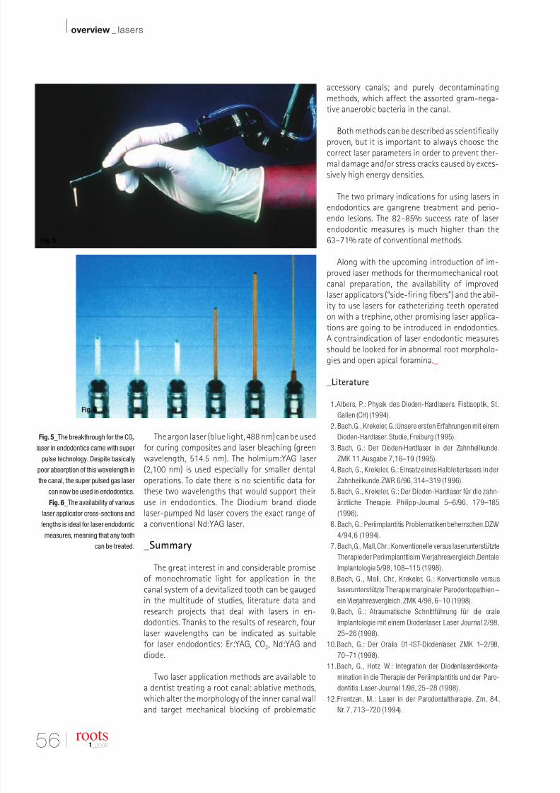
8/13/2019 Dente de Roots 1
http://slidepdf.com/reader/full/dente-de-roots-1 50/58
56 I
I overview _ lasers
roots1_2006
The argon laser (blue light, 488 nm) can be usedfor curing composites and laser bleaching (greenwavelength, 514.5 nm). The holmium:YAG laser(2,100 nm) is used especially for smaller dentaloperations. To date there is no scientific data forthese two wavelengths that would support theiruse in endodontics. The Diodium brand diodelaser-pumped Nd laser covers the exact range ofa conventional Nd:YAG laser.
_Summary
The great interest in and considerable promiseof monochromatic light for application in thecanal system of a devitalized tooth can be gaugedin the multitude of studies, literature data andresearch projects that deal with lasers in en-dodontics. Thanks to the results of research, fourlaser wavelengths can be indicated as suitablefor laser endodontics: Er:YAG, CO2, Nd:YAG anddiode.
Two laser application methods are available toa dentist treating a root canal: ablative methods,which alter the morphology of the inner canal walland target mechanical blocking of problematic
accessory canals; and purely decontaminatingmethods, which affect the assorted gram-nega-tive anaerobic bacteria in the canal.
Both methods can be described as scientifically
proven, but it is important to always choose thecorrect laser parameters in order to prevent ther-mal damage and/or stress cracks caused by exces-sively high energy densities.
The two primary indications for using lasers inendodontics are gangrene treatment and perio-endo lesions. The 82–85% success rate of laserendodontic measures is much higher than the63–71% rate of conventional methods.
Along with the upcoming introduction of im-proved laser methods for thermomechanical rootcanal preparation, the availability of improvedlaser applicators (“side-firing fibers”) and the abil-ity to use lasers for catheterizing teeth operatedon with a trephine, other promising laser applica-tions are going to be introduced in endodontics.A contraindication of laser endodontic measuresshould be looked for in abnormal root morpholo-gies and open apical foramina._
_Literature
1.Albers, P.: Physik des Dioden-Hardlasers. Fisbaoptik, St.
Gallen (CH) (1994).2. Bach,G., Krekeler,G.:Unsere ersten Erfahrungen mit einem
Dioden-Hardlaser. Studie, Freiburg (1995).
3. Bach, G.: Der Dioden-Hardlaser in der Zahnheilkunde.
ZMK 11,Ausgabe 7,16–19 (1995).
4. Bach, G., Krekeler, G.: Einsatz eines Halbleiterlasers in der
Zahnheilkunde.ZWR 6/96,314–319 (1996).
5. Bach, G., Krekeler, G.: Der Dioden-Hardlaser für die zahn-
ärztliche Therapie. Philipp-Journal 5–6/96, 179–185
(1996).
6. Bach, G.: Periimplantitis Problematiken beherrschen.DZW
4/94,6 (1994).
7. Bach,G., Mall,Chr.:Konventionelle versus laserunterstützte
Therapie der Periimplantitis im Vierjahresvergleich.DentaleImplantologie 5/98, 108–115 (1998).
8.Bach, G., Mall, Chr., Krekeler, G.: Konventionelle versus
laserunterstützte Therapie marginaler Parodontopathien –
ein Vierjahresvergleich. ZMK 4/98, 6–10 (1998).
9. Bach, G.: Atraumatische Schnittführung für die orale
Implantologie mit einem Diodenlaser. Laser Journal 2/98,
25–26 (1998).
10.Bach, G.: Der Oralia 01-IST-Diodenlaser. ZMK 1–2/98,
70–71 (1998).
11.Bach, G., Hotz, W.: Integration der Diodenlaserdekonta-
mination in die Therapie der Periimplantitis und der Paro-
dontitis. Laser-Journal 1/98, 25–28 (1998).
12.Frentzen, M.: Laser in der Parodontaltherapie. Zm, 84,
Nr. 7, 713–720 (1994).
Fig. 5_The breakthrough for the CO2
laser in endodontics came with super
pulse technology. Despite basically
poor absorption of this wavelength in
the canal, the super pulsed gas laser
can now be used in endodontics.
Fig. 6_The availability of various
laser applicator cross-sections and
lengths is ideal for laser endodontic
measures, meaning that any tooth
can be treated.
Fig. 5
Fig. 6
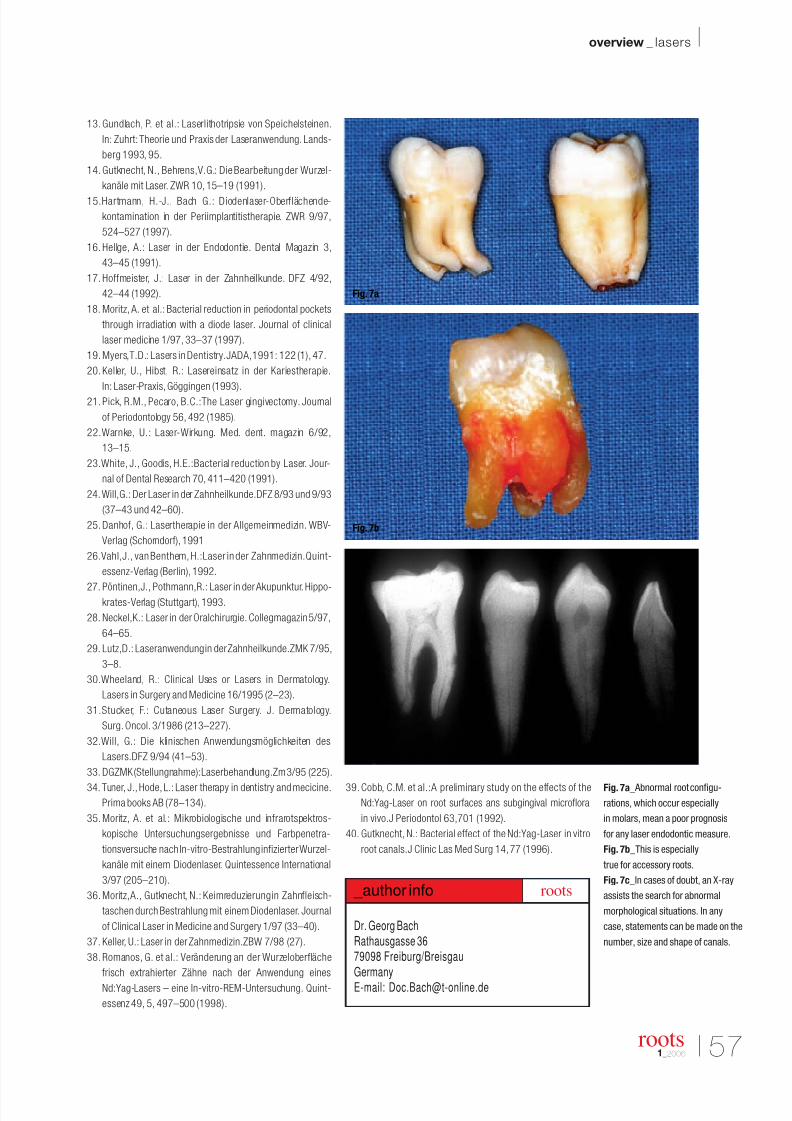
8/13/2019 Dente de Roots 1
http://slidepdf.com/reader/full/dente-de-roots-1 51/58
I 57
overview _ lasers I
roots1_2006
13. Gundlach, P. et al.: Laserlithotripsie von Speichelsteinen.
In: Zuhrt: Theorie und Praxis der Laseranwendung. Lands-
berg 1993, 95.
14. Gutknecht, N., Behrens,V.G.: Die Bearbeitung der Wurzel-
kanäle mit Laser. ZWR 10,15–19 (1991).
15.Hartmann, H.-J., Bach G.: Diodenlaser-Oberflächende-
kontamination in der Periimplantitistherapie. ZWR 9/97,
524–527 (1997).
16. Hellge, A.: Laser in der Endodontie. Dental Magazin 3,
43–45 (1991).
17. Hoffmeister, J.: Laser in der Zahnheilkunde. DFZ 4/92,
42–44 (1992).
18. Moritz, A. et al.: Bacterial reduction in periodontal pockets
through irradiation with a diode laser. Journal of clinical
laser medicine 1/97, 33–37 (1997).
19. Myers,T.D.: Lasers in Dentistry.JADA,1991: 122 (1), 47.
20. Keller, U., Hibst, R.: Lasereinsatz in der Kariestherapie.
In: Laser-Praxis,Göggingen (1993).
21. Pick, R.M., Pecaro, B.C.:The Laser gingivectomy. Journal
of Periodontology 56, 492 (1985).
22.Warnke, U.: Laser-Wirkung. Med. dent. magazin 6/92,
13–15.
23.White, J., Goodis, H.E.:Bacterial reduction by Laser. Jour-
nal of Dental Research 70, 411–420 (1991).
24.Will,G.: Der Laser in der Zahnheilkunde.DFZ 8/93 und 9/93
(37–43 und 42–60).
25. Danhof, G.: Lasertherapie in der Allgemeinmedizin. WBV-
Verlag (Schorndorf),1991
26.Vahl,J., van Benthem, H.:Laser in der Zahnmedizin.Quint-
essenz-Verlag (Berlin),1992.
27. Pöntinen,J., Pothmann,R.: Laser in der Akupunktur. Hippo-krates-Verlag (Stuttgart),1993.
28. Neckel,K.: Laser in der Oralchirurgie. Collegmagazin 5/97,
64–65.
29. Lutz,D.: Laseranwendung in der Zahnheilkunde.ZMK 7/95,
3–8.
30.Wheeland, R.: Clinical Uses or Lasers in Dermatology.
Lasers in Surgery and Medicine 16/1995 (2–23).
31.Stucker, F.: Cutaneous Laser Surgery. J. Dermatology.
Surg. Oncol. 3/1986 (213–227).
32.Will, G.: Die klinischen Anwendungsmöglichkeiten des
Lasers.DFZ 9/94 (41–53).
33. DGZMK (Stellungnahme):Laserbehandlung.Zm 3/95 (225).
34. Tuner, J., Hode,L.: Laser therapy in dentistry and mecicine.Prima books AB (78–134).
35. Moritz, A. et al.: Mikrobiologische und infrarotspektros-
kopische Untersuchungsergebnisse und Farbpenetra-
tionsversuche nach In-vitro-Bestrahlung infizierter Wurzel-
kanäle mit einem Diodenlaser. Quintessence International
3/97 (205–210).
36. Moritz,A., Gutknecht, N.: Keimreduzierung in Zahnfleisch-
taschen durch Bestrahlung mit einem Diodenlaser. Journal
of Clinical Laser in Medicine and Surgery 1/97 (33–40).
37. Keller, U.: Laser in der Zahnmedizin.ZBW 7/98 (27).
38. Romanos, G. et al.: Veränderung an der Wurzeloberfläche
frisch extrahierter Zähne nach der Anwendung eines
Nd:Yag-Lasers – eine In-vitro-REM-Untersuchung. Quint-
essenz 49, 5, 497–500 (1998).
39. Cobb, C.M. et al.:A preliminary study on the effects of theNd:Yag-Laser on root surfaces ans subgingival microflora
in vivo.J Periodontol 63,701 (1992).
40. Gutknecht, N.: Bacterial effect of the Nd:Yag-Laser in vitro
root canals.J Clinic Las Med Surg 14,77 (1996).
Fig. 7a_ Abnormal root configu-
rations, which occur especially
in molars, mean a poor prognosis
for any laser endodontic measure.
Fig. 7b_This is especially
true for accessory roots.
Fig. 7c_In cases of doubt, an X-ray
assists the search for abnormal
morphological situations. In any
case, statements can be made on the
number, size and shape of canals.
Dr. Georg BachRathausgasse 3679098 Freiburg/Breisgau
Germany
E-mail: [email protected]
_author info roots
Fig. 7a
Fig. 7b
Fig. 7c
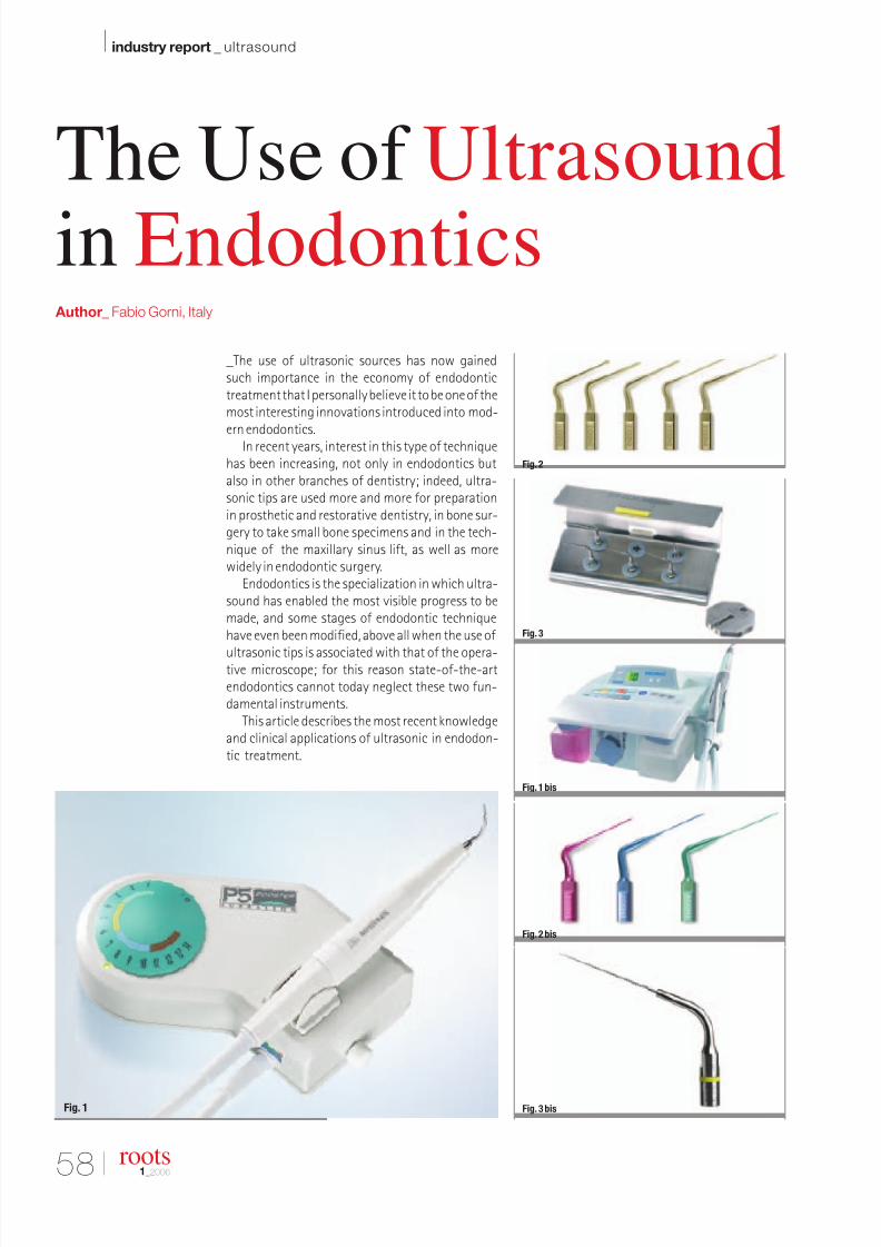
8/13/2019 Dente de Roots 1
http://slidepdf.com/reader/full/dente-de-roots-1 52/58
_The use of ultrasonic sources has now gainedsuch importance in the economy of endodontictreatment that I personally believe it to be one of themost interesting innovations introduced into mod-ern endodontics.
In recent years, interest in this type of techniquehas been increasing, not only in endodontics butalso in other branches of dentistry; indeed, ultra-sonic tips are used more and more for preparationin prosthetic and restorative dentistry, in bone sur-gery to take small bone specimens and in the tech-nique of the maxillary sinus lift, as well as morewidely in endodontic surgery.
Endodontics is the specialization in which ultra-sound has enabled the most visible progress to bemade, and some stages of endodontic techniquehave even been modified, above all when the use of
ultrasonic tips is associated with that of the opera-tive microscope; for this reason state-of-the-artendodontics cannot today neglect these two fun-damental instruments.
This article describes the most recent knowledgeand clinical applications of ultrasonic in endodon-tic treatment.
58 I
I industry report _ ultrasound
roots1_2006
The Use of Ultrasound
in Endodontics Author_ Fabio Gorni, Italy
Fig. 1
Fig. 2
Fig. 3
Fig. 1 bis
Fig. 2 bis
Fig. 3 bis
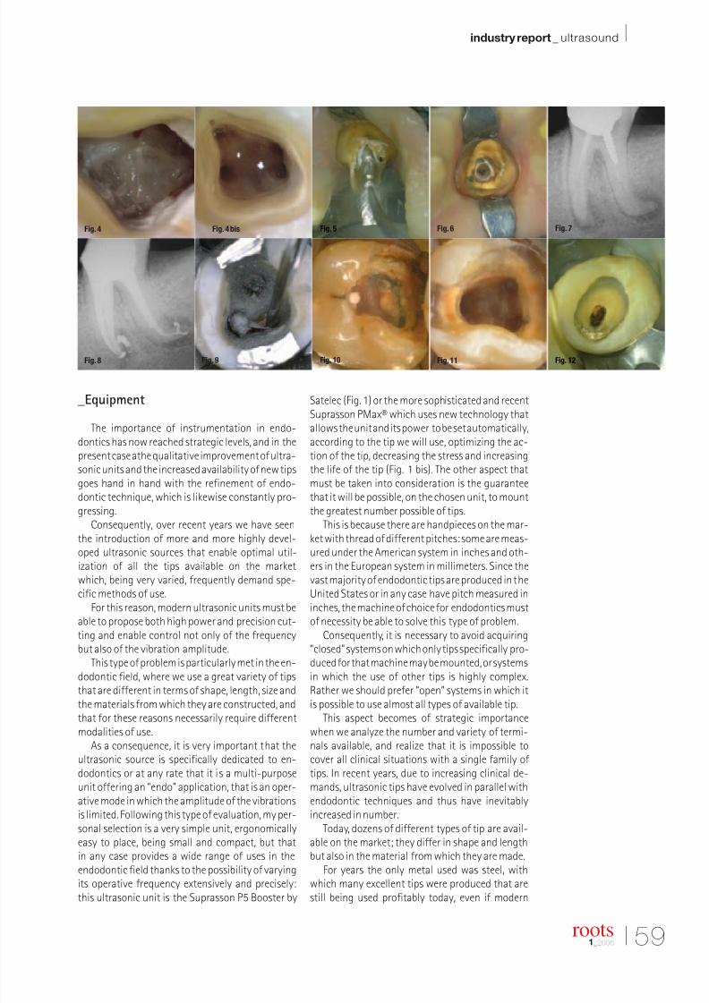
8/13/2019 Dente de Roots 1
http://slidepdf.com/reader/full/dente-de-roots-1 53/58
I 59
industry report _ ultrasound I
roots1_2006
_Equipment
The importance of instrumentation in endo-dontics has now reached strategic levels, and in thepresent case athe qualitative improvement of ultra-sonic units and the increased availability of new tipsgoes hand in hand with the refinement of endo-dontic technique, which is likewise constantly pro-gressing.
Consequently, over recent years we have seenthe introduction of more and more highly devel-oped ultrasonic sources that enable optimal util-ization of all the tips available on the marketwhich, being very varied, frequently demand spe-cific methods of use.
For this reason, modern ultrasonic units must beable to propose both high power and precision cut-ting and enable control not only of the frequencybut also of the vibration amplitude.
This type of problem is particularly met in the en-dodontic field, where we use a great variety of tipsthat are different in terms of shape, length, size and
the materials from which they are constructed, andthat for these reasons necessarily require differentmodalities of use.
As a consequence, it is very important that theultrasonic source is specifically dedicated to en-dodontics or at any rate that it is a multi-purposeunit offering an “endo” application, that is an oper-ative mode in which the amplitude of the vibrationsis limited. Following this type of evaluation, my per-sonal selection is a very simple unit, ergonomicallyeasy to place, being small and compact, but thatin any case provides a wide range of uses in theendodontic field thanks to the possibility of varyingits operative frequency extensively and precisely:this ultrasonic unit is the Suprasson P5 Booster by
Satelec (Fig. 1) or the more sophisticated and recentSuprasson PMax® which uses new technology thatallows the unit and its power to be set automatically,according to the tip we will use, optimizing the ac-tion of the tip, decreasing the stress and increasingthe life of the tip (Fig. 1 bis). The other aspect thatmust be taken into consideration is the guaranteethat it will be possible, on the chosen unit, to mountthe greatest number possible of tips.
This is because there are handpieces on the mar-ket with thread of different pitches: some are meas-ured under the American system in inches and oth-ers in the European system in millimeters. Since thevast majority of endodontic tips are produced in theUnited States or in any case have pitch measured ininches, the machine of choice for endodontics mustof necessity be able to solve this type of problem.
Consequently, it is necessary to avoid acquiring“closed” systems on which only tips specifically pro-duced for that machine may be mounted, or systemsin which the use of other tips is highly complex.Rather we should prefer “open” systems in which it
is possible to use almost all types of available tip.This aspect becomes of strategic importance
when we analyze the number and variety of termi-nals available, and realize that it is impossible tocover all clinical situations with a single family of tips. In recent years, due to increasing clinical de-mands, ultrasonic tips have evolved in parallel withendodontic techniques and thus have inevitablyincreased in number.
Today, dozens of different types of tip are avail-able on the market; they differ in shape and lengthbut also in the material from which they are made.
For years the only metal used was steel, withwhich many excellent tips were produced that arestill being used profitably today, even if modern
Fig. 4 bisFig. 4 Fig. 5 Fig. 6 Fig. 7
Fig. 8 Fig. 9 Fig. 10 Fig. 11 Fig. 12
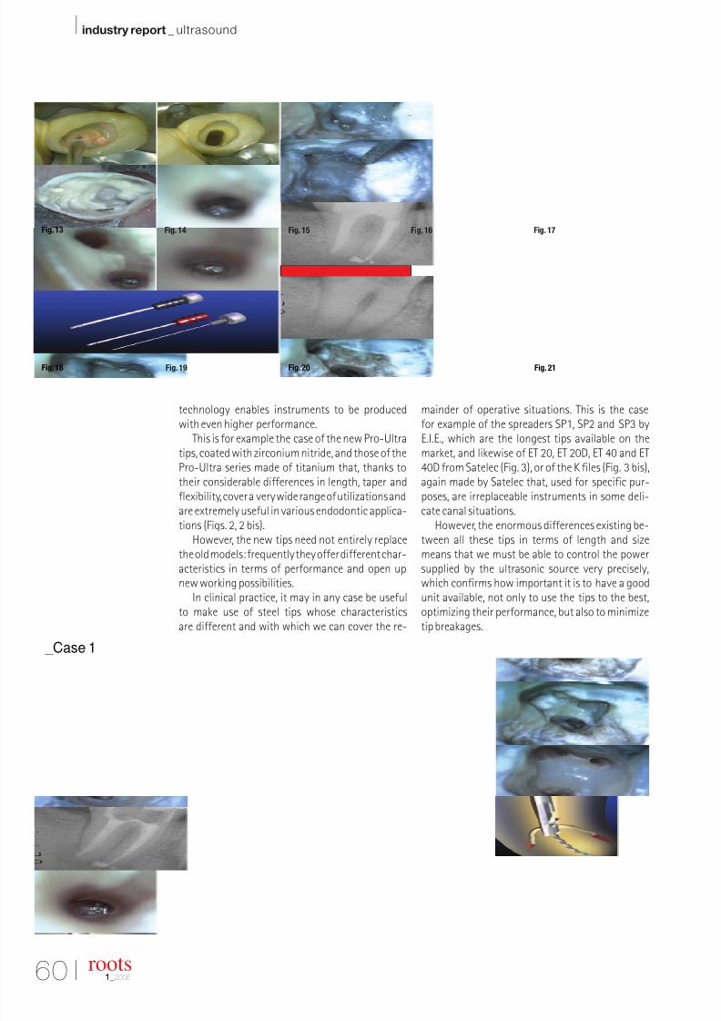
8/13/2019 Dente de Roots 1
http://slidepdf.com/reader/full/dente-de-roots-1 54/58
60 I
I industry report _ ultrasound
technology enables instruments to be producedwith even higher performance.
This is for example the case of the new Pro-Ultratips, coated with zirconium nitride, and those of thePro-Ultra series made of titanium that, thanks totheir considerable differences in length, taper andflexibility, cover a very wide range of utilizations andare extremely useful in various endodontic applica-tions (Figs. 2, 2 bis).
However, the new tips need not entirely replacethe old models: frequently they offer different char-acteristics in terms of performance and open upnew working possibilities.
In clinical practice, it may in any case be usefulto make use of steel tips whose characteristicsare different and with which we can cover the re-
mainder of operative situations. This is the casefor example of the spreaders SP1, SP2 and SP3 byE.I.E., which are the longest tips available on themarket, and likewise of ET 20, ET 20D, ET 40 and ET40D from Satelec (Fig. 3), or of the K files (Fig. 3 bis),again made by Satelec that, used for specific pur-poses, are irreplaceable instruments in some deli-cate canal situations.
However, the enormous differences existing be-
tween all these tips in terms of length and sizemeans that we must be able to control the powersupplied by the ultrasonic source very precisely,which confirms how important it is to have a goodunit available, not only to use the tips to the best,optimizing their performance, but also to minimizetip breakages.
roots1_2006
_Case 1
Fig. 13 Fig. 14 Fig. 15 Fig. 16 Fig. 17
Fig. 18 Fig. 19 Fig. 20 Fig. 21
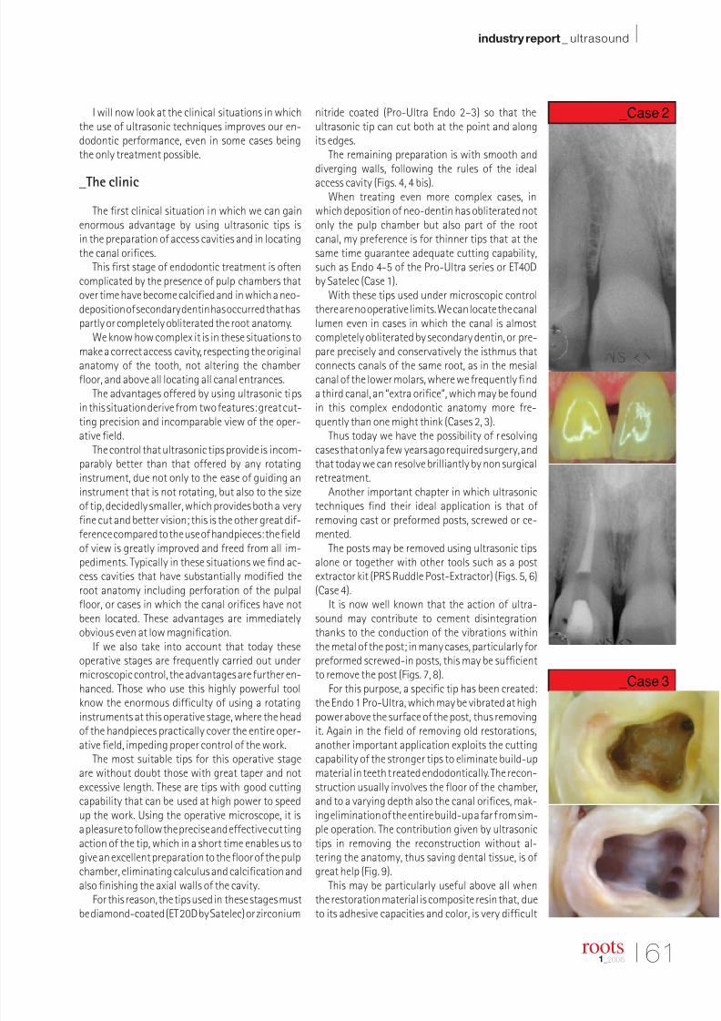
8/13/2019 Dente de Roots 1
http://slidepdf.com/reader/full/dente-de-roots-1 55/58
I 61
industry report _ ultrasound I
roots1_2006
I will now look at the clinical situations in whichthe use of ultrasonic techniques improves our en-dodontic performance, even in some cases beingthe only treatment possible.
_The clinic
The first clinical situation in which we can gainenormous advantage by using ultrasonic tips isin the preparation of access cavities and in locatingthe canal orifices.
This first stage of endodontic treatment is often
complicated by the presence of pulp chambers that
over time have become calcified and in which a neo-
deposition of secondary dentin has occurred that has
partly or completely obliterated the root anatomy.
We know how complex it is in these situations tomake a correct access cavity, respecting the originalanatomy of the tooth, not altering the chamberfloor, and above all locating all canal entrances.
The advantages offered by using ultrasonic tipsin this situation derive from two features: great cut-ting precision and incomparable view of the oper-ative field.
The control that ultrasonic tips provide is incom-parably better than that offered by any rotatinginstrument, due not only to the ease of guiding aninstrument that is not rotating, but also to the sizeof tip, decidedly smaller, which provides both a veryfine cut and better vision; this is the other great dif-
ference compared to the use of handpieces: the fieldof view is greatly improved and freed from all im-pediments. Typically in these situations we find ac-cess cavities that have substantially modified theroot anatomy including perforation of the pulpalfloor, or cases in which the canal orifices have notbeen located. These advantages are immediatelyobvious even at low magnification.
If we also take into account that today theseoperative stages are frequently carried out undermicroscopic control, the advantages are further en-hanced. Those who use this highly powerful toolknow the enormous difficulty of using a rotating
instruments at this operative stage, where the headof the handpieces practically cover the entire oper-ative field, impeding proper control of the work.
The most suitable tips for this operative stageare without doubt those with great taper and notexcessive length. These are tips with good cuttingcapability that can be used at high power to speedup the work. Using the operative microscope, it isa pleasure to follow the precise and effective cuttingaction of the tip, which in a short time enables us togive an excellent preparation to the floor of the pulpchamber, eliminating calculus and calcification andalso finishing the axial walls of the cavity.
For this reason, the tips used in these stages mustbe diamond-coated (ET 20D by Satelec) or zirconium
nitride coated (Pro-Ultra Endo 2–3) so that theultrasonic tip can cut both at the point and alongits edges.
The remaining preparation is with smooth anddiverging walls, following the rules of the ideal
access cavity (Figs. 4, 4 bis).When treating even more complex cases, in
which deposition of neo-dentin has obliterated notonly the pulp chamber but also part of the rootcanal, my preference is for thinner tips that at thesame time guarantee adequate cutting capability,such as Endo 4-5 of the Pro-Ultra series or ET40Dby Satelec (Case 1).
With these tips used under microscopic controlthere are no operative limits. We can locate the canallumen even in cases in which the canal is almostcompletely obliterated by secondary dentin, or pre-pare precisely and conservatively the isthmus thatconnects canals of the same root, as in the mesialcanal of the lower molars, where we frequently finda third canal, an “extra orifice”, which may be foundin this complex endodontic anatomy more fre-quently than one might think (Cases 2, 3).
Thus today we have the possibility of resolvingcases that only a few years ago required surgery, andthat today we can resolve brilliantly by non surgicalretreatment.
Another important chapter in which ultrasonictechniques find their ideal application is that ofremoving cast or preformed posts, screwed or ce-
mented.The posts may be removed using ultrasonic tips
alone or together with other tools such as a postextractor kit (PRS Ruddle Post-Extractor) (Figs. 5, 6)(Case 4).
It is now well known that the action of ultra-sound may contribute to cement disintegrationthanks to the conduction of the vibrations withinthe metal of the post; in many cases, particularly forpreformed screwed-in posts, this may be sufficientto remove the post (Figs. 7, 8).
For this purpose, a specific tip has been created:the Endo 1 Pro-Ultra, which may be vibrated at high
power above the surface of the post, thus removingit. Again in the field of removing old restorations,another important application exploits the cuttingcapability of the stronger tips to eliminate build-upmaterial in teeth treated endodontically. The recon-struction usually involves the floor of the chamber,and to a varying depth also the canal orifices, mak-ing elimination of the entire build-up a far from sim-ple operation. The contribution given by ultrasonictips in removing the reconstruction without al-tering the anatomy, thus saving dental tissue, is of great help (Fig. 9).
This may be particularly useful above all whenthe restoration material is composite resin that, dueto its adhesive capacities and color, is very difficult
_Case 3
_Case 2
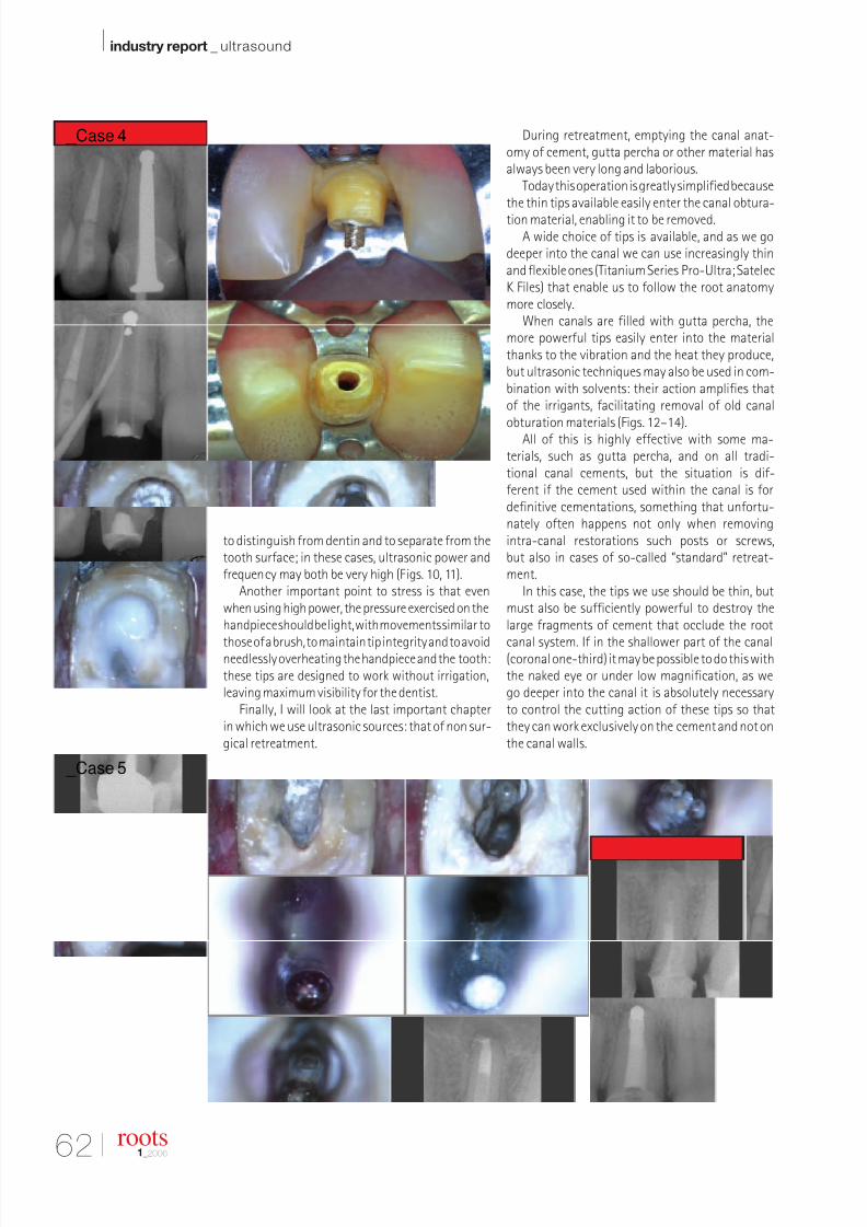
8/13/2019 Dente de Roots 1
http://slidepdf.com/reader/full/dente-de-roots-1 56/58
62 I
I industry report _ ultrasound
to distinguish from dentin and to separate from thetooth surface; in these cases, ultrasonic power andfrequency may both be very high (Figs. 10, 11).
Another important point to stress is that even
when using high power, the pressure exercised on the
handpiece should be light, with movements similar to
those of a brush, to maintain tip integrity and to avoidneedlessly overheating the handpiece and the tooth:
these tips are designed to work without irrigation,
leaving maximum visibility for the dentist.
Finally, I will look at the last important chapterin which we use ultrasonic sources: that of non sur-gical retreatment.
During retreatment, emptying the canal anat-omy of cement, gutta percha or other material hasalways been very long and laborious.
Today this operation is greatly simplified becausethe thin tips available easily enter the canal obtura-
tion material, enabling it to be removed.A wide choice of tips is available, and as we go
deeper into the canal we can use increasingly thinand flexible ones (Titanium Series Pro-Ultra; SatelecK Files) that enable us to follow the root anatomymore closely.
When canals are filled with gutta percha, themore powerful tips easily enter into the materialthanks to the vibration and the heat they produce,but ultrasonic techniques may also be used in com-bination with solvents: their action amplifies thatof the irrigants, facilitating removal of old canalobturation materials (Figs. 12–14).
All of this is highly effective with some ma-terials, such as gutta percha, and on all tradi-tional canal cements, but the situation is dif-ferent if the cement used within the canal is fordefinitive cementations, something that unfortu-nately often happens not only when removingintra-canal restorations such posts or screws,but also in cases of so-called “standard” retreat-ment.
In this case, the tips we use should be thin, butmust also be sufficiently powerful to destroy thelarge fragments of cement that occlude the root
canal system. If in the shallower part of the canal(coronal one-third) it may be possible to do this withthe naked eye or under low magnification, as wego deeper into the canal it is absolutely necessaryto control the cutting action of these tips so thatthey can work exclusively on the cement and not onthe canal walls.
roots1_2006
_Case 4
_Case 5
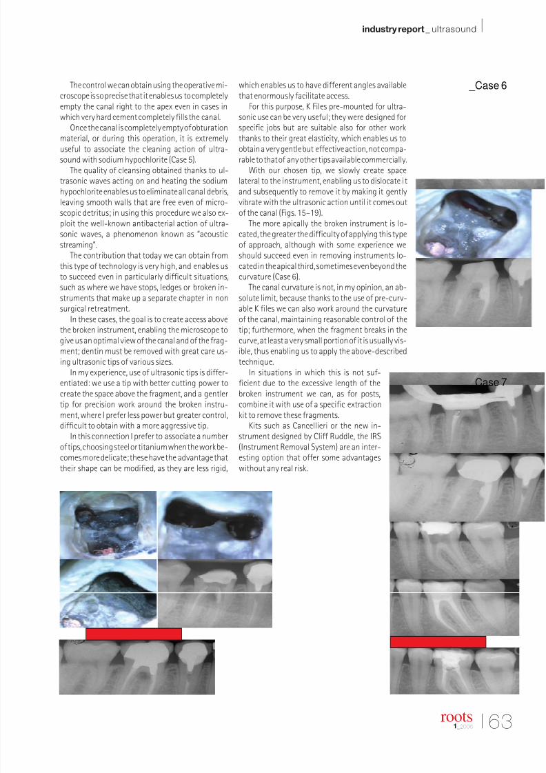
8/13/2019 Dente de Roots 1
http://slidepdf.com/reader/full/dente-de-roots-1 57/58
I 63
industry report _ ultrasound I
roots1_2006
The control we can obtain using the operative mi-
croscope is so precise that it enables us to completely
empty the canal right to the apex even in cases in
which very hard cement completely fills the canal.
Once the canal is completely empty of obturation
material, or during this operation, it is extremelyuseful to associate the cleaning action of ultra-sound with sodium hypochlorite (Case 5).
The quality of cleansing obtained thanks to ul-trasonic waves acting on and heating the sodiumhypochlorite enables us to eliminate all canal debris,leaving smooth walls that are free even of micro-scopic detritus; in using this procedure we also ex-ploit the well-known antibacterial action of ultra-sonic waves, a phenomenon known as “acousticstreaming”.
The contribution that today we can obtain fromthis type of technology is very high, and enables usto succeed even in particularly difficult situations,such as where we have stops, ledges or broken in-struments that make up a separate chapter in nonsurgical retreatment.
In these cases, the goal is to create access abovethe broken instrument, enabling the microscope togive us an optimal view of the canal and of the frag-ment; dentin must be removed with great care us-ing ultrasonic tips of various sizes.
In my experience, use of ultrasonic tips is differ-entiated: we use a tip with better cutting power tocreate the space above the fragment, and a gentler
tip for precision work around the broken instru-ment, where I prefer less power but greater control,difficult to obtain with a more aggressive tip.
In this connection I prefer to associate a numberof tips, choosing steel or titanium when the work be-comes more delicate; these have the advantage thattheir shape can be modified, as they are less rigid,
which enables us to have different angles availablethat enormously facilitate access.
For this purpose, K Files pre-mounted for ultra-
sonic use can be very useful; they were designed for
specific jobs but are suitable also for other work
thanks to their great elasticity, which enables us to
obtain a very gentle but effective action, not compa-
rable to that of any other tips available commercially.
With our chosen tip, we slowly create spacelateral to the instrument, enabling us to dislocate itand subsequently to remove it by making it gentlyvibrate with the ultrasonic action until it comes outof the canal (Figs. 15–19).
The more apically the broken instrument is lo-cated, the greater the difficulty of applying this typeof approach, although with some experience weshould succeed even in removing instruments lo-cated in the apical third, sometimes even beyond thecurvature (Case 6).
The canal curvature is not, in my opinion, an ab-solute limit, because thanks to the use of pre-curv-able K files we can also work around the curvatureof the canal, maintaining reasonable control of thetip; furthermore, when the fragment breaks in thecurve, at least a very small portion of it is usually vis-ible, thus enabling us to apply the above-describedtechnique.
In situations in which this is not suf-ficient due to the excessive length of thebroken instrument we can, as for posts,
combine it with use of a specific extractionkit to remove these fragments.
Kits such as Cancellieri or the new in-strument designed by Cliff Ruddle, the IRS(Instrument Removal System) are an inter-esting option that offer some advantageswithout any real risk.
_Case 7
_Case 6
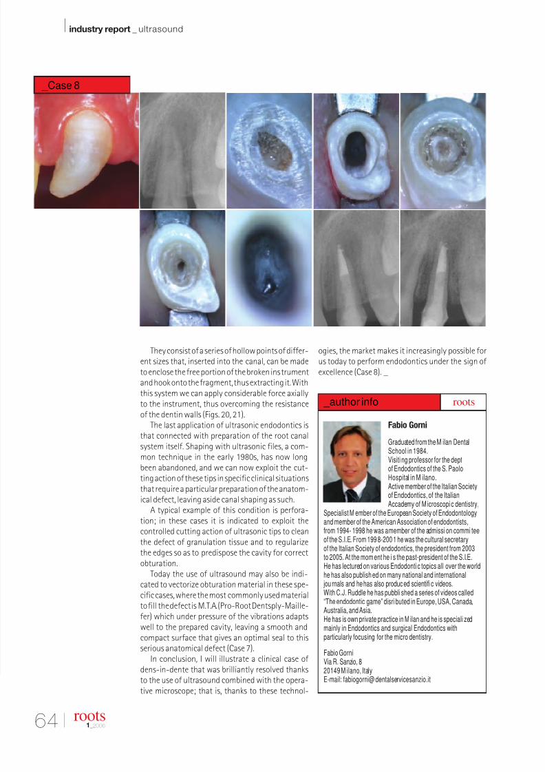
8/13/2019 Dente de Roots 1
http://slidepdf.com/reader/full/dente-de-roots-1 58/58
I industry report _ ultrasound
They consist of a series of hollow points of differ-ent sizes that, inserted into the canal, can be madeto enclose the free portion of the broken instrumentand hook onto the fragment, thus extracting it. Withthis system we can apply considerable force axially
to the instrument, thus overcoming the resistanceof the dentin walls (Figs. 20, 21).
The last application of ultrasonic endodontics isthat connected with preparation of the root canalsystem itself. Shaping with ultrasonic files, a com-mon technique in the early 1980s, has now longbeen abandoned, and we can now exploit the cut-ting action of these tips in specific clinical situationsthat require a particular preparation of the anatom-ical defect, leaving aside canal shaping as such.
A typical example of this condition is perfora-tion; in these cases it is indicated to exploit thecontrolled cutting action of ultrasonic tips to clean
the defect of granulation tissue and to regularizethe edges so as to predispose the cavity for correctobturation.
Today the use of ultrasound may also be indi-cated to vectorize obturation material in these spe-cific cases, where the most commonly used materialto fill the defect is M.T.A. (Pro-Root Dentsply-Maille-fer) which under pressure of the vibrations adapts
ell to the prepared ca it lea ing a smooth and
ogies, the market makes it increasingly possible forus today to perform endodontics under the sign of excellence (Case 8). _
Fabio Gorni
Graduated from the M ilan DentalSchool in 1984.Visiti ng professor for the deptof Endodontics of the S. PaoloHospital in M ilano.Active member of the Italian Societyof Endodontics, of the ItalianAccademy of M icroscopi c dentistry,
Specialist M ember of the European Society of Endodontologyand member of the American Association of endodontists,from 1994- 1998 he was a member of the admissi on commi tee
of the S.I.E. From 199 8-200 1 he was the cultural secretaryof the Italian Society of endodontics, the president from 2003to 2005. At the mom ent he i s the past-president of the S.I.E.He has lectured on various Endodonti c topics all over the worldhe has also publish ed on many national and international
jou rnals and he has also produced scientifi c videos.With C.J. Ruddle he has pubbli shed a series of videos called“The endodontic game” disri buted in Europe, USA, Canada,Australia, and Asia.He has is own private practice in M ilan and he is speciali zed
i l i E d d ti d i l E d d ti ith
_author inforoots
_Case 8