CRISTINA LUÍSA CONCEIÇÃO DE OLIVEIRA ANÁLISE …
122
CRISTINA LUÍSA CONCEIÇÃO DE OLIVEIRA ANÁLISE COMPARADA DA ULTRAESTRUTURA DOS ESPERMATOZÓIDES E MORFOLOGIA DA GLÂNDULA BRANQUIAL EM ESPÉCIES DE CHEIRODONTINAE (CHARACIFORMES: CHARACIDAE) UNIVERSIDADE FEDERAL DO RIO GRANDE DO SUL PORTO ALEGRE 2007 Tese apresentada ao Programa de Pós-Graduação em Biologia Animal, Instituto de Biociências, Universidade Federal do Rio Grande do Sul, como requisito parcial à obtenção do título de Doutora em Biologia Animal. Área de Concentração: Biologia Comparada Orientador: Prof. Dr. Luiz Roberto Malabarba Co-orientador: Prof. Dr. John Robert Burns
Transcript of CRISTINA LUÍSA CONCEIÇÃO DE OLIVEIRA ANÁLISE …
CRISTINA LUÍSA CONCEIÇÃO DE OLIVEIRA
ANÁLISE COMPARADA DA ULTRAESTRUTURA DOS
ESPERMATOZÓIDES E MORFOLOGIA DA GLÂNDULA
BRANQUIAL EM ESPÉCIES DE CHEIRODONTINAE
(CHARACIFORMES: CHARACIDAE)
UNIVERSIDADE FEDERAL DO RIO GRANDE DO SUL
PORTO ALEGRE
2007
Tese apresentada ao Programa de Pós-Graduação
em Biologia Animal, Instituto de Biociências,
Universidade Federal do Rio Grande do Sul, como
requisito parcial à obtenção do título de Doutora em
Biologia Animal.
Área de Concentração: Biologia Comparada
Orientador: Prof. Dr. Luiz Roberto Malabarba
Co-orientador: Prof. Dr. John Robert Burns

ii
ANÁLISE COMPARADA DA ULTRAESTRUTURA DOS
ESPERMATOZÓIDES E MORFOLOGIA DA GLÂNDULA
BRANQUIAL EM ESPÉCIES DE CHEIRODONTINAE
(CHARACIFORMES: CHARACIDAE)
CRISTINA LUÍSA CONCEIÇÃO DE OLIVEIRA
___________________________________
Profa. Dra. Irani Quagio Grassiotto
___________________________________
Dr. Marco Aurélio Azevedo
___________________________________
Prof. Dra. Clarice Bernhardt Fialho
___________________________________
Prof. Dr. Luiz Roberto Malabarba

iii
AGRADECIMENTOS
Ao Prof. Dr. Luiz Roberto Malabarba que, mesmo tendo muitos
compromissos, mostrou-se presente, prestativo e paciente. Muito
obrigada pela orientação, incentivo, empolgação, ensinamentos e
por ser um exemplo de pesquisador.
Ao Prof. Dr. John Robert Burns, por me receber na George
Washington University (GWU). Obrigada pelos cinco meses de
orientação, empolgação e ensinamentos de histologia, microscopia
eletrônica de transmissão e inglês.
Ao CNPq pela bolsa e a taxa de bancada que proporcionaram a
realização de estágios importantes, participação de eventos
científicos e a compra materiais para o desenvolvimento da
pesquisa.
Ao Dr. Stanley Weitzman por me receber no National Museum
of Natural History (USNM), Washington D.C., pela atenção e pelo
empréstimo dos materiais.
À Profa Dra. Irani Quagio-Grassiotto por me receber na
Universidade Estadual Paulista em Botucatu (UNESP) e por ensinar
a analisar as imagens de microscopia eletrônica de transmissão.
Às técnicas Maria Helena Moreno e Claudete dos Santos
Tardivo da UNESP, pelo treinamento de Microscopia eletrônica de
transmissão.
Ao técnico Vicente Salvador da UNESP, pelos ensinamentos de
histologia em resina, pelas caronas e pela amizade durante a
minha estada em Botucatu.
Às colegas da UNESP, Fernanda e Clariana pela atenciosa
recepção em Botucatu.
À técnica Circe Machado da Universidade Federal do Rio
Grande do Sul (UFRGS) pela auxilio na histologia e amizade.
Aos funcionários dos Centros de Microscopia eletrônica da
UFRGS e Pontifícia Universidade Católica do Rio Grande do Sul
(PUCRS) pelo auxílio na preparação das amostras, em especial a
Carmem e a Miriam da PUCRS pela paciência e disposição.
Ao Suresh Benjamim da GWU, pelo auxilio na preparação das
amostras de microscopia eletrônica de varredura e operação do
microscópio.
Ao professor Dr. Gavin Riordan da GWU pelas dicas na
preparação das amostras de TEM.
Aos professores e funcionários do Programa de Pós-Graduação
em Biologia Animal, em especial aos professores Marta Fabián,
iv
Dr.Ludwig Buckup, Inga Mendes que acompanharam o meu trabalho e
deram sugestões nos seminários anuais.
Aos professores Dr. Marco Azevedo, Dra. Clarice Bernhardt
Fialho e Dra. Laura Verrastro pelas sugestões e leitura crítica
do meu exame de qualificação.
À amiga Dra. Cristina Motta Bührnheim pelo empréstimo do
material e pela divisão dos peixes escassos. Pela troca de
informações e por criado a tribo Odontostilbini.
Ao amigo Alexandre Charcansky da PUCRS, pela ajuda em
assuntos de informática.
Ao André Baptista pelo incentivo e dedicação em todos
momentos.
Ao meu irmão Eduardo Oliveira e ao André Baptista por
resolverem os problemas enquanto estive em Washington.
Aos colegas do laboratório de ictiologia da UFRGS pela
ajuda no laboratório, nas coletas e no transporte dos reagentes
até o Centro de Microscopia Eletrônica da UFRGS e por muitos
momentos alegres.
Aos colegas da GWU. Em especial ao Robert Javonillo pela
ajuda com os micrótomos, a Kamali Carroll pela estada em Silver
Spring, e ao Sivasubramanian Balasubramanian pela amizade e pelos
cafés e muffins nos intervalos de trabalho nos finais de semanas.
A todas as pessoas que de alguma maneira contribuíram para
a realização deste trabalho.
SUMÁRIO
Agradecimentos ..............................................................................................iii
Resumo............................................................................................................vi
Abstract ...........................................................................................................viii
Introdução........................................................................................................1

v
Objetivos.........................................................................................................10
Capítulo I: Sperm ultrastructure of the species of Cheirodontinae
(Teleostei: Characidae): description and implications on the phylogeny
of the inseminating compsurins……………………………………………….
Capítulo II: Sperm ultrastructure in the inseminating Macropsobrycon
uruguayanae (Teleostei: Characidae: Cheirodontinae)……………………
Capítulo III: Gill-derived glands morphology in Cheirodontinae
(Teleostei: Characidae) ……………………………………………………….
Conclusões ..............................................................................................
Anexo 1: Instruções para os autores da Journal of Morphology ..............
17 55 76 102 105
vi
RESUMO
A tese é divida em três capítulos, seguindo as regras da revista Journal of
Morphology. O primeiro capítulo descreve a ultraestrutura dos espermatozóides de seis
queirodontíneos inseminadores pertencentes à tribo Compsurini (Kolpotocheirodon
theloura, Compsura heterura, Acinocheirodon melanogramma, Saccoderma hastatus,
"Odontostilbe" dialeptura e "Odontostilbe" mitoptera), e de quatro espécies de
fertilização externa, três destas pertencentes à tribo Cheirodontini (Cheirodon
interruptus, Serrapinnus calliurus, e Serrapinnus heterodon) e uma espécie incertae
sedis na subfamília (Odontostilbe pequira). Os espermatozóides de espécies de
fertilização externa mostram estruturas conservativas e os espermatozóides de
espécies inseminadoras apresentam mudanças estruturais, principalmente no núcleo
espermático. As espécies da tribo Compsurini apresentam espermatozóides com peças
intermediárias mais longas, que se estreitam progressivamente no sentido distal, e
vesículas mais largas e em menor número. Em espécies de fertilização externa as
peças intermediárias são mais curtas e terminam abruptamente, e as vesículas são
menores e em maior número. Uma matriz foi construída baseada em dez caracteres da
ultraestrutura dos espermatozóides. Obteve-se uma hipótese de relações parcialmente
resolvida entre espécies inseminadoras. A análise somente de caracteres de
ultraestrutura de espermatozóides, no entanto, não foi informativa para a hipótese de
relações entre espécies de fertilização externa. As relações entre os membros de
Cheirodontinae e a evolução da morfologia dos espermatozóides são discutidas. O
segundo capítulo descreve a ultraestrutura dos espermatozóides da espécie
inseminadora Macropsobrycon uruguayanae. Os espermatozóides apresentam núcleos
moderadamente alongados e cromatina elétron-densa. Durante a espermiogênese a
rotação nuclear acontece, deixando o flagelo posterior ao núcleo. Os centríolos são
vii
paralelos, e o centríolo proximal é ligeiramente anterior ao distal. A ponta do centríolo
proximal encontra-se dentro da rasa fossa nuclear. Estrias centriolares denominadas de
rootlets partem de ambos centríolos. Nove microtúbulos acessórios circulam o axonema
externamente. O flagelo tem axonema com a configuração típica (9+2). Além dos
espermatozóides normais, são encontrados no lúmen testicular espermatozóides
atípicos denominados paraespermatozóides. Estas células se assemelham ao
espermatozóide em muitos aspectos, mas seu núcleo tem forma irregular e a cromatina
é menos elétron-densa. Discutem-se as especializações vistas nos espermatozóides,
as possíveis adaptações relacionadas ao hábito de inseminação e o fato de que a
origem e função dos paraespermatozóides permanecem indeterminadas em
Macropsobrycon uruguayanae. O terceiro capítulo descreve a glândula branquial de 17
espécies de Cheirodontinae. A glândula branquial está localizada na região anterior da
cavidade branquial em ambos os lados do corpo. Esta estrutura foi encontrada em
todos machos maduros. A glândula é pequena em espécies de fertilização externa
ocupando no máximo 10 filamentos branquiais e em espécies inseminadoras, ela ocupa
uma grande extensão ou o arco inteiro. Em algumas partes da glândula de A
Aphyocheirodon hemigrammus, C. heterura, K. theloura, M. uruguayanae e S. hastatus,
as lamelas não permanecem, ficando somente as células secretoras da glândula
branquial. Um material não celular foi observado dentro das câmaras da glândula de K.
theloura. Tanto espécies inseminadoras quanto de fertilização externa de
Cheirodontinae apresentam glândula, não existindo relação entre a presença de
glândula branquial e inseminação. A função da glândula não é conhecida, mas pela
presença desta estrutura somente em machos maduros, esta pode ser usada para
produção e liberação de secreção para a atração da fêmea durante o período
reprodutivo ou na inibição de outros machos. As glândulas branquiais de
queirodontíneos e de outros caracídeos são comparadas.
viii
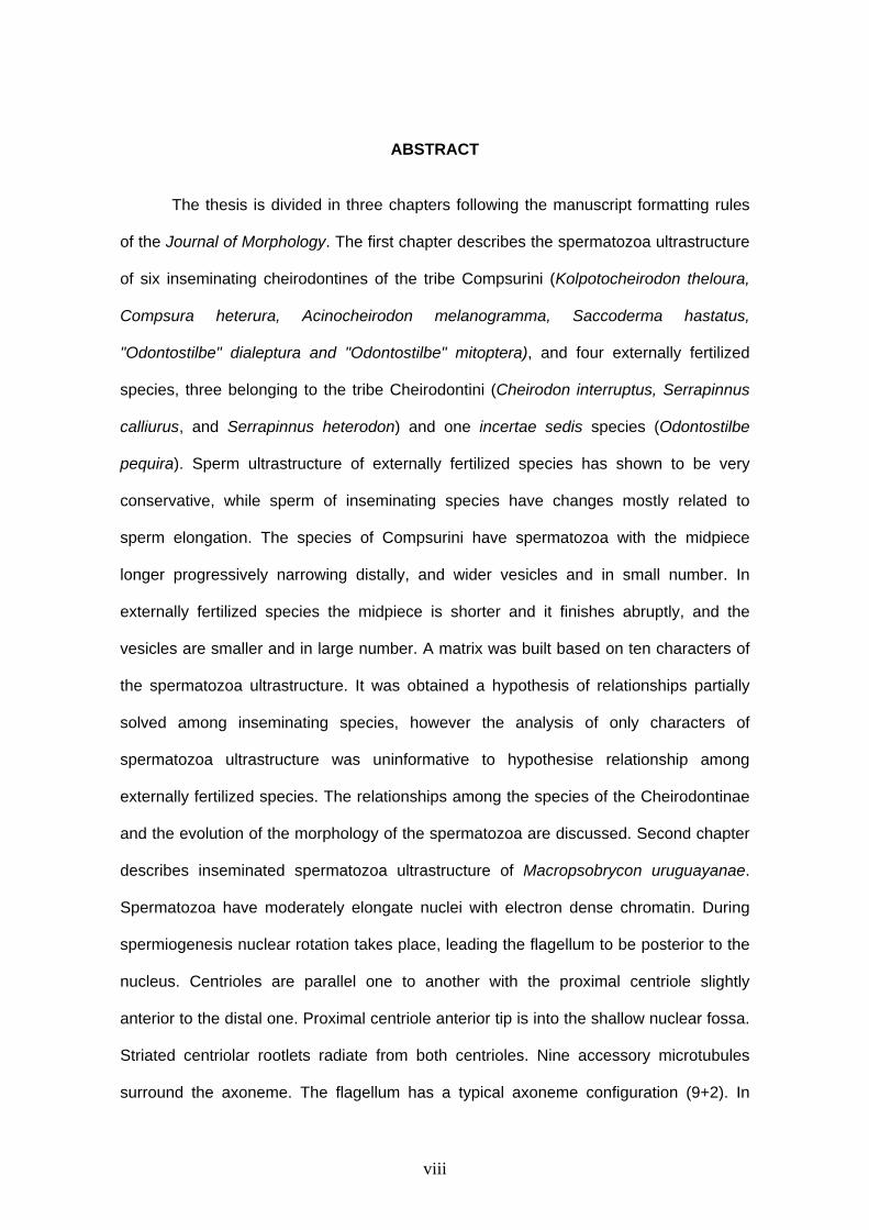
ABSTRACT
The thesis is divided in three chapters following the manuscript formatting rules
of the Journal of Morphology. The first chapter describes the spermatozoa ultrastructure
of six inseminating cheirodontines of the tribe Compsurini (Kolpotocheirodon theloura,
Compsura heterura, Acinocheirodon melanogramma, Saccoderma hastatus,
"Odontostilbe" dialeptura and "Odontostilbe" mitoptera), and four externally fertilized
species, three belonging to the tribe Cheirodontini (Cheirodon interruptus, Serrapinnus
calliurus, and Serrapinnus heterodon) and one incertae sedis species (Odontostilbe
pequira). Sperm ultrastructure of externally fertilized species has shown to be very
conservative, while sperm of inseminating species have changes mostly related to
sperm elongation. The species of Compsurini have spermatozoa with the midpiece
longer progressively narrowing distally, and wider vesicles and in small number. In
externally fertilized species the midpiece is shorter and it finishes abruptly, and the
vesicles are smaller and in large number. A matrix was built based on ten characters of
the spermatozoa ultrastructure. It was obtained a hypothesis of relationships partially
solved among inseminating species, however the analysis of only characters of
spermatozoa ultrastructure was uninformative to hypothesise relationship among
externally fertilized species. The relationships among the species of the Cheirodontinae
and the evolution of the morphology of the spermatozoa are discussed. Second chapter
describes inseminated spermatozoa ultrastructure of Macropsobrycon uruguayanae.
Spermatozoa have moderately elongate nuclei with electron dense chromatin. During
spermiogenesis nuclear rotation takes place, leading the flagellum to be posterior to the
nucleus. Centrioles are parallel one to another with the proximal centriole slightly
anterior to the distal one. Proximal centriole anterior tip is into the shallow nuclear fossa.
Striated centriolar rootlets radiate from both centrioles. Nine accessory microtubules
surround the axoneme. The flagellum has a typical axoneme configuration (9+2). In
ix
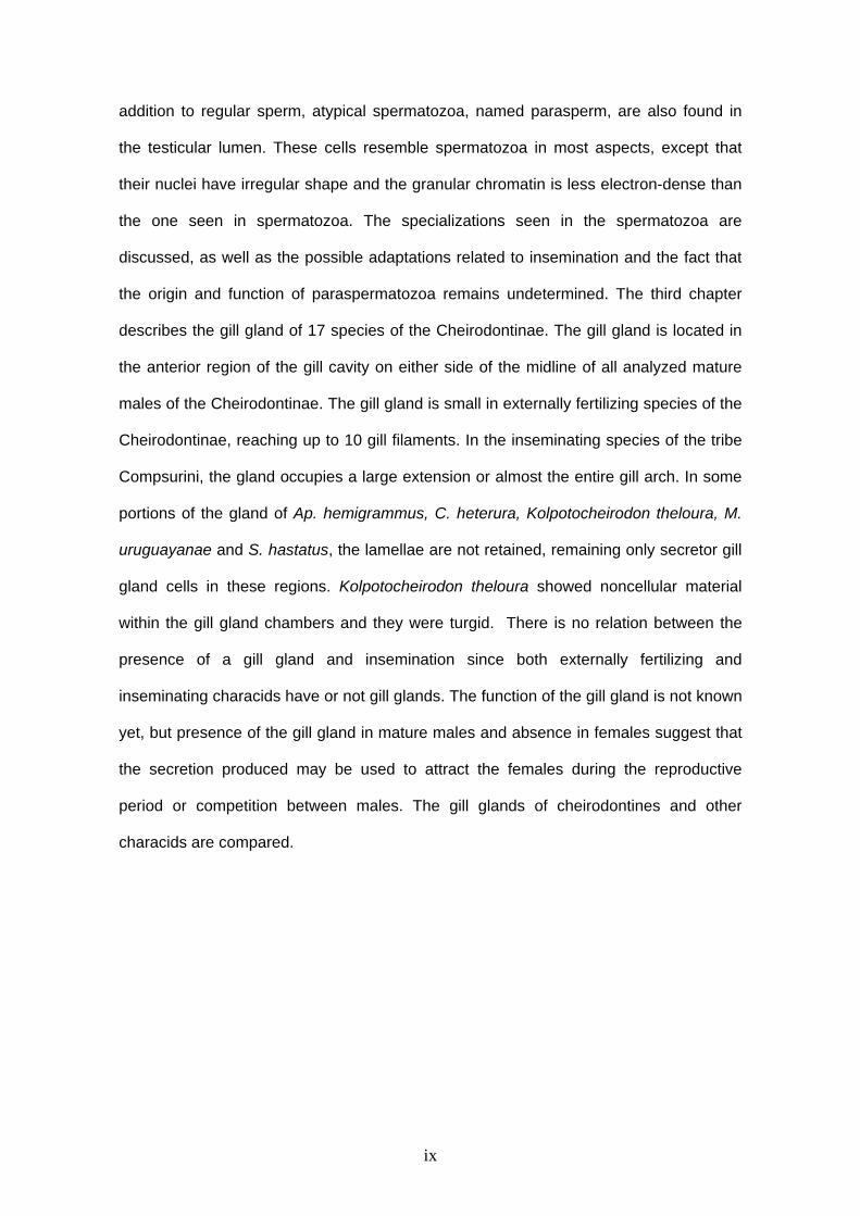
addition to regular sperm, atypical spermatozoa, named parasperm, are also found in
the testicular lumen. These cells resemble spermatozoa in most aspects, except that
their nuclei have irregular shape and the granular chromatin is less electron-dense than
the one seen in spermatozoa. The specializations seen in the spermatozoa are
discussed, as well as the possible adaptations related to insemination and the fact that
the origin and function of paraspermatozoa remains undetermined. The third chapter
describes the gill gland of 17 species of the Cheirodontinae. The gill gland is located in
the anterior region of the gill cavity on either side of the midline of all analyzed mature
males of the Cheirodontinae. The gill gland is small in externally fertilizing species of the
Cheirodontinae, reaching up to 10 gill filaments. In the inseminating species of the tribe
Compsurini, the gland occupies a large extension or almost the entire gill arch. In some
portions of the gland of Ap. hemigrammus, C. heterura, Kolpotocheirodon theloura, M.
uruguayanae and S. hastatus, the lamellae are not retained, remaining only secretor gill
gland cells in these regions. Kolpotocheirodon theloura showed noncellular material
within the gill gland chambers and they were turgid. There is no relation between the
presence of a gill gland and insemination since both externally fertilizing and
inseminating characids have or not gill glands. The function of the gill gland is not known
yet, but presence of the gill gland in mature males and absence in females suggest that
the secretion produced may be used to attract the females during the reproductive
period or competition between males. The gill glands of cheirodontines and other
characids are compared.

INTRODUÇÃO
Cheirodontinae
As espécies pertencentes à subfamília Cheirodontinae habitam ambientes lênticos e
regiões de planície. Os Cheirodontinae são encontrados na maioria das bacias
hidrográficas da América do Sul e Central, como bacia Amazônica, Orinoco, Paraná-
Paraguai e São Francisco. Quatro espécies de Cheirodon são os únicos Characiformes
encontrados no oeste dos Andes no Chile (Malabarba, 1998, 2003).
Quatro sinapomorfias são apresentadas por Malabarba (1998) para definir
Cheirodontinae: a presença de dentes pedunculados e expandidos na região distal, a
presença de uma única série de dentes na premaxila, a ausência de mancha umeral e a
existência de uma falha da cobertura muscular na região umeral em ambos lados do
corpo na região anterior da bexiga natatória, denominada pseudotímpano – supõe-se
que esta ausência de musculatura na região umeral facilite a transmissão de ondas
sonoras do ambiente para a bexiga natatória e desta para o ouvido interno através do
aparelho de Weber (Malabarba 1998). Recentemente, a monofilia de Cheirodontinae
apresentada por Malabarba (1998) foi confirmada por Bührnheim (2006, dados não
publicados), baseando-se em uma nova analise filogenética com 53 táxons e 169
caracteres. A subfamília é redefinida por 15 sinapomorfias, duas correspondem as
sinapomorfias encontradas por Malabarba (1998): a ausência da mancha umeral e a
presença do pseudotímpano e cinco novas unicamente derivadas como o contorno do
processo anteromedial do mesetmóide; o primeiro infraorbital subretangular com a
porção anteroventral estendida; a porção posterior edêntula da maxila
aproximadamente com tamanho equivalente ao da porção anterior com dentes da
maxila; o perfil da nadadeira anal dos machos com o lobo anterior agudo e o perfil distal
côncavo; e em geral a presença de dois ou três dentes na maxila (Bührnheim, 2006).
2
A subfamília Cheirodontinae compreende duas tribos, Cheirodontini e Compsurini,
além de alguns gêneros considerados incertae sedis.
Tribo Cheirodontinae
A tribo Cheirodontini é reconhecida baseando-se principalmente em caracteres
relacionados ao dimorfismo sexual secundário observado nos raios procorrentes
ventrais da nadadeira caudal e nos raios da nadadeira anal dos machos. A tribo
Cheirodontini é composta por 22 espécies (Malabarba, 2003) distribuídos nos seguintes
seis gêneros: Cheirodon Girard, 1855 (distribuído no Sul do Brasil, Uruguai, Argentina e
Chile); Serrapinnus Malabarba, 1998 (distribuído na bacia Amazônica, bacia do rio
Uruguai, bacia do rio São Francisco, laguna dos Patos, bacia Paraná Paraguai);
Nanocheirodon Malabarba, 1998 (distribuído na Colômbia no lago Maracaibo e bacia do
rio Magdalena); Heterocheirodon Malabarba, 1999 (distribuído na bacia do rio Uruguai e
laguna dos Patos); Spintherobolus Eigenmann, 1911 (distribuído no alto rio Tietê e nos
rios costeiros do Sudeste do Brasil) e um novo gênero, ainda não descrito (denominado
novo gênero e espécie C em Malabarba, 1998).
Tribo Compsurini
A tribo Compsurini inclui os queirodontíneos com inseminação, sendo caracterizada
pela transferência de esperma dos testículos dos machos maduros para os ovários das
fêmeas, como descrito em Burns et al. (1997). Os membros da tribo Compsurini
apresentam também órgãos especializados na nadadeira caudal dos machos, que
variam desde a presença de escamas modificadas até a presença de tecidos
hipertrofiados aparentemente de função glandular (Malabarba & Weitzman, 1999,
2000). A tribo Compsurini é formada por nove espécies distribuídas nos seguintes cinco
3
gêneros: Acinocheirodon Malabarba & Weitzman, 1999 (distribuído nas bacias dos rios
São Francisco e Jequitinhonha); Compsura Eigenmann, 1915 (gênero encontrado no
Panamá, na bacia do rio São Francisco e rios costeiros do Nordeste); Kolpotocheirodon
Malabarba & Weitzman, 2000 (cabeceiras do rio Paraná e São Francisco em Brasília e
Bahia); Macropsobrycon Eigenmann, 1915 (laguna dos Patos, bacia do rio Uruguai,
bacia do rio Tramandaí); e Saccoderma Schultz, 1944 (bacia do rio Maracaibo na
Venezuela, bacia do rio Magdalena e rio Sinú na Colômbia). “Odontostilbe” dialeptura
(Fink & Weitzman, 1974) encontrado em rios da Costa Rica e Panamá, e “O.” mitoptera
(Fink & Weitzman, 1974) distribuído no Panamá, são considerados pertencentes à tribo
Compsurini e uma nova designação de gênero é necessária para estas espécies
(Malabarba, 1998; Malabarba & Weitzman, 1999; Malabarba, 2003).
Gêneros incertae sedis
Os gêneros Aphyocheirodon Eigenmann, 1915 (distribuído no alto rio Paraná);
Cheirodontops Schultz, 1944 (encontrado na bacia do rio Orinoco, Venezuela);
Odontostilbe Cope, 1870 (distribuído na bacia Amazônica, bacia dos rios Mana, Maroni
e Comté na Guiana Francesa, bacia do Panamá, bacia do rio Pilcomayo na Bolivia,
bacia do rio Napo no Equador, bacia Paraná Paraguai e bacia do rio Uruguai);
Prodontocharax Eigenmann & Pearson, 1953 (encontrado na bacia Amazônica, na
Bolívia e no Peru); e Pseudocheirodon Meek & Hildebrand, 1916 (distribuído na costa
rica e Panamá) não foram classificados em nenhuma das duas tribos, pois
apresentavam caracteres de dimorfismo sexual pouco evidentes. Recentemente
Bührnheim (2006) propôs a criação da tribo Odontostilbini. Esta tribo foi baseada em
treze sinapomorfias encontradas nos canais sensoriais do parietal e do primeiro
infraorbital, na forma do segundo e sexto infraorbitais, no bordo anterodorsal da maxila,
no palatino, na protuberância lateral da maxila inferior, na parte lateral exposta do ramo

4
inferior do ângulo-articular, no branquiostegal mais posterior, no comprimento do raio
não ramificado da nadadeira pélvica, no perfil da nadadeira anal e na extensão da linha
lateral. Bührnheim propôs duas novas sinonímias, Aphyocheirodon e Cheirodontops,
como sinônimos de Holoshesthes. Além disso, três gêneros são revalidados:
Holoshesthes e Lobodeuterodon saem da sinonímia de Odontostilbe, e Amblystilbe da
sinonímia de Prodontocharax. A tribo Odontostilbini é composta por 26 espécies
distribuídas nos seguintes 6 gêneros: Amblystilbe Fowler, 1940; Holoshesthes
Eigenmann 1903; Lobodeuterodon Fowler, 1945; Odontostilbe Cope 1870,
Prodontocharax Pearson, 1924 e Pseudocheirodon Meek & Hildebrand, 1916.
Estudos realizados em Cheirodontinae
Entre os estudos recentes sobre sistemática, reprodução e caracteres sexuais
primários e secundários em Cheirodontinae podemos citar: Burns et al. (1997)
descrevem a morfologia dos espermatozóides; Malabarba (1998) apresenta definição e
relações filogenéticas; Burns et al. (1998) descrevem a ultraestrutura de M.
uruguayanae Eigenmann 1915; Gelain et al. (1999), estudam aspectos da reprodução
de Serrapinnus calliurus (Boulenger, 1900); Weitzman & Malabarba (1999) revisam
Spintherobolus; Malabarba & Weitzman (1999) descrevem Acinocheirodon; Malabarba
& Bertaco (1999) descrevem Heterocheirodon; Malabarba & Weitzman (2000)
descrevem Kolpotocheirodon; Braun et al. (2000), descrevem a biologia reprodutiva de
Cheirodon ibicuhiensis Eigenmann, 1915 da lagoa Fortaleza, RS; Oliveira et al. (2002),
estimam o período reprodutivo, o tipo de desova e a fecundidade de C. ibicuhiensis do
arroio Ribeiro, rio Grande do Sul; Malabarba (2003) caracteriza a subfamília e lista as
espécies; Oliveira (2003) estima o período reprodutivo e fecundidade e também
compara o desenvolvimento de caracteres sexuais secundários (glândula branquial e
ganchos) com a maturação gonadal em duas espécies Compsura heterura Eigenmann,
1915 e Odontostilbe sp.; Silvano et al. (2003) estudam o período reprodutivo e
fecundidade para Serrapinnus piaba (Lütken, 1874) do rio Ceará-Mirim, Rio Grande do

5
Norte; Gusmão-Pompiani (2003) descreve a ultraestrutura dos espermatozóides de
Serrapinnus notomelas (Eigenmann 1915); Malabarba et al. (2004) descrevem
Kolpotocheirodon figueiredoi; Azevedo (2004) estuda a biologia reprodutiva de M.
uruguayanae do rio Ibicui Mirim-RS; Burns & Weitzman (2005) descrevem a
ultraestrutura dos espermatozóides de Serrapinnus kriegi (Schindler, 1937); Bührnheim
& Malabarba (2006) redescrevem a espécie tipo de Odontostilbe e descrevem três
espécies novas O. ecuadorensis, O. nareuda e O. pareci; Bührnheim (2006) estuda a
sistemática de Odontostilbe com a proposição de uma nova tribo Odontostilbini e
redefinição dos gêneros incertae sedis.
Ultraestrutura dos espermatozóides
A análise da ultraestrutura de espermatozóides e da espermatogênese em
teleósteos tem revelado importantes características morfológicas (Mattei, 1991;
Spadella, 2004) e tem ajudado a esclarecer questões a respeito da filogenia das
espécies de peixes (Jamieson, 1991; Mattei, 1991).
A forma, o comprimento e a largura do núcleo espermático apresentam variação
entre as espécies e são freqüentemente associadas ao tipo de fecundação. A maioria
das espécies de fecundação externa possui espermatozóides com núcleo arredondado,
sendo denominado de aquasperma (Jamieson, 1991). O alongamento do núcleo é
observado em espécies de fecundação interna ou inseminadoras (presença de
espermatozóides nos ovários, sem conhecimento do exato momento da fecundação)
(Burns et al., 1995, 1997; Burns & Weitzman, 2005). Algumas hipóteses são
apresentadas para as possíveis vantagens seletivas do alongamento do núcleo.
Núcleos espermáticos alongados são mais aerodinâmicos e podem facilitar a
penetração na micrópila do ovócito (Fawcett, 1970; Jamieson, 1991) e podem passar
pelo gonoporo da fêmea mais facilmente, aumentando o número de espermatozóides
transferidos num determinado momento (Burns & Weitzman, 2005).
Outro aspecto relacionado ao núcleo é a posição deste em relação ao flagelo e isto
6
depende do tipo de espermiogênese, que pode ser do tipo 1 ou do tipo 2 segundo a
classificação de Mattei (1970). Em ambos, o início do desenvolvimento do flagelo
ocorre lateralmente ao núcleo. Na espermiogênese tipo I as mitocôndrias estão
dispersas no citoplasma, o complexo centriolar está disposto lateralmente ao núcleo e
preso à membrana plasmática e o centríolo distal forma o corpúsculo basal e
desenvolve o flagelo. No processo tipo I os centríolos migram em direção ao núcleo e
trazem junto à membrana plasmática e o flagelo inicial. Ocorre uma depressão no
contorno nuclear sendo denominado de fossa nuclear. O núcleo faz uma rotação de 90°
em relação ao eixo flagelar. No final do processo o flagelo fica perpendicular ao núcleo.
A rotação nuclear tem sido observada em todas as espécies de Curimatidae e em
quase todos os Characiformes (Quagio-Grassiotto et al., 2003) com exceção de
Acestrorhynchus falcatus (Bloch 1794) em Acestrorhynchidae (Matos et al., 2000), dos
Glandulocaudinae (Burns et al., 1998; Pecio & Rafiński, 1999), Stevardiinae (Burns et
al., 1998, Pecio et al., 2005), Bryconamericus stramineus Eigenmann 1908 em
Characidae (Gusmão-Pompiani, 2003, Gusmão-Pompiani et al., submetido), em
Cheirodontinae (Burns et al., 1997) e em três espécies consideradas incertae sedis em
Characidae, Bryconadenos tanaothoros (Weitzman et al., 2005) e Brittanichthys axelrodi
(Javonillo et al., 2007).
O processo de espermiogênese tipo II é semelhante ao tipo I, mas difere deste por
não ocorrer rotação nuclear em relação ao flagelo, resultando num flagelo paralelo ao
núcleo. Também ocorre a formação da fossa nuclear, mas os centríolos encontram-se
fora da fossa nuclear. A espermiogênese do tipo III foi descrita recentemente para
Siluriformes da família Pimelodidae (Quagio-Grassiotto & Carvalho, 2000). A
espermátide jovem apresenta núcleo central e complexo centriolar medial ao núcleo e
preso à membrana. Durante a espermiogênese a rotação nuclear não ocorre, os
centríolos não migram permanecendo junto à membrana plasmática e o canal
citoplasmático e a fossa nuclear não se formam. O espermatozóide apresenta flagelo
7
perpendicular ao núcleo. A espermiogênese do tipo III foi observada algumas espécies
de Callichthyidae e Loricariidae (Spadella, 2004).
As espécies de fertilização externa geralmente apresentam uma peça intermediária
pequena, enquanto que nas espécies inseminadoras esta estrutura é normalmente
mais alongada (Jamieson, 1991; Mattei; 1991). Uma peça intermediária maior poderia
abrigar um número maior de mitocôndrias, aumentando a capacidade de geração de
energia da célula e viabilidade dos espermatozóides (Fawcett, 1970; Pecio & Rafiński,
1994).
A ultraestrutura de espermatozóides foi estuda em Glandulocaudinae [Mimagoniates
barberi Regan 1907, por Pecio & Rafinski (1994); Mimagoniates barberi e M. microlepis
(Steindachner 1877) por Burns et al. (1998)]; Stevardiinae [Corynopoma riisei Gill 1858,
Pseudocorynopoma doriae Perugia 1891, Diapoma speculiferum Cope 1894, Diapoma
sp. por Burns et al. (1998); Tyttocharax cochui (Ladiges 1950), T. tambopatensis
Weitzman & Ortega 1995 e Scopaeocharax rhinodus (Böhlke 1958) por Pecio et al.
(2005)]; Aphyocharacinae [Aphyocharax anisitsi Eigenmann & Kennedy, 1903 por Burns
et al. (1998) e por Gusmão-Pompiani (2003)]; Tetragonopterinae [Tetragonopterus
argenteus Cuvier 1816 por Gusmão-Pompiani (2003)]; Stethaprioninae [Poptella
paraguayensis (Eigenmann 1907) por Gusmão-Pompiani (2003)]; Characinae
[Roeboides bonariensis (Reinhardt, 1851), Galeocharax humeralis (Valenciennes, 1834)
e Galeocharax knerii (Steindachner, 1879) por Gusmão-Pompiani (2003)]; espécies
incertae sedis em Characidae [Triportheus paranensis (Kner, 1858), Bryconops affinis
(Günther, 1864), Hyphessobrycon eques (Steindachner 1882), Moenkhausia
sanctaefilomenae (Steindachner, 1907), Bryconamericus stramineus Eigenmann 1908,
Salminus maxillosus (Cuvier, 1816), Brycon microlepis Perugia 1897 e B. orbignyanus
(Valenciennes, 1850) por Gusmão-Pompiani (2003); Hollandichthys (Eigenmann, 1909)
por Azevedo (2004); S. maxillosus, B. microlepis e B. orbignyanus por Veríssimo-
Silveira et al. (2006); Brittanichthys axelrodi Géry 1965, por Javonillo et al. (2007). Em
Cheirodontinae as informações são escassas, sendo disponíveis somente para três
8
espécies, M. uruguayanae (Burns & Weitzman, 1998, 2005), S. notomelas (Gusmão-
Pompiani, 2003) e S. kriegi (Burns & Weitzman, 2005).
Glândula branquial
Burns & Weitzman (1996) descreveram uma nova estrutura encontrada nas
brânquias de Corynopoma riisei, um stevardiine. Esta estrutura foi denominada de
glândula branquial e é formada pela união e modificação funcional dos filamentos mais
ventrais da hemibranquia externa do primeiro arco branquial em ambos lado do corpo.
Os filamentos da glândula branquial estão unidos externamente através de um tecido
epitelial estratificado, mantendo a individualidade interiormente e formando câmaras,
por onde a secreção é conduzida ao exterior (Burns & Weitzman, 1996; Bushmann et
al., 2002). Entre as lamelas são encontradas células cilíndricas.
O desenvolvimento da glândula branquial parece iniciar com a multiplicação das
células epiteliais que revestem os filamentos primários mais anteriores da porção mais
ventral, estendendo-se para os filamentos seguintes. Esta expansão celular parece
iniciar nas células que revestem a base dos filamentos, próximo ao arco, estendendo-se
sobre os filamentos até quase a sua extremidade distal (Oliveira, 2003).
Os filamentos branquiais envolvidos na formação da glândula branquial
provavelmente perdem a função respiratória. A função da glândula braquial ainda não
está definida, mas por iniciar o seu desenvolvimento em machos em maturação
gonadal, ser bem desenvolvida em machos maduros e estar ausente em fêmeas, esta
pode estar relacionada à reprodução. A secreção produzida por esta glândula pode ser
utilizada para atrair as fêmeas durante o período reprodutivo (Burns & Weitzman, 1996;
Bushmann et al., 2002) ou pode estar associada à competição entre machos
(Bushmann et al., 2002).
A glândula branquial foi encontrada em outras espécies de Stevardiinae
(Bushmann et al., 2002) e em outras subfamílias de Characidae, como Cheirodontinae
(Oliveira, 2000, 2003; Azevedo, 2004; Bührnheim, 2006); Aphyocharacinae (Gonçalves
9
et al., 2005), e algumas espécies consideradas incertae sedis em Characidae
(Weitzman et al., 2005).
Esta tese é divida em três capítulos que tratam da descrição da ultraestrutura
dos espermatozóides e descrição da glândula branquial de representantes de diferentes
gêneros de Cheirodontinae. Os capítulos seguem as regras da revista Journal of
Morphology.
10
OBJETIVOS
- Descrever a ultraestrutura dos espermatozóides em espécies de Cheirodontinae
utilizando o método de microscopia eletrônica de transmissão (MET);
- Comparar a morfologia dos espermatozóides ao tipo de estratégia reprodutiva
apresentada pelas espécies (inseminação ou fecundação externa);
- Comparar e analisar possíveis homologias na ultraestrutura dos espermatozóides
entre as espécies de queirodontíneos e outros caracídeos, formulando hipóteses acerca
da evolução destes caracteres e da filogenia dos táxons envolvidos;
- Investigar a presença e comparar a morfologia da glândula branquial em espécies de
Cheirodontinae;
- Comparar e analisar possíveis homologias entre a glândula branquial nos
queirodontíneos e outros caracídeos, formulando hipóteses acerca da evolução destes
caracteres em Characidae e mais especificamente em Cheirodontinae.
11
REFERÊNCIAS BIBLIOGRÁFICAS
Azevedo MA 2004. Análise comparada de caracteres reprodutivos em três linhagens de
Characidae (Teleostei: Ostariophysi) com inseminação. Tese de doutorado,
Universidade Federal do Rio Grande do Sul, Porto Alegre, Brasil. 238p.
Braum AS, Lewis DS, Fontoura NF. 2000. Biologia reprodutiva de Cheirodon
ibicuhiensis (Eigenmann, 1915) na lagoa Fortaleza, Cidreira, Rio Grande do Sul,
Brasil (Teleostei: Characidae: Cheirodontinae). Comum. Mus. Ciênc. Tecnol.
PUCRS, sér. Zool.13 (2):159-166.
Buhrnheim CM. 2006. Sistemática de Odontostilbe Cope, 1870 com a proposição de
uma nova tribo Odontostilbini e redefinição dos gêneros incertae sedis de
Cheirodontinae (Ostariophysi: Characiformes: Characidae). Tese de doutorado.
Pontifícia Universidade Católica de Porto Alegre, Porto Alegre, Brazil. 315p.
Buhrnheim CM, Malabarba LR. 2006. Redescription of Odontostilbe fugitiva, type
species of Odontostilbe Cope, 1870 (Teleostei: Characidae: Cheirodontinae), and
description of three new species from the Amazon basin. Neotrop Ichthyol 4(2):167-
196
Burns JR, Weitzman SH. 1996. Novel gill-derived gland in the male swordtail characin,
Corynopoma riisei (Teleostei: Characidae: Glandulocaudinae). Copeia 1996 (3):627-
633.
Burns JR, Weitzman SH. 2005. Insemination in ostariophysan fishes. In: Uribe MC,
Grier HJ, editors. Viviparous Fishes. Homestead, FL: New Life Publications. p 107-
134.
12
Burns JR, Weitzman SH, Grier HJ, Menezes NA. 1995. Internal fertilization, testis and
sperm morphology in glandulocaudine fishes (Teleostei: Characidae:
Glandulocaudinae). J Morphol 224:131-145.
Burns JR, Weitzman SH, Lange KR, Malabarba LR. 1998. Sperm ultrastructure in
characid fishes (Teleostei: Ostariophysi). In: Malabarba LR, Reis RE, Vari RP,
Lucena ZMS, Lucena CAS, editors. Phylogeny and Classification of Neotropical
Fishes. Porto Alegre, Brazil: EDIPUCRS. pp 235-244.
Burns JR, Weitzman SH, Malabarba LR. 1997. Insemination in eight species of
Cheirodontine fishes (Teleostei: Characidae: Cheirodontinae). Copeia 1997(2): 433-
438.
Bushmann PJ, Burns JR, Weitzman SH. 2002. Gill-derived glands in glandulocaudine
fishes (Teleostei: Characidae: Glandulocaudinae). J. Morphol. 253:187-195.
Fawcett DW. 1970. A comparative view of sperm ultrastructure. Biol. Reprod. Suppl.
2:90-127.
Gelain D, Fialho CB, Malabarba LR. 1999. Biologia reprodutiva de Serrapinnus calliurus
(Boulenger, 1900) (Characidae, Cheirodontinae) do arroio Ribeiro, Barra do Ribeiro,
RS, Brasil. Comun. Mus. Cienc. Tecnol. PUCRS sér. Zool. 12:72-82.
Gonçalves TK, Azevedo MA, Malabarba LR. 2005. Reproductive biology and
development of sexually dimorphic structures in Aphyocharax anisitsi (Ostariophysi:
Characidae). Neotrop Ichthyol 3(3):433-438.
Gusmão-Pompiani P. 2003. Ultraestrutura da espermiogênese e dos espermatozóides
de peixes da ordem Characiformes, família Characidae (Teleostei, Ostariophysi):
uma abordagem filogenética. Tese de doutorado, Instituto de Biociências da
Universidade Estadual Paulista, São Paulo. 86 p.
13
Gusmão-Pompiani P, Malabarba LR, Oliveira C, Quagio-Grassiotto I. Spermatozoa
ultrastructure of representatives of Tetragonopterinae, Stethaprioninae and some
incertae sedis in Characidae (Teleostei: Ostariophysi: Characiformes). In press
Jamieson BGM 1991. Fish evolution and systematics: evidence from spermatozoa.
Cambridge, Cambridge University Press, 319 p.
Javonillo R, Burns JR, Weitzman SH. 2007. Reproductive morphology of Brittanichthys
axelrodi (Teleostei: Characidae), a miniature inseminating fish from South America. J
Morphol 268:23-32.
Malabarba LR. 1998. Monophyly of the Cheirodontinae, characters and major clades
(Ostariophysi: Characidae). In: Malabarba LR, Reis RE, Vari RP, Lucena ZMS,
Lucena CAS, editors. Phylogeny and Classification of Neotropical Fishes. Porto
Alegre, Brazil: EDIPUCRS. p 193-233.
Malabarba LR. 2003. Subfamily Cheirodontinae (Characins, tetra) In: Reis RE,
Kullander SO, Ferraris CJ, editors. Check list of the freshwater fishes of South and
Central America. Porto Alegre, Brazil: EDIPUCRS. p 215- 221.
Malabarba LR, Bertaco VA. 1999. Description of a new species of Heterocheirodon
Malabarba (Teleostei: Characidae: Cheirodontinae: Cheirodontini), with further
comments on the diagnosis of the genus. Comun. Mus. Cienc. Tecnol. PUCRS sér.
Zool. 12:83-109.
Malabarba LR, Lima FCT, Weitzman SH. 2004. A new species of Kolpotocheirodon
(Teleostei: Characidae: Cheirodontinae: Compsurini) from Bahia, Northeastern
Brazil, with a new diagnosis of the genus. Proc Biol Soc Wash 117:317-329.
Malabarba LR, Weitzman SH. 1999. A new genus species of south american fishes
(Teleostei: Characidae: Cheirodontinae) with a derived caudal fin, incluing comments

14
about inseminating Cheirodontinae. Proc. Biol. Soc. Wash. 112 (2): 411-432.
Malabarba LR, Weitzman SH. 2000. A new genus and species of inseminating fishes
(Teleostei: Characidae: Compsurini) from South America with uniquely derived
caudal-fin dermal papillae. Proc. Biol. Soc. Wash. 113 (2):269-283.
Matos E, Matos P, Corral L, Azevedo C. 2000. Estrutura fina do espermatozóide de
Acestrorhyncus falcatus Bloch (Teleostei, Characidae) da região norte do Brasil.
Rev. Bras. Zoologia. v.17, p.747-52.
Mattei X. 1970. Spermiogenése des poisson. In Comparative Spermatology Baccetti B,
editor, New York: Academic Press. pp. 57-72.
Mattei X. 1991. Spermatozoon ultrastructure and its systematic implications in fishes.
Can. J. Zoo., 69:3038-3055.
Oliveira CLC, Fialho CB, Malabarba LR. 2002. Período reprodutivo, desova e
fecundidade de Cheirodon ibicuhiensis Eigenmann, 1915 (Ostariophysi: Characidae)
do arroio Ribeiro, Rio Grande do Sul, Brasil. Comun. Mus. Ciênc. Tecnol. PUCRS
sér. Zool. 15 (1):3-14.
Oliveira CLC 2003. Análise comparada de caracteres reprodutivos e da glândula
branquial de duas espécies de Cheirodontinae (Teleostei: Characidae). Dissertação
de Mestrado, Programa de Pós-Graduação em Biologia Animal. Universidade
Federal do Rio Grande do Sul, Porto Alegre, Brasil. 80p.
Pecio A, Burns JR, Weitzman SH. 2005. Sperm and spermatozeugma ultrastructure in
the inseminating species Tyttocharax cochui, T. tambopatensis, and Scopaeocharax
rhinodus (Pisces: Teleostei: Characidae: Glandulocaudinae: Xenurobryconini). J.
Morphol 263:216-226.
15
Pecio A, Rafiński J. 1994. Structure of the testes, spermatozoa and spermatozeugmata
of Mimagoniates barberi Regan, 1907 (Teleostei: Characidae), an internally
fertilizing, oviparous fish. Acta Zool 75:179-185.
Pecio A, Rafiński J. 1999. Spermiogenesis in Mimagoniates barberi (Teleostei:
Ostariophysi: Characidae), an oviparous, internally fertilizing fish. Acta Zool 80: 35-
45.
Quagio-Grassiotto I. 2003. Spermiogenesis and spermatozoa ultrastructure in five
species of the Curimatidae with some considerations on spermatozoal ultrastructure
in Characiformes. Neotrop Ichthyol1(1):35-45.
Quagio-Grassiotto I, Carvalho ED. 2000. Ultrastructure of Sorubim lima (Teleostei,
Siluriformes, Siluriformes) spermiogenesis. J. Submicrosc. Cytol. Pathol., 32:654-
659.
Silvano J, Oliveira CLC, Fialho CB, Gurgel HCB. 2003. Reproductive period and
fecundity of Serrapinnus piaba (Lütken, 1874) (Characidae: Cheirodontinae) from rio
Ceará Mirim, Rio Grande do Norte, Brazil. Neotrop Ichthyol 1(1):61-66.
Spadella MA. 2004. Estudo filogenético na Superfamília Loricarioidea (Teleostei:
Siluriformes) com base na ultraestrutura dos espermatozóides. Dissertação de
mestrado da Universidade Estadual de Campinas. 175p.
Veríssimo-Silveira R, Gusmão-Pompiani P, Vicentini CA, Quagio-Grassiotto I. 2006.
Spermiogenesis and spermatozoa ultrastruture in Salminus and Brycon, two
primitive genera in Characidae (Teleostei: Ostariophysi: Characiformes). Acta Zool
87: 305-313.
Weitzman SH, Malabarba LR. 1999. Systematics of Spintherobolus (Teleostei:
Characidae: Cheirodontinae) from eastern Brazil. Ichthyol. Explor. Freshwaters 10
16
(1): 1-43.
Weitzman SH, Menezes NA, Evers H-G, Burns JR. 2005. Putative relationships among
inseminating and externally fertilizing characids, with a description of a new genus
and species of Brazilian inseminating fish bearing an anal-fin gland in males
(Characiformes: Characidae). Neotrop Ichthyol 3:329-360.
17
Capítulo 1:
Sperm ultrastructure of the species of Cheirodontinae (Teleostei: Characidae): description and implications on the phylogeny of the inseminating compsurins
Cristina L. C. de Oliveira 1*, Luiz R. Malabarba 1, 2, John R. Burns 1, 3
Author’s names and addresses:
1 Departamento de Zoologia, Universidade Federal do Rio Grande do Sul, Porto Alegre,
Rio Grande do Sul, Brazil. 2 Museu de Ciências e Tecnologia, Pontifícia Universidade Católica de Porto Alegre, Rio
Grande do Sul, Brazil. 3 Department of Biological Sciences, George Washington University, Washington, DC,
USA, 20052.
*Correspondence to: Cristina Luísa Conceição de Oliveira. Av. Bento Gonçalves 9500
prédio 43435, sala 104, Departamento de Zoologia, Universidade Federal do Rio
Grande do Sul, Porto Alegre, Rio Grande do Sul, Brazil, CEP 91501-970. E-mail:
Number of text pages: 14
Number of figures: 12
Abbreviated title (running headline): Sperm ultrastructure of Cheirodontinae
Send proofs to Cristina Luísa Conceição de Oliveira, Av. Bento Gonçalves 9500 prédio
43435, sala 104, Departamento de Zoologia, Universidade Federal do Rio Grande do
Sul, Porto Alegre, Rio Grande do Sul, Brazil, CEP 91501-970. Phone: (55 51) 3225
8881. E-mail: [email protected]
Keywords: sperm ultrastructure, insemination, Compsurini, Cheirodontinae
18
ABSTRACT
The spermatozoa ultrastructure is described in six inseminating cheirodontines of the
tribe Compsurini (Kolpotocheirodon theloura, Compsura heterura, Acinocheirodon
melanogramma, Saccoderma hastatus, “Odontostilbe” dialeptura and “Odontostilbe”
mitoptera), and four externally fertilized species, three belonging to the tribe
Cheirodontini (Cheirodon interruptus, Serrapinnus calliurus, and Serrapinnus heterodon)
and one incertae sedis species (Odontostilbe pequira). Sperm ultrastructure of
externally fertilized species has shown to be very conservative, while sperm of
inseminating species presented several changes mostly related to sperm elongation.
The aquasperm of the inseminating species of Kolpotocheirodon is also differentiated
regarding the aquasperm of the externally fertilized ones. A phylogenetic analysis is
performed using ten characters derived from sperm ultrastructural analysis, resulting in
a partially solved hypothesis of relationships among inseminating species, but being
uninformative to hypothesize relationships among externally fertilized species.
Hypotheses of character evolution in the sperm morphology are discussed based on the
resulting phylogeny.
19
INTRODUCTION
Cheirodontines are small characid fishes largely distributed in freshwater
drainages from Central and South America, constituting the only characiform group
found in the western slope of the Andes, in Chile (Malabarba, 1998; 2003). This
subfamily is characterized by the presence of pedunculate jaw teeth largely compressed
and expanded distally, absence of a humeral spot and presence of a gap on the
muscles covering the anterior portion of the swim bladder, referred to as
pseudotympanum (Malabarba, 1998).
Cheirodontinae contains two tribes, the Cheirodontini and Compsurini, plus some
genera considered incertae sedis in the subfamily by Malabarba (1998). These Incertae
Sedis genera, however, were hypothesized to belong to a single tribe, the Odontostilbini,
in the unpublished work of Bührnheim (2006). The species of Cheirodontini are
characterized by the presence of secondary sexually dimorphic characters associated
with the anal-fin rays and ventral procurrent caudal-fin rays of males. The species of
Compsurini are characterized by the presence of modified anal-fin hooks, modified
caudal-fin rays and/or scales in mature males, and transference of sperm from the testis
to ovaries, that has been denominated insemination (Burns et al., 1997, 1998). The tribe
Odontostilbini as proposed by Bührnheim (2006) is characterized by thirteen
synapomorphies, related to osteological modifications of the cranial bones, the
elongation of the unbranched pelvic-fin ray, the absence of sexual dimorphism in the
anal-fin profile; and the completeness of the lateral line.
Previous studies on the sperm ultrastructure of cheirodontines are limited to two
externally fertilized species of the tribe Cheirodontini, Serrapinnus notomelas (from
Gusmão-Pompiani, 2003) and S. kriegi (from Burns and Weitzman, 2005), and one
inseminating species of the tribe Compsurini, Macropsobrycon uruguayanae (Burns et
20
al., 1998; Burns and Weitzman, 2005; Oliveira et al., this volume).
Characters of spermatozoa ultrastructure have been demonstrated useful in
phylogenetic studies (Jamieson, 1991; Mattei, 1991). The purpose of this study is to
provide a comparative analysis of sperm ultrastructure among the species of all
cheirodontine genera and discuss their putative relationships.
MATERIAL AND METHODS
Examined specimens belong to ANSP - Academy of Natural Sciences,
Philadelphia, USA; MCP - Museu de Ciências e Tecnologia, Porto Alegre, Brazil; MNRJ
- Museu Nacional, Rio de Janeiro, Brazil; UFRGS - Universidade Federal do Rio Grande
do Sul, Porto Alegre, Brazil; and USNM - National Museum of Natural History,
Washington D.C., USA. Testis of mature males of Acinocheirodon melanogramma
Malabarba and Weitzman, 1999 (MCP 19142, 34.97mm SL); Cheirodon interruptus
(Jenyns, 1842) (UFRGS 8977, 33. 50 mm SL; USNM 176077, 31.2 mm SL); Compsura
heterura Eigenmann, 1915 (UFRGS 8979, 27.20 mm SL); Kolpotocheirodon theloura
Malabarba and Weitzman, 2000 (MNRJ 18081, 26.11 mm SL); Macropsobrycon
uruguayanae Eigenmann, 1915 (MCP 18588, 39.0 mm SL; UFRGS 8792, SL 29 mm
UFRGS 8791, 31 mm); “Odontostilbe” dialeptura (Fink and Weitzman, 1974) (USNM
348763, 35.5 mm SL; USNM 209500, 27.27 mm SL); “O”. mitoptera (Fink and
Weitzman, 1974) (USNM 208541, 34.33 mm SL; USNM 348762, 35.5 mm SL);
Odontostilbe pequira (Steindachner, 1882) (UFRGS 8990, 36.9 mm SL); Saccoderma
hastatus (Eigenmann, 1913) (ANSP 139487, 30.0 mm SL); Serrapinnus calliurus
(Boulenger, 1900) (MCP 18500, 28.5 mm SL) and Serrapinnus heterodon (Eigenmann,
1915) (UFRGS 8793, 29.7 mm SL) were removed for transmission electron microscopy
(TEM) and scanning electron microscopy (SEM) analysis. The specimens of K. theloura,
S. hastatus and A. melanogramma were obtained from museum collections; therefore
the whole fishes had been fixed in formalin. Testis of C. heterura, C. interruptus, M.
21
uruguayanae, “O”. dialeptura, “O”. mitoptera, C. interruptus, O. pequira, S. calliurus, S.
heterodon and were removed from fresh collected specimens and fixed in 2.5%
glutaraldehyde and 2% paraformaldehyde in 0.1 M phosphate buffer, pH 7.4. The testis
of Macropsobrycon uruguayanae is described and discussed in a separate article
(Oliveira et al., this volume).
For SEM, testes were dehydrated in ethanol series, critical-point dried, crushed
with needle and spread on stubs with carbon double-stick tape and sputter-coated with
carbon and gold. Samples obtained were viewed in a Philips XL 30 scanning electron
microscopy from Pontificia Universidade Católica do Rio Grande do Sul, Brazil; in a Jeol
6060 scanning electron microscopy from Universidade Federal do Rio Grande do Sul,
Brazil or in LEO 1430VP scanning electron microscopy from George Washington
University, USA.
For TEM, testes were cut in small pieces (1 mm3), rinsed in phosphate buffer and
post-fixed in 1% osmium tetroxide in phosphate buffer. Afterwards, these samples were
dehydrated in acetone series, infiltrated and embedded in Araldite-Epon (Embed-812) or
in Araldite 502. Ultrathin sections were stained with a saturated of uranyl acetate in 50%
alcohol and lead citrate. The resulting sections were viewed in a JEOL 1200 from
Universidade Federal do Rio Grande do Sul, Brazil, in a Philips CM100 transmission
electron microscope from Universidade Estadual Paulista, Botucatu, Brazil and in a
JEOL JEM 1200 from George Washington University, Washington DC, USA.
The phylogenetic analysis was performed with Hennig86, and the data entered
with Tree Gardener. A matrix was constructed with a total of 10 characters based on
spermatozoa ultrastructure. The ingroup included 7 species and all genera of the tribe
Compsurini (A. melanogramma, C. heterura, K. theloura, M. uruguayanae, “O”.
dialeptura, “O”. mitoptera and S. hastatus), 1 species of Odontostilbini (O. pequira) and
3 species of Cheirodontini (C. interruptus, S. calliurus and S. heterodon). Character
polarity was determined through outgroup comparison. One hypothetical outgroup was
established based on the analysis of the sperm ultrastructure of the following taxa:

22
Aphyocharax anisitsi Eigenmann and Kennedy 1903 (MCP 18583, 33.0 mm SL); Charax
stenopterus (Cope, 1894) (MCP 18474, 77.0 mm SL); Cyanocharax itaimbe Malabarba
and Weitzman, 2003 (MCP 18496, 36.5 mm SL); Bryconamericus stramineus
(Eigenmann, 1908) (data from Gusmão-Pompiani, 2003) and Brycon microlepis
(Perugia, 1897) (data from Veríssimo-Silveira et al., 2006). Aphyocharax anisitsi, C.
stenopterus and C. itaimbe were fixed with the same fixative. Character states found in
both the ingroup and outgroup were considered primitive and assigned state 0 in the
hypothetical outgroup. Character states found in the ingroup and absent in the outgroup
were considered derived and assigned state 1 (or 1+n). In the case of multiple character
states present in both ingroup and outgroup, it was considered uninformed in the
hypothetical outgroup, except when noted. All characters have the same weight and
were considered unordered, except when noted.
Measurements of the nucleus were taken from TEM micrographs, being
considered the longest longitudinal axis and the longest transversal length.
RESULTS
Sperm morphology of inseminating species
Kolpotocheirodon theloura
Spermatozoa are typical aquasperm with a spherical nucleus (Figs. 1A, 2). There
is no acrosomal vesicle. Nuclei measure approximately 1.6 μm in diameter. The
chromatin is homogeneous and electron-dense. A narrow cytoplasmic strip surrounds
the nucleus and organelles are absent in this thin area. The cytoplasmic canal length
measures approximately 1.5 μm (Figs. 2B, D, E). There is a shallow nuclear fossa (Figs.
2A, B) and both centrioles are located outside it. Distal and proximal centrioles are
parallel (Figs. 2A, C) and the proximal centriole is slightly anterior to the distal one. The
midpiece is elongated and narrow. There are at least five mitochondria. They are

23
spherical, small and are located posterior to the nucleus, in the anterior and medial
portion of the midpiece (Fig. 2). It is possible to identify vesicles in the posterior end of
the midpiece (Figs. 2B, E). There are few and large vesicles. A single flagellum is
present posterior to the nucleus, lacking fins or membranous compartment. It was not
possible to see more details of sperm ultrastructure due to fixation with formalin.
Compsura heterura
Compsura heterura has spermatozoa with a slightly elongate nucleus (Figs. 1B,
3). Acrosomal vesicle is absent. Nucleus reaches approximately 2.7 μm in length and
1.4 μm in width. Chromatin is homogeneous and highly electron dense. A narrow
cytoplasmic strip surrounds the nucleus and no organelles are viewed in this area. The
cytoplasmic canal length measures approximately 1.6 μm in length. The anterior portion
of the midpiece contains six large globular mitochondria, which surround the proximal
portion of the flagellum (Figs. 3A, C, E). Large vesicles are found in the posterior region
of the midpiece in small number (Figs. 3A, C, F). Centrioles are parallel one to another
and the anterior tip of distal centriole is inside a shallow nuclear fossa. The flagellum is
posterior to the nucleus and it does not have fins or membranous compartment.
Axoneme has the usual 9+2 arrangement.
Acinocheirodon melanogramma and Saccoderma hastatus
Acinocheirodon melanogramma and Saccoderma hastatus have similar
spermatozoa. Both have elongate nuclei and no acrosomal vesicle. In A.
melanogramma reaches approximately 3.1 μm in length and 1.3 μm in width (Figs. 1C,
4A) and in S. hastatus, the nucleus reaches approximately 3.5 μm in length and 0.9 μm
in width (Figs. 1D, 5A). The nucleus is lateral and has condensed chromatin. A long
lateral cytoplasmic canal remains attached along of the cell in both species and reach
approximately 2.9 μm in A. melanogramma and 4.5 μm in S. hastatus (Figs. 4A ,B, 5A-
24
C). The distal centriole is outside of the nuclear fossa and the anterior tip of the proximal
centriole is located inside. This centriole is anterior and oblique to the distal one (Figs.
4A, 5B). Large globular mitochondria are located in the end of the nucleus (Figs. 4A, C,
5A). Acinocheirodon melanogramma has three mitochondria (Fig. 4C). It was not
possible to count the mitochondria in S. hastatus. Details of mitochondria, axoneme and
vesicles were not viewed because of formalin fixation.
“Odontostilbe” dialeptura and “O”. mitoptera
“Odontostilbe” dialeptura and “O”. mitoptera exhibit very similar spermatozoa.
Both species have elongate nucleus reaching approximately 2.6 μm in length and 1.2
μm in width (Figs. 1E,F, 6, 7). Acrosome is absent. Chromatin of the nucleus is
condensed. The nucleus is lateral and parallel to the flagellum (Figs. 6A, 7A). The
cytoplasmic canal is very long, measuring approximately 3.3 μm in length in “O”.
dialeptura and 3.2 μm in “O”. mitoptera. Centriolar complex is located in the anterior tip
of the nucleus. The proximal centriole is anterior and oblique to the distal one and both
are located outside the nuclear fossa (Figs. 6B, 7B). The anterior border of the nucleus
is clearly oblique to the longest axis of the nucleus. Mitochondria are located on the
anterior tip of the nucleus. “Odontostilbe” dialeptura has at least two big globular
mitochondria (Fig. 6A) and “O”. mitoptera has three (Fig. 7C). The mitochondria show
electron dense crystals. Vesicles are found at one side of the nucleus, attached to the
flagellum and along all nucleus extension, but they are more concentrated at the anterior
tip of the cell, after mitochondria region. Flagellar axoneme has typical 9 + 2
microtubules doublets. There is no fins or membranous compartment.
Sperm morphology of externally fertilization species
Spermatozoa of C. interruptus, S. heterodon, S. calliurus and O. pequira are very
25
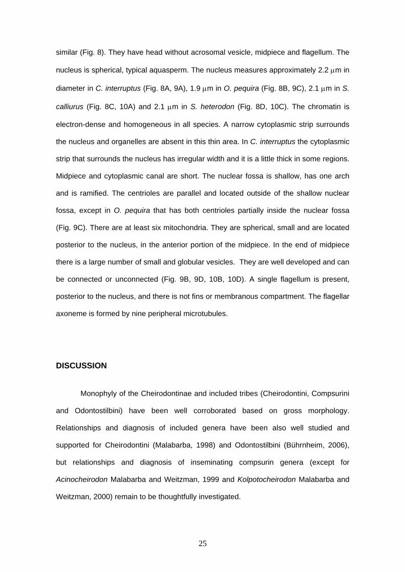
similar (Fig. 8). They have head without acrosomal vesicle, midpiece and flagellum. The
nucleus is spherical, typical aquasperm. The nucleus measures approximately 2.2 μm in
diameter in C. interruptus (Fig. 8A, 9A), 1.9 μm in O. pequira (Fig. 8B, 9C), 2.1 μm in S.
calliurus (Fig. 8C, 10A) and 2.1 μm in S. heterodon (Fig. 8D, 10C). The chromatin is
electron-dense and homogeneous in all species. A narrow cytoplasmic strip surrounds
the nucleus and organelles are absent in this thin area. In C. interruptus the cytoplasmic
strip that surrounds the nucleus has irregular width and it is a little thick in some regions.
Midpiece and cytoplasmic canal are short. The nuclear fossa is shallow, has one arch
and is ramified. The centrioles are parallel and located outside of the shallow nuclear
fossa, except in O. pequira that has both centrioles partially inside the nuclear fossa
(Fig. 9C). There are at least six mitochondria. They are spherical, small and are located
posterior to the nucleus, in the anterior portion of the midpiece. In the end of midpiece
there is a large number of small and globular vesicles. They are well developed and can
be connected or unconnected (Fig. 9B, 9D, 10B, 10D). A single flagellum is present,
posterior to the nucleus, and there is not fins or membranous compartment. The flagellar
axoneme is formed by nine peripheral microtubules.
DISCUSSION
Monophyly of the Cheirodontinae and included tribes (Cheirodontini, Compsurini
and Odontostilbini) have been well corroborated based on gross morphology.
Relationships and diagnosis of included genera have been also well studied and
supported for Cheirodontini (Malabarba, 1998) and Odontostilbini (Bührnheim, 2006),
but relationships and diagnosis of inseminating compsurin genera (except for
Acinocheirodon Malabarba and Weitzman, 1999 and Kolpotocheirodon Malabarba and
Weitzman, 2000) remain to be thoughtfully investigated.
26
Phylogenetic analyses based on ten ultrastructural sperm morphology characters
(Appendix I) further contributes to the recognition of the Compsurini as monophyletic
and allows hypothesize the relationships among included taxa. Sperm morphology of
the externally fertilized species of Cheirodontini and Odontostilbini, however, seems to
be very conservative (based on the analysis of the specimens available) and has shown
uninformative to hypothesize relationships among included species (Fig. 11).
Although conservative, the aquasperm of the species of Cheirodontini and
Odontostilbini is clearly distinct of those of other externally fertilized characiforms. The
vesicles found in the midpiece present a large variability in shape and size among the
Characiformes, or even are lacking in some of it representatives. Among externally
fertilized cheirodontines, there is a pattern found in the externally examined species that
comprises a large number of small globular vesicles fulfilling most of the midpiece
(Character 10, state 1). Such a pattern has not been found in other Characiformes,
except for a similar pattern described for Citharinus sp. (Mattei et al., 1995), but with
clearly smaller vesicles. The sperm cells of the inseminating species of the Compsurini
(including the aquasperm of Kolpotocheirodon) have a small number of larger and
somewhat elongated vesicles, but there is no clear recognized pattern different of those
of other characiforms (e.g. Aphyocharax anisitsi – Burns et al., 1998:237, fig. 1, and our
observation).
The positioning of Kolpotocheirodon species within the Compsurini has been
supported previously only by the presence of insemination, lacking any morphological
evidence of their affinity to the remaining species of the tribe (Malabarba, 1998).
Regardless the fact Kolpotocheirodon species have an aquasperm with a spherical
nucleus, the longer cytoplasmic canal and elongated midpiece (Character 4, state 1)
support a close relationship with the inseminating compsurins. Aquasperm of externally
fertilized cheirodontines have a short midpiece, truncated posteriorly. Aquasperm of
Kolpotocheirodon species and introsperm of other compsurins have the midpiece

27
elongated with longer cytoplasmic canal, progressively narrow posteriorly (not truncate
posteriorly). Thus, although classified as an aquasperm due to the spherical nucleus,
the spermatozoon of Kolpotocheirodon is derived and different of those externally
fertilized species.
Elongate midpiece is a characteristic associated with insemination (Jamieson,
1991; Mattei, 1991). The resulting enlargement of midpiece is believed to make possible
an increase in the sperm capacity of generate energy, especially because of the greater
amount of mitochondria (Koya et al., 2002). This could provide energy for sperm
dispersal throughout the ovary and could also prolong viable sperm storage within the
ovary (Pecio and Rafinski, 1994; Yao et al, 1995). Most of the externally fertilizing
teleosts have a short midpiece (Burns and Weitzman, 2005). Although Brycon and
Salminus considered two primitive genera in Characidae have long midpiece and have
externally fertilizing (Veríssimo-Silveira et al., 2006).
Two of the three putative synapomorphies that supports a monophyletic clade
containing C. heterura, M. uruguayanae, A. melanogramma, S. hastatus, “Odontostilbe”
mitoptera and “O.” dialeptura are related to nuclear elongation (Characters 1 and 2).
Compsura heterura and M. uruguayanae exhibit very similar bullet shaped spermatozoa
(Character 1, state 1), while S. hastatus, A. melanogramma, “O”. mitoptera and “O”.
dialeptura have a clearly elongated nucleus (Character 1, state 2). Regardless the fact
this character was considered unordered in the analysis, the resulting hypothesis
supports the bullet shaped spermatozoa as a synapomorphy to the clade containing C.
heterura, M. uruguayanae, A. melanogramma, S. hastatus, “Odontostilbe” mitoptera and
“O.” dialeptura and an intermediary stage in the evolution of the elongate spermatozoa
of the clade containing A. melanogramma, S. hastatus, “Odontostilbe” mitoptera and
“O.” dialeptura.
Nucleus size and shape, related to the relation between its length and width
28
(Character 2) also allowed the recognition of three different stages in spermatozoa
elongation. The primitive condition in Cheirodontinae seems to be that found in all
examined externally fertilized species and in Kolpotocheirodon theloura, that presents
an spherical nucleus, whose width mean ranges from 1.67 to 2.17 μm and nucleus
length mean ranges from 1.59 to 2.18μm (Character 2, state 0). Two discrete derived
states related to nucleus elongation were found, corresponding to nucleus width mean
ranging from 1.03 to 1.40 μm and nucleus length mean ranging from 2.40 to 2.96μm
(Character 2, state 1) found in C. heterura, M. uruguayanae, A. melanogramma,
“Odontostilbe” mitoptera and “O.” dialeptura, and nucleus width mean 0.7 μm and
nucleus length mean 3.17μm (Character 2, state 2) observed in S. hastatus. Again, even
considered unordered in the analysis, the elongation of the sperm cells (Character 2,
state 1) consists in a synapomorphy of the Clade containing C. heterura, M.
uruguayanae, A. melanogramma, S. hastatus, “Odontostilbe” mitoptera and “O.”
dialeptura and an intermediary stage in the evolution of the extremely elongate and
narrow spermatozoa of S. hastatus (Character 2, state 2).
Nuclear elongation is the spermatozoon morphological specialization most
associated with insemination (Jamieson, 1991; Burns et al., 1995, 1997; Burns and
Weitzman, 2005). There are many possibilities why insemination and nuclear
elongation could be an adaptive advantage (Ginzburg, 1968; Gardiner, 1978; Jamieson,
1991; Burns and Weitzman, 2005) all emphasizing the less energy spend in fertilization.
According to Burns and Weitzman (2005), the elongate nuclei allows sperm cells to be
thinner, streamlined and to have lesser head area, being able to pass through female
gonopore in more number at the same time, moving easily through female reproductive
tract and suffering less resistance to cross fluids, tending to move at forward direction
(Gardiner, 1978). In addition, it enables side-to-side sperm alignment and clumping,
facilitating spermatozoa to flow together (Burns and Weitzman, 2005), possibly
facilitating flow and reducing losses of cells to the environment (Ginzburg, 1968).
29
The third character supporting the monophyly of the clade C. heterura, M.
uruguayanae, A. melanogramma, S. hastatus, “Odontostilbe” mitoptera and “O.”
dialeptura is related to the position of the centriolar complex in relation to the nuclear
fossa. In externally fertilized species and Kolpotocheirodon theloura both centrioles are
located outside the nuclear fossa (Character 5, state 0), while C. heterura, M.
uruguayanae, A. melanogramma and S. hastatus possess the anterior tip of the
proximal centriole located inside the nuclear fossa and the distal centriole outside
(Character 5, state 1). “Odontostilbe” mitoptera and “O.” dialeptura have both centrioles
located outside the nuclear fossa, but this is considered under parsimony a secondary
reversal and a synapomorphy of these two species.
Acinocheirodon melanogramma, “O”. dialeptura, “O”. mitoptera and S. hastatus
constitute a monophyletic subclade of the Compsurini, supported by four characters
shared by all species: the shape of the nucleus (Character 1, state 2, discussed above),
the absence of nuclear rotation (Character 3), the relative position of the centrioles
(Character 6), and the position of the mitochondria (Character 7).
Mattei (1970) has classified two types of spermiogenesis, according to the
presence or absence of nuclear rotation. In both types, the flagellum develops laterally
to the nucleus. In the type I, nuclear rotation takes place and the flagellum is located
perpendicular to the nucleus (Character 3, state 0). In the type II, nucleus does not
rotate and flagellum remains lateral to the nucleus (Character 3, state 1). The nuclear
rotation occurs at most of Characiformes. Spermiogenesis type I was observed in the
externally fertilized cheirodontins and odontostilbins and some compsurins species as
C. heterura, M. uruguayanae and K. theloura. Spermiogenesis type II is found in S.
hastatus, A. melanogramma, “O”. mitoptera and “O”. dialeptura and constitutes a
synapomorphy of those species. Glandulocaudinae (Burns et al., 1998; Pecio and
Rafinski, 1999), Stevardiinae (Burns et al., 1998; Pecio et al., 2005), one
Acestrorhynchidae Acestrorhyncus falcatus (Matos et al., 2000), one Tetragonopterinae,
30
Bryconamericus stramineus (Gusmão-Pompiani et al., in press) and three species
incertae sedis in Characidae, “Cheirodon” stenodon (Gusmão-Pompiani et al., in press)
Bryconadenos tanaothoros (Weitzman et al., 2005) and Brittanichthys axelrodi (Javonillo
et al., 2007) also have type II spermiogenesis, but independently acquired from those of
some inseminating compsurins.
The perpendicular position of the centrioles (Character 6, state 1) and the
mitochondria lodged in the posterior tip of the nucleus aside a small portion near the
midlength and distant from the origin of the flagellum (Character 7, state 1) are also
synapomorphic for the clade containing A. melanogramma, “O”. dialeptura, “O”.
mitoptera and S. hastatus. The mitochondria position in these species is not a
consequence and redundant with the absence of nuclear rotation. In glandulocaudines
and stevardiines without nuclear rotation, the mitochondria are usually located in a
certain extension along the flagellum, aside and behind the nucleus.
The last clade supported by sperm ultrastructural characters is formed by “O”.
dialeptura and “O”. mitoptera. Both species are provisionally allocated in Odontostilbe,
awaiting for a new generic allocation in the Compsurini (Malabarba, 1998). The results
obtained herein supports their recognition in a new and monophyletic genus, but
information on the ultrastructure of the spermatozoa are still lacking in four species of
the compsurins. Monophyly of this clade is supported by the position of both centrioles
outside the nuclear fossa (character 5, state 0 - see discussion above), by the shape of
the anterior border of the nucleus, asymmetric, clearly oblique to the longest axis of the
nucleus (Character 8, state 1), and in the position of the vesicles, located after the
mitochondria, in the basal region of the midpiece and along the cytoplasmic collar in one
side of the nucleus (Character 10, state 1). The anterior border of the nucleus is
semicircular or emarginated in other Cheirodontinae, inseminating or not. The oblique
anterior anterior border of the nucleus observed in “O”. dialeptura and “O”. mitoptera is
unique among described spermatozoa of characiforms. Likewise, the presence of
31
vesicles along the nucleus inside the cytoplasmic collar was observed only in “O”.
dialeptura and “O”. mitoptera, and has not been found in other characiforms.
The analysis presented herein supported the monophyly of the Compsurini and
allowed hypothesize relationships among representatives of all genera, as well as with
“O”. dialeptura and “O”. mitoptera. These results conflict the preliminary hypothesis of
relationships among compsurins presented in Malabarba (1998), suggesting further
investigation is needed in gross morphology of these taxa.
ACKNOWLEDGMENTS
Irani Quagio-Grassiotto and technicians from Universidade Estadual Paulista,
Botucatu, Brazil and technicians from Microscopia da Universidade Federal do Rio
Grande do Sul, Porto Alegre, Brazil for help with transmission electron microscopy
(TEM). We thank technicians from Centro de Microscopia da Pontifícia Universidade
Católica do Rio Grande do Sul, Porto Alegre, Brazil and Suresh Benjamim from George
Washington University (GWU) for assistance with the critical-point dried and scanning
electron microscopy, Gavin Riordan from GWU for help with TEM stains techniques and
Cristina Motta Buhrnheim from Universidade Federal do Amazonas, Manaus, Brazil for
provide some specimens used. This research was supported by Conselho Nacional de
Desenvolvimento Científico e Tecnológico (CNPq; Proc. 476821/2003-7; Proc.
478002/2006-8) from Brazil.
32
LITERATURE CITED
Buhrnheim CM. 2006. Sistemática de Odontostilbe Cope, 1870 com a proposição de
uma nova tribo Odontostilbini e redefinição dos gêneros incertae sedis de
Cheirodontinae (Ostariophysi: Characiformes: Characidae). Tese de doutorado.
Pontifícia Universidade Católica de Porto Alegre, Porto Alegre, Brazil. 315p.
Burns JR, Weitzman SH. 2005. Insemination in ostariophysan fishes. In: Uribe MC,
Grier HJ, editors. Viviparous Fishes. Homestead, FL: New Life Publications. p 107-
134.
Burns JR, Weitzman SH, Grier HJ, Menezes NA. 1995. Internal fertilization, testis and
sperm morphology in glandulocaudine fishes (Teleostei: Characidae:
Glandulocaudinae). J Morphol 224:131-145.
Burns JR, Weitzman SH, Lange KR, Malabarba LR. 1998. Sperm ultrastructure in
characid fishes (Teleostei: Ostariophysi). In: Malabarba LR, Reis RE, Vari RP,
Lucena ZMS, Lucena CAS, editors. Phylogeny and Classification of Neotropical
Fishes. Porto Alegre, Brazil: EDIPUCRS. pp 235-244.
Burns JR, Weitzman SH, Malabarba LR. 1997. Insemination in eight species of
cheirodontine fishes (Teleostei: Characidae: Cheirodontinae). Copeia 1997: 433-
438.
Gardiner DM. 1978. Fine structure of the spermatozoon of the viviparous teleost
Cymatogaster aggregata. J Fish Biol 13:435-438.
Ginzburg AS. 1968. Fertilization in Fishes and the Problem of Polyspermy. Moscow:
Academy of Science USSR. 366 pp.
33
Gusmão-Pompiani P. 2003. Ultraestrutura da espermiogênese e dos espermatozóides
de peixes da ordem Characiformes, família Characidae (Teleostei, Ostariophysi):
uma abordagem filogenética. Tese de doutorado, Instituto de Biociências da
Universidade Estadual Paulista, São Paulo. 86 pp.
Jamieson BGM. 1991. Fish evolution and systematics: evidence from spermatozoa.
Cambridge, Cambridge University Press, 319 pp.
Koya Y, Munehara H, Takano K. 2002. Sperm storage and motility in the ovary of the
marine sculpin Alcichthys alcicornis (Teleostei: Scorpaeniformes), with internal
gametic association. J Exp Zool 292:145-155.
Malabarba LR. 1998. Monophyly of the Cheirodontinae, characters and major clades
(Ostariophysi: Characidae). In: Malabarba LR, Reis RE, Vari RP, Lucena ZMS,
Lucena CAS, editors. Phylogeny and Classification of Neotropical Fishes. Porto
Alegre, Brazil: EDIPUCRS. pp 193-233.
Malabarba LR. 2003. Subfamily Cheirodontinae (Characins, tetra) In: Reis RE,
Kullander SO, Ferraris CJ. Check list of the freshwater fishes of South and Central
America. Porto Alegre, Brazil: EDIPUCRS. pp 215- 221.
Malabarba LR, Weitzman SH. 1999. A new genus species of south american fishes
(Teleostei: Characidae: Cheirodontinae) with a derived caudal fin, incluing comments
about inseminating Cheirodontinae. Proc. Biol. Soc. Wash. 112 (2): 411-432.
Malabarba LR, Weitzman SH. 2000. A new genus and species of inseminating fishes
(Teleostei: Characidae: Compsurini) from South America with uniquely derived
caudal-fin dermal papillae. Proc. Biol. Soc. Wash. 113 (2): 269-283.
Mattei X. 1970. Spermiogenése des poisson. In Comparative Spermatology Baccetti B,
editor, New York: Academic Press. pp. 57-72.
34
Mattei X. 1991. Spermatozoon ultrastructure and its systematic implications in fishes.
Can J Zool 69: 3038-3055.
Mattei X, Marchand B, Thiaw OT. 1995. Unusual midpiece in the spermatozoon of the
teleost fish, Citharinus sp. J. Submicrosc. Cytol. Pathol., 27, p.189-191.
Matos E, Matos P, Corral L, Azevedo C. 2000. Estrutura fina do espermatozóide de
Acestrorhyncus falcatus Bloch (Teleostei, Characidae) da região norte do Brasil.
Revista Brasileira de Zoologia 17, 747-752.
Oliveira CLC, Malabarba LR, Burns JR. Chapter 3, this volume. Gill-derived glands
morphology in Cheirodontinae (Teleostei: Characidae).
Pecio A, Rafiński J. 1994. Structure of the testes, spermatozoa and spermatozeugmata
of Mimagoniates barberi Regan, 1907 (Teleostei: Characidae), an internally
fertilizing, oviparous fish. Acta Zool 75:179-185.
Pecio A, Rafiński, J. 1999. Spermiogenesis in Mimagoniates barberi (Teleostei:
Ostariophysi: Characidae), an oviparous, internally fertilizing fish. Acta Zool 80: 35-
45.
Pecio A, Burns JR, Weitzman SH. 2005. Sperm and spermatozeugmata ultrastructure in
the inseminating species Tyttocharax cochui, T. tambopatensis, and Scopaeocharax
rhinodus (Pisces: Teleostei: Characidae: Glandulocaudinae: Xenurobryconini). J
Morphol 263:216-226.
Veríssimo-Silveira R, Gusmão-Pompiani P, Vicentini CA, Quagio-Grassiotto I. 2006.
Spermiogenesis and spermatozoa ultrastruture in Salminus and Brycon, two
primitive genera in Characidae (Teleostei: Ostariophysi: Characiformes). Acta Zool
87: 305-313.

35
Weitzman SH, Menezes NA, Evers H-G, Burns JR. 2005. Putative relationships among
inseminating and externally fertilizing characids, with a description of a new genus
and species of Brazilian inseminating fish bearing an anal-fin gland in males
(Characiformes: Characidae). Neotrop Ichthyol 3:329-360.
Yao Z, Emerson CJ, Crim LW. 1995. Ultrastructure of the spermatozoa and eggs of the
ocean pout (Macrozoarces americanus), an internally fertilizing marine fish. Mol
Reprod Dev 42: 58-64.
36
Appendix A
List of Characters
Two autapomorphies of M. uruguayanae, presence of accessory microtubules and
striated rootlet (see Oliveira et al., this volume) were not used in analyses, because
autapomorphies are not informative to hypothesize relationships.
Character 1 (CI = 1.00) - Nucleus shape (unordered) [modified from character 70 in
Malabarba, 1998]:
0= spherical
1= bullet shaped
2= elongated
The spherical nucleus that constitutes the typical aquasperm (State 0) was assigned
to the hypothetical outgroup since a spherical nucleus represent the primitive condition
among externally fertilized characiforms. The elongate nucleus found in some other
representatives of the Characidae have been considered as non-homologous to the
condition found in Compsurini (Malabarba, 1998; Weitzman and Menezes, 1998;
Weitzman et al., 2005). This character was considered unordered because we consider
the bullet shaped cannot be assumed as an intermediary step in the evolution of the
elongate nucleus.
Character 2 (CI = 1.00) - Relation between length and width of nucleus (ordered).
0= nucleus width mean ranging from 1.67 to 2.17 μm; and nucleus length mean
ranging from 1.59 to 2.18 μm.
1= nucleus width mean ranging from 1.03 to 1.40 μm; and nucleus length mean
ranging from 2.40 to 2.96 μm.
2= nucleus width mean 0.7 μm; and nucleus length mean 3.17 μm.

37
The analysis of the means of the length and width of the nucleus per species
allowed the recognition of three distinct classes of spermatozoa shape, based on both
three distinct and discontinuous ranges of nucleus length and three distinct and
discontinuous ranges of nucleus width (Fig. 12). The first class includes the typical
aquasperm with similar measurements of length and width, shared by all externally
fertilized species and the inseminating species of Kolpotocheirodon. The second class
includes elongated sperm shared by Compsura heterura, Acinocheirodon
melanogramma, M. uruguayanae uruguayanae, “Odontostilbe” dialeptura and “O”.
mitoptera. The third class includes only Saccoderma that possess an even more
elongate and narrow sperm nucleus.
The character was considered ordered in the analysis and state 0 corresponding
to aquasperm assigned to the hypothetical outgroup.
Character 3 (CI = 1.00) - Nuclear rotation (ordered):
0= present with flagellum posterior to the nucleus
1= absent with flagellum attached laterally to the nucleus
The character was considered ordered in the analysis and state 0 corresponding
to aquasperm assigned to the hypothetical outgroup.
Characters 4 (CI = 1.00) - Shape of midpiece and length of citoplasmatic canal
(ordered):
0 = midpiece roundish nearly truncated posteriorly and citoplasmatic canal length
approximately half nucleus length
1 = midpiece elongated not truncated posteriorly; citoplasmatic canal length is longer
than half nucleus length
The character was considered ordered in the analysis, and state 0,
corresponding to aquasperm, was assigned to the hypothetical outgroup.
38
Character 5 (CI = 1.00) - Position of centriolar complex in relation to the nuclear fossa
(unordered):
0= both centrioles are outside the nuclear fossa.
1= anterior tip of the proximal centriole is located inside the nuclear fossa and the
distal centriole is outside.
2= both centrioles are partially inside the nuclear fossa.
The position of the centriolar complex was found in two states in the ingroup, but it is
also variable in the outgroup. In the outgroup, the state 0 was observed in Knodus, and
state 1 in Charax. Brycon microlepis (=Brycon hilarii) has both centrioles inside the
nuclear fossa and Bryconamericus stramineus has proximal centriole is inside the
nuclear fossa and distal centriole is outside. It is not possible to order this character and
it was considered uninformed in the hypothetical outgroup.
Character 6 (CI = 0.50) - Centrioles (unordered):
0= parallel
1= perpendicular
This Character is variable in the outgroup and was considered uninformed in the
hypothetical outgroup. It is not possible to order this character.
Character 7 (CI = 1.00) - Mitochondria position (ordered):
0= in the midpiece, posterior to the nucleus and around the origin of the flagellum
1= near the tip of nucleus (without nuclear rotation) and distant from the origin of the
flagellum
The mitochondria location in A. melanogramma, “O”. dialeptura, O mitoptera and
Saccoderma (state 1) is clearly derived and not similar to the mitochondria position in
other inseminating characid species that do not present nuclear rotation. In these
species mitochondria is lodged in the posterior tip of the nucleus aside a small portion
near the midlength of flagellum. In glandulocaudines and stevardiines without nuclear
39
rotation, the mitochondria are usually located in a certain extension along the flagellum,
aside and behind the nucleus.
Character 8 (CI = 1.00) - Anterior border of the nucleus (ordered):
0= semicircular or emarginated
1= asymmetric, clearly oblique to the longest axis of the nucleus
The anterior border of the nucleus in “O”. dialeptura and “O”. mitoptera is oblique,
with the lateral of the nucleus opposite to the flagellum clearly longer than the lateral of
the nucleus where the flagellum is attached. In comparative taxa it is observed only the
state 0.
Character 9 (CI = 1.00) - Vesicles (ordered):
0= elongated vesicles
1= large number of small and globular vesicles
The vesicles found in the midpiece present a large variability in shape and size
among the Characiformes, or even are lacking in some of it representatives. Among
cheirodontines, there is a pattern found in the externally examined species that
comprises a large number of small and globular vesicles fulfilling most of the midpiece.
Such a pattern has not been found in other Characiformes, except for a similar pattern
described for Citharinus sp. (Mattei et al., 1995), but with clearly smaller vesicles.
The inseminating species of the Cheirodontinae have a small number of larger
and somewhat elongated vesicles. There is no clear recognized pattern common to all
inseminating species of the Cheirodontinae and different of those of all outgroup
characiforms (e.g. Aphyocharax anisitsi - Burns et al., 1998:237, fig. 1) being assigned
State 0 for these taxa.
Character 10 (CI = 1.00) - Vesicles position (ordered):
0 = located only after the mitochondria, in the basal region of the midpiece.
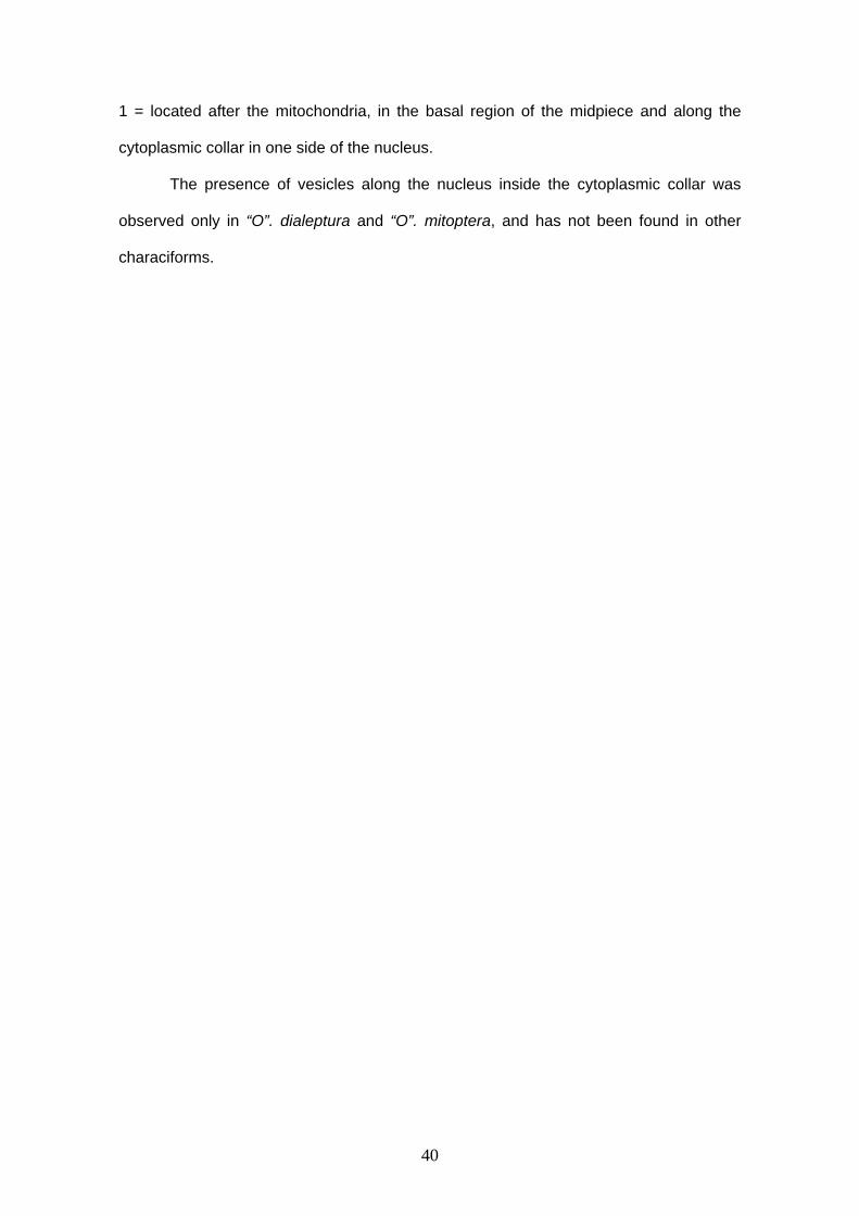
40
1 = located after the mitochondria, in the basal region of the midpiece and along the
cytoplasmic collar in one side of the nucleus.
The presence of vesicles along the nucleus inside the cytoplasmic collar was
observed only in “O”. dialeptura and “O”. mitoptera, and has not been found in other
characiforms.

41
Appendix B. Characters matrix of 10 characters for the 11 taxa of Cheirodontinae.
1 2 3 4 5 6 7 8 9 10
outgroup 0 0 0 0 0 0 0 0 0 0
Kolpotocheirodon theloura 0 0 0 1 0 0 0 0 0 0
Compsura heterura 1 1 0 1 1 0 0 0 0 0
Macropsobrycon uruguayanae 1 1 0 1 1 0 0 0 0 0
Acinocheirodon melanogramma 2 1 1 1 1 1 1 0 0 0
Saccoderma hastatus 2 2 1 1 1 1 1 0 0 0
“Odontostilbe” dialeptura 2 1 1 1 0 1 1 1 0 1
“Odontostilbe” mitoptera 2 1 1 1 0 1 1 1 0 1
Cheirodon interruptus 0 0 0 0 0 0 0 0 1 0
Odontostilbe pequira 0 0 0 0 2 0 0 0 1 0
Serrapinnus calliurus 0 0 0 0 0 0 0 0 1 0
Serrapinnus heterodon 0 0 0 0 0 0 0 0 1 0
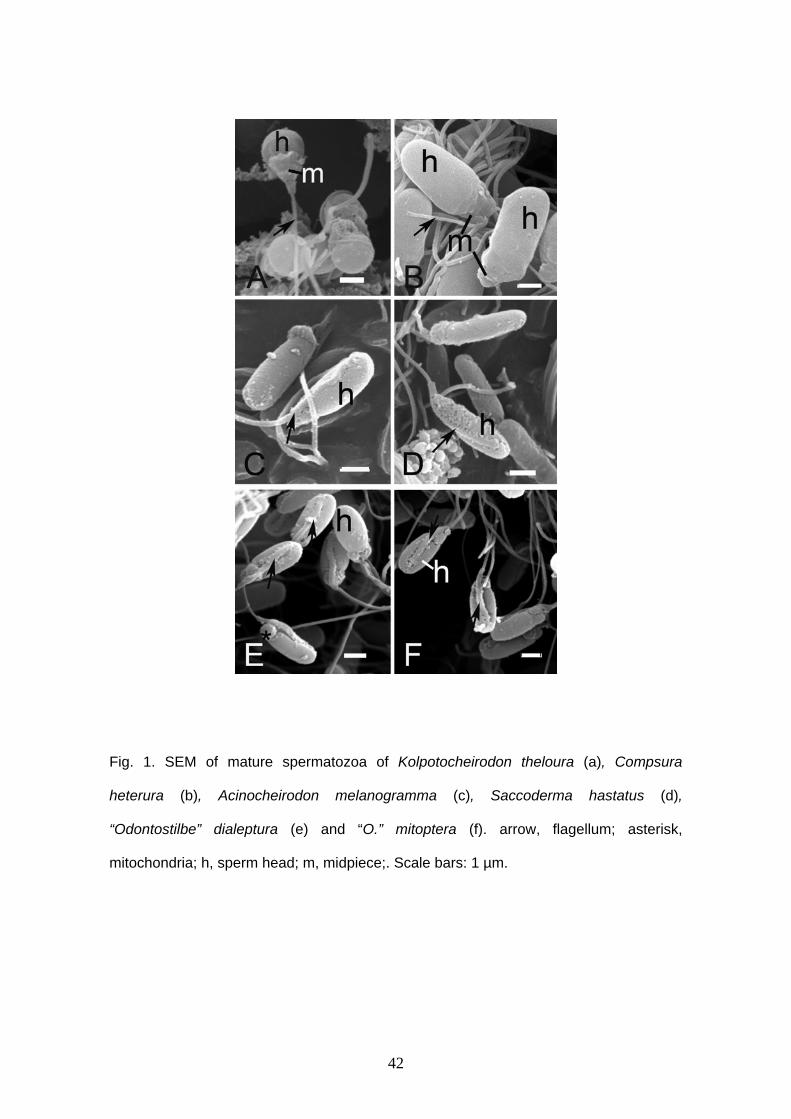
42
Fig. 1. SEM of mature spermatozoa of Kolpotocheirodon theloura (a), Compsura
heterura (b), Acinocheirodon melanogramma (c), Saccoderma hastatus (d),
“Odontostilbe” dialeptura (e) and “O.” mitoptera (f). arrow, flagellum; asterisk,
mitochondria; h, sperm head; m, midpiece;. Scale bars: 1 µm.
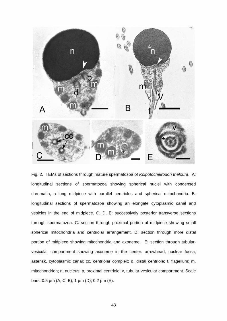
43
Fig. 2. TEMs of sections through mature spermatozoa of Kolpotocheirodon theloura. A:
longitudinal sections of spermatozoa showing spherical nuclei with condensed
chromatin, a long midpiece with parallel centrioles and spherical mitochondria. B:
longitudinal sections of spermatozoa showing an elongate cytoplasmic canal and
vesicles in the end of midpiece. C, D, E: successively posterior transverse sections
through spermatozoa. C: section through proximal portion of midpiece showing small
spherical mitochondria and centriolar arrangement. D: section through more distal
portion of midpiece showing mitochondria and axoneme. E: section through tubular-
vesicular compartment showing axoneme in the center. arrowhead, nuclear fossa;
asterisk, cytoplasmic canal; cc, centriolar complex; d, distal centriole; f, flagellum; m,
mitochondrion; n, nucleus; p, proximal centriole; v, tubular-vesicular compartment. Scale
bars: 0.5 µm (A, C; B); 1 µm (D); 0.2 µm (E).
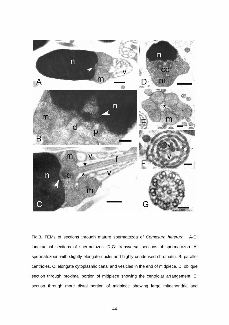
44
Fig.3. TEMs of sections through mature spermatozoa of Compsura heterura. A-C:
longitudinal sections of spermatozoa. D-G: transversal sections of spermatozoa. A:
spermatozoon with slightly elongate nuclei and highly condensed chromatin. B: parallel
centrioles. C: elongate cytoplasmic canal and vesicles in the end of midpiece. D: oblique
section through proximal portion of midpiece showing the centriolar arrangement. E:
section through more distal portion of midpiece showing large mitochondria and

45
axoneme. F: section through tubular-vesicular compartment showing axoneme in the
center. G: section through typical axoneme. arrowhead, nuclear fossa; asterisk,
cytoplasmic canal; cc, centriolar complex; d, distal centriole; f, flagellum; m,
mitochondrion; n, nucleus; p, proximal centriole; v, tubular-vesicular compartment.
Scale bars: 0.5 µm (A, C, D); 0.2 µm (B, E, F); 50 nm (G).
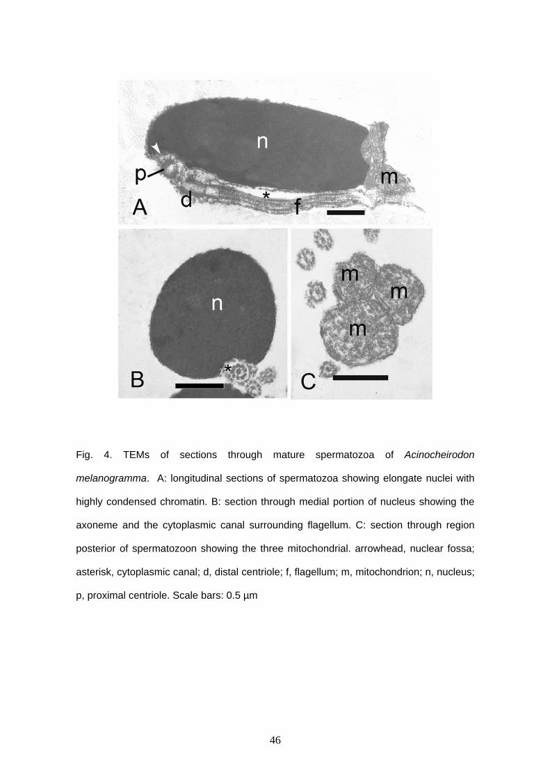
46
Fig. 4. TEMs of sections through mature spermatozoa of Acinocheirodon
melanogramma. A: longitudinal sections of spermatozoa showing elongate nuclei with
highly condensed chromatin. B: section through medial portion of nucleus showing the
axoneme and the cytoplasmic canal surrounding flagellum. C: section through region
posterior of spermatozoon showing the three mitochondrial. arrowhead, nuclear fossa;
asterisk, cytoplasmic canal; d, distal centriole; f, flagellum; m, mitochondrion; n, nucleus;
p, proximal centriole. Scale bars: 0.5 µm
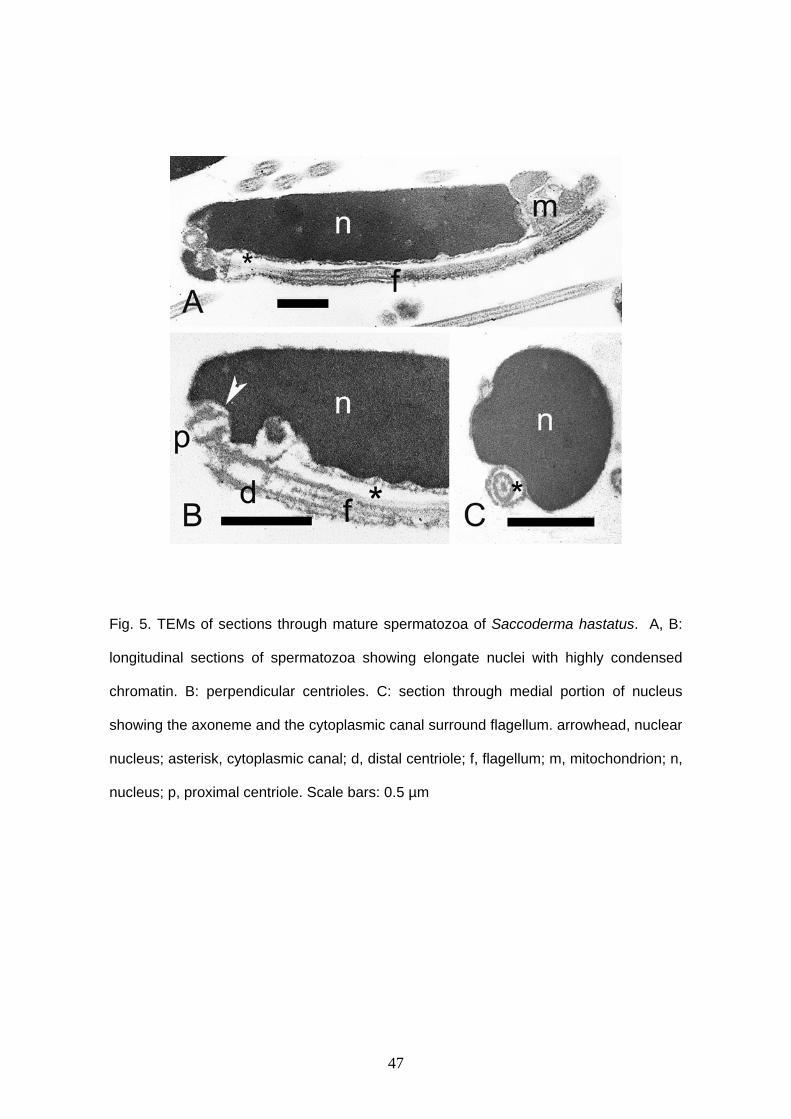
47
Fig. 5. TEMs of sections through mature spermatozoa of Saccoderma hastatus. A, B:
longitudinal sections of spermatozoa showing elongate nuclei with highly condensed
chromatin. B: perpendicular centrioles. C: section through medial portion of nucleus
showing the axoneme and the cytoplasmic canal surround flagellum. arrowhead, nuclear
nucleus; asterisk, cytoplasmic canal; d, distal centriole; f, flagellum; m, mitochondrion; n,
nucleus; p, proximal centriole. Scale bars: 0.5 µm
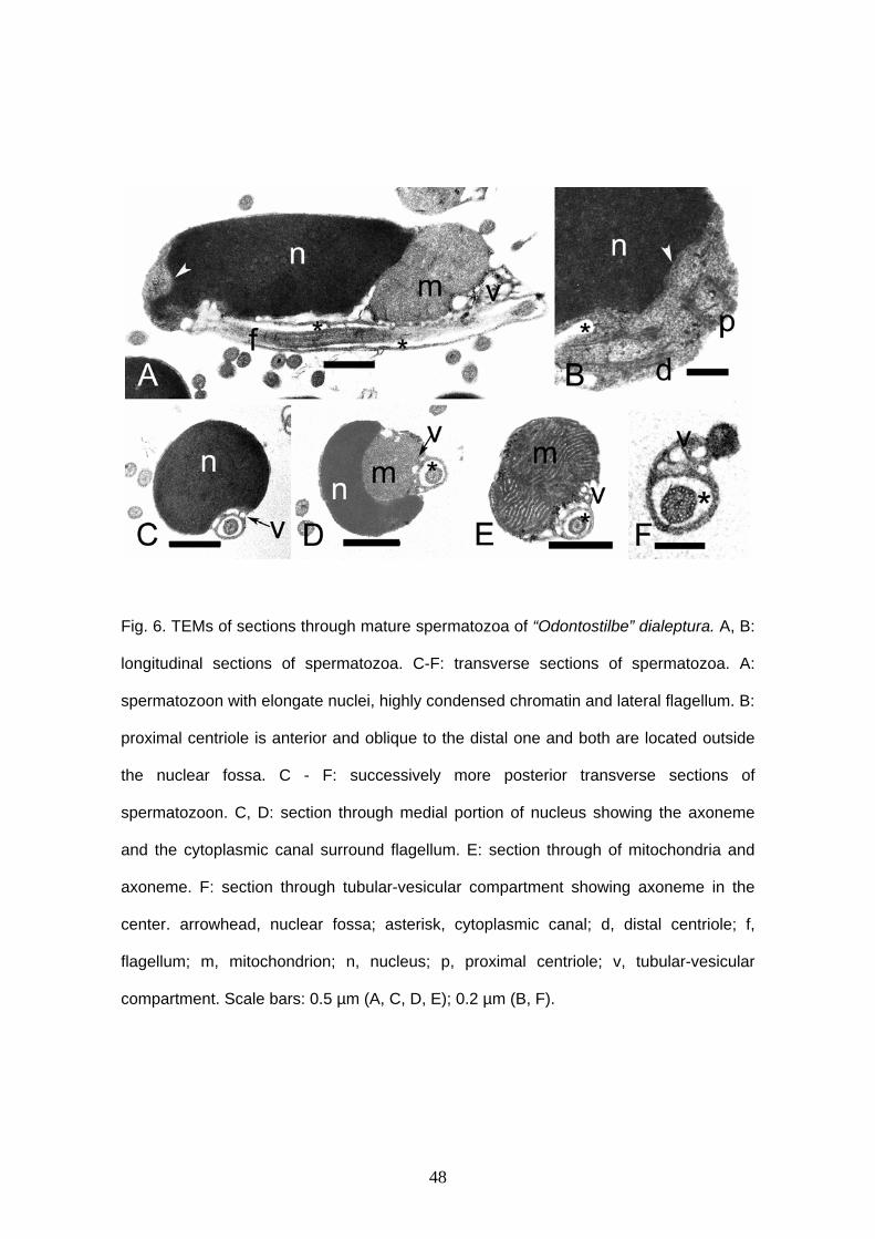
48
Fig. 6. TEMs of sections through mature spermatozoa of “Odontostilbe” dialeptura. A, B:
longitudinal sections of spermatozoa. C-F: transverse sections of spermatozoa. A:
spermatozoon with elongate nuclei, highly condensed chromatin and lateral flagellum. B:
proximal centriole is anterior and oblique to the distal one and both are located outside
the nuclear fossa. C - F: successively more posterior transverse sections of
spermatozoon. C, D: section through medial portion of nucleus showing the axoneme
and the cytoplasmic canal surround flagellum. E: section through of mitochondria and
axoneme. F: section through tubular-vesicular compartment showing axoneme in the
center. arrowhead, nuclear fossa; asterisk, cytoplasmic canal; d, distal centriole; f,
flagellum; m, mitochondrion; n, nucleus; p, proximal centriole; v, tubular-vesicular
compartment. Scale bars: 0.5 µm (A, C, D, E); 0.2 µm (B, F).

49
Fig. 7. TEMs of sections through mature spermatozoa of “Odontostilbe” mitoptera. A, B,
C: longitudinal sections of spermatozoa. D, E: transverse sections of spermatozoa. A:
elongate lateral nucleus with highly condensed chromatin, a big mitochondrion and
vesicles in one side of the nucleus and surround the mitochondrion. B: proximal
centriole is anterior and oblique to the distal one and both are located outside the
nuclear fossa. C: three mitochondria located after the tip of nucleus, lateral flagellum
and vesicles. D: section through medial portion of nucleus showing the axoneme and
the cytoplasmic canal surround flagellum. E: section through tubular-vesicular
compartment showing axoneme in the center. arrowhead, nuclear fossa; asterisk,
cytoplasmic canal; d, distal centriole; f, flagellum; m, mitochondrion; n, nucleus; p,
proximal centriole; v, tubular-vesicular compartment. Scale bars: 0.5 µm (A, B, C, D); 0.2
µm (E).
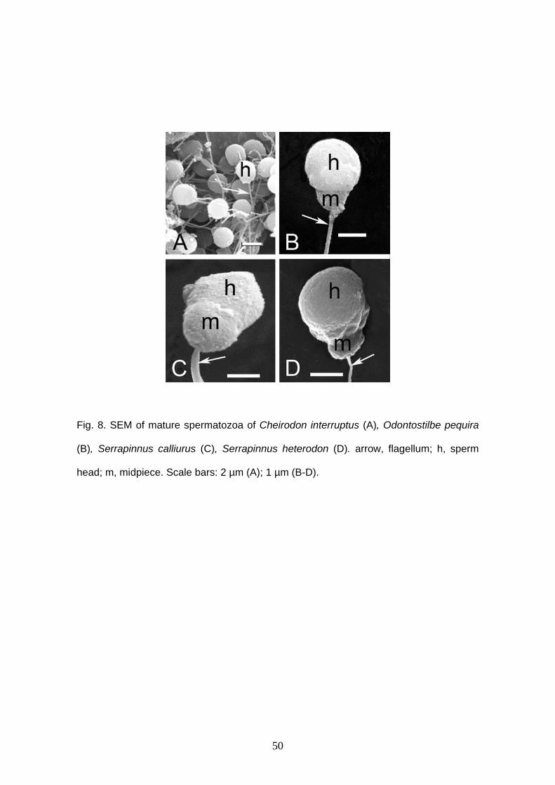
50
Fig. 8. SEM of mature spermatozoa of Cheirodon interruptus (A), Odontostilbe pequira
(B), Serrapinnus calliurus (C), Serrapinnus heterodon (D). arrow, flagellum; h, sperm
head; m, midpiece. Scale bars: 2 µm (A); 1 µm (B-D).
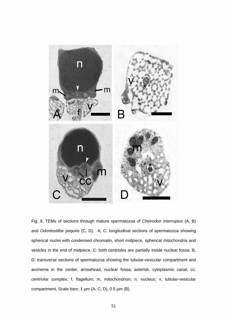
51
Fig. 9. TEMs of sections through mature spermatozoa of Cheirodon interruptus (A, B)
and Odontostilbe pequira (C, D). A, C: longitudinal sections of spermatozoa showing
spherical nuclei with condensed chromatin, short midpiece, spherical mitochondria and
vesicles in the end of midpiece. C: both centrioles are partially inside nuclear fossa. B,
D: transverse sections of spermatozoa showing the tubular-vesicular compartment and
axoneme in the center. arrowhead, nuclear fossa; asterisk, cytoplasmic canal; cc,
centriolar complex; f, flagellum; m, mitochondrion; n, nucleus; v, tubular-vesicular
compartment. Scale bars: 1 µm (A, C, D), 0.5 µm (B).
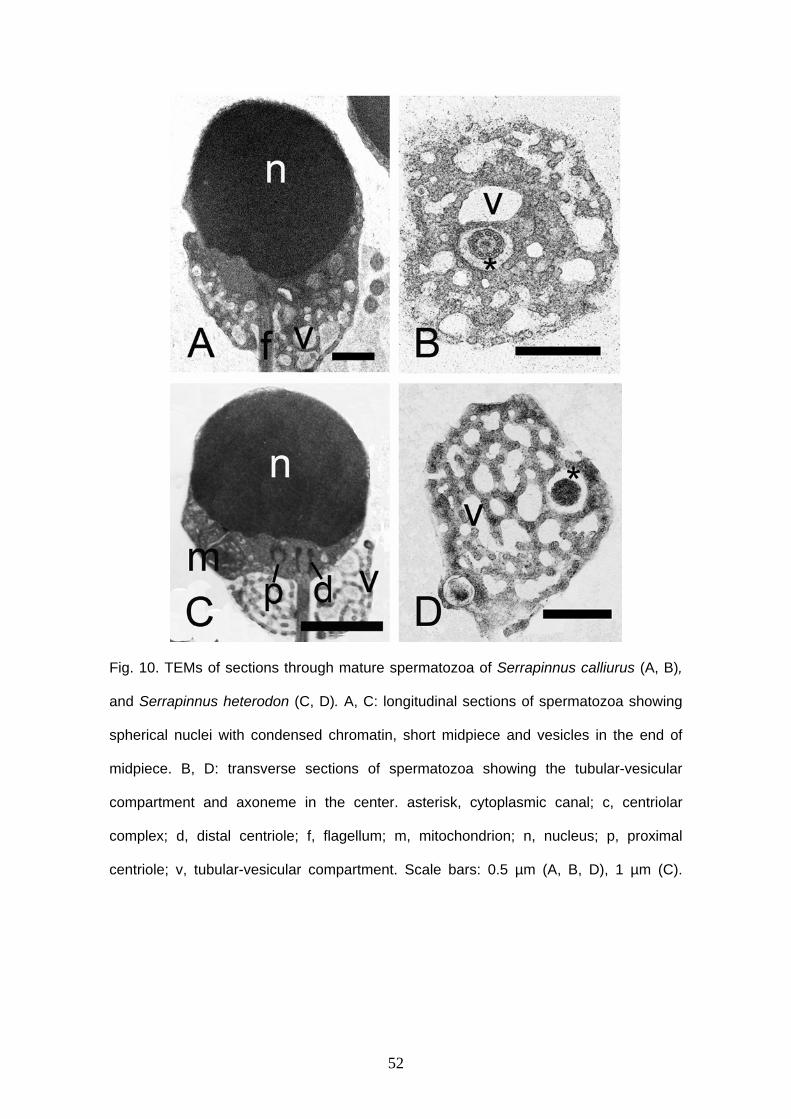
52
Fig. 10. TEMs of sections through mature spermatozoa of Serrapinnus calliurus (A, B),
and Serrapinnus heterodon (C, D). A, C: longitudinal sections of spermatozoa showing
spherical nuclei with condensed chromatin, short midpiece and vesicles in the end of
midpiece. B, D: transverse sections of spermatozoa showing the tubular-vesicular
compartment and axoneme in the center. asterisk, cytoplasmic canal; c, centriolar
complex; d, distal centriole; f, flagellum; m, mitochondrion; n, nucleus; p, proximal
centriole; v, tubular-vesicular compartment. Scale bars: 0.5 µm (A, B, D), 1 µm (C).
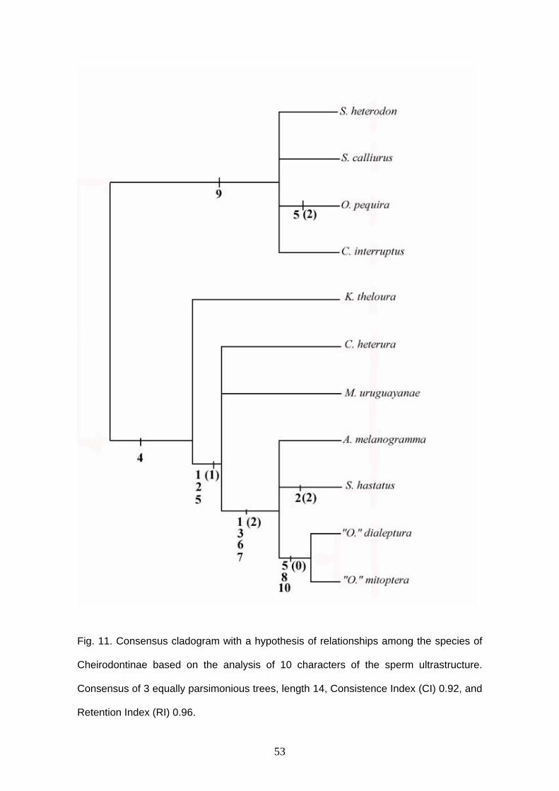
53
Fig. 11. Consensus cladogram with a hypothesis of relationships among the species of
Cheirodontinae based on the analysis of 10 characters of the sperm ultrastructure.
Consensus of 3 equally parsimonious trees, length 14, Consistence Index (CI) 0.92, and
Retention Index (RI) 0.96.
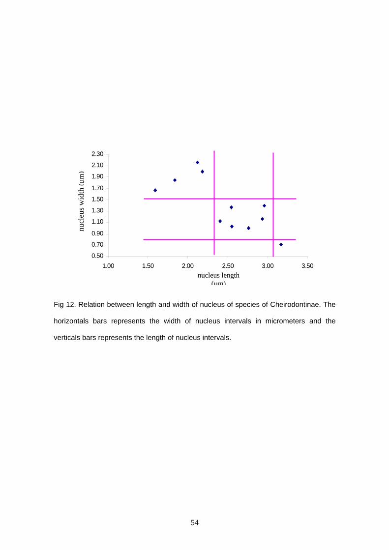
54
Fig 12. Relation between length and width of nucleus of species of Cheirodontinae. The
horizontals bars represents the width of nucleus intervals in micrometers and the
verticals bars represents the length of nucleus intervals.
0.50
0.70
0.90
1.10
1.30
1.50
1.70
1.90
2.10
2.30
1.00 1.50 2.00 2.50 3.00 3.50nucleus length
(µm)
nucl
eus w
idth
(µm
)
55
Capítulo 2: Sperm ultrastructure in the inseminating Macropsobrycon uruguayanae
(Teleostei: Characidae: Cheirodontinae) Author’s names and addresses:
Cristina L. C. de Oliveira 1*, John R. Burns 2, Luiz R. Malabarba 1, 3 and Stanley H.
Weitzman4
1 Departamento de Zoologia, Universidade Federal do Rio Grande do Sul, Porto Alegre,
Rio Grande do Sul, Brazil 2 Department of Biological Sciences, George Washington University, Washington, D.C.
20052, USA 3 Museu de Ciências e Tecnologia, Pontifícia Universidade Católica de Porto Alegre, Rio
Grande do Sul, Brazil. 4 Division of Fishes, Department of Vertebrate Zoology, National Museum of Natural
History, Smithsonian Institution, Washington, DC 20560, USA.
*Correspondence to: Cristina Luísa Conceição de Oliveira. Av. Bento Gonçalves 9500
prédio 43435, sala 104, Departamento de Zoologia, Universidade Federal do Rio
Grande do Sul, Porto Alegre, Rio Grande do Sul, Brazil, CEP 91501-970. E-mail:
Number of text pages: 8
Number of figures: 5
Abbreviated title (running headline): Sperm ultrastructure of Macropsobrycon
Send proofs to Cristina Luísa Conceição de Oliveira, Av. Bento Gonçalves 9500 prédio
43435, sala 104, Departamento de Zoologia, Universidade Federal do Rio Grande do
Sul, Porto Alegre, Rio Grande do Sul, Brazil, CEP 91501-970. Phone: (55 51) 3225
8881. E-mail: [email protected]
Keywords: insemination, sperm ultrastructure, parasperm, accessory microtubules,
striated rootlet.
56
ABSTRACT
Macropsobrycon uruguayanae is a small inseminating characid (tetra) of the tribe
Compsurini. Although spermatozoa can be found within the ovarian cavity close to
oocytes, the exact moment of fertilization has not yet been determined. Mature
spermatozoa have moderately elongate nuclei with electron dense chromatin. During
spermiogenesis nuclear rotation takes place. Elongate mitochondria with lamellar cristae
are found posterior to the nucleus. Centrioles are parallel to one another with the
proximal centriole slightly anterior to the longer distal one. The anterior tip of the
proximal centriole is located within a shallow nuclear fossa. Electron-dense spurs are
associated within the anterior and posterior ends of the distal centriole. Striated
centriolar rootlets radiate both anteriorly and posteriorly from the distal centriole. Nine
longitudinal accessory microtubules surround the axoneme in the proximal flagellum.
The flagellum has a typical 9+2 axoneme with no intratubular differentiation. Atypical
spermatozoa are also found in the testicular lumen. These cells resemble spermatozoa
in most aspects, except that their nuclei are variable in shape, with the granular
chromatin less electron-dense than that seen in spermatozoa. The origin and function of
these cells could not be determined. The specializations seen in the spermatozoa are
discussed as possible adaptations related to the habit of insemination.

57
INTRODUCTION
Species of the subfamily Cheirodontinae, family Characidae, are characterized
by the presence of pedunculated jaw teeth, absence of a humeral spot and a gap
between the muscles covering the anterior portion of the swim bladder, referred to as
the pseudotympanum. This subfamily comprises two tribes, the Cheirodontini and the
Compsurini, plus several incertae sedis genera. Macropsobrycon uruguayanae belongs
to the Compsurini whose species are characterized by the presence of modified anal-fin
hooks, and modified caudal-fin rays and/or scales in mature males, and the ability of
males to transfer sperm to the ovary of the female, i.e. insemination (Burns et al., 1997;
Malabarba, 1998, 2003). Insemination has often been associated with modifications of
the sperm cells that appear to relate to this unique mode of reproduction (Jamieson,
1991; Burns and Weitzman, 2005). Additionally, since insemination is a necessary first
step toward viviparity, examination of the morphological specializations of the
spermatozoa of inseminating species may be useful in understanding the initial steps
toward live-bearing.
Characters obtained from sperm ultrastructure have been useful in hypothesizing
phylogenetic relationships among fishes (Jamieson, 1991; Mattei, 1991). Prior to
Jamieson (1991), no information on sperm ultrastructure was available for any species
of the family Characidae, which is estimated to contain over 962 species of freshwater
fishes (Nelson, 2006). Since that time at least some information on sperm ultrastructure
for over 35 characid species has become available and the data indicate marked
morphological variation, particularly among those species that are inseminating (Burns
and Weitzman, 2005). For cheirodontine species, data are available for two externally
fertilizing species, Serrapinnis notomelas (from Gusmão-Pompiani, 2003) and S. kriegi
(from Burns and Weitzman, 2005), and very limited data for one inseminating species,
Macropsobrycon uruguayanae (Burns et al., 1998; Burns and Weitzman, 2005). The
main purpose of the present study is to provide a detailed analysis of sperm

58
ultrastructure in Macropsobrycon uruguayanae. The morphological specializations found
are discussed as possible adaptations related to the reproductive habit of insemination.
MATERIALS AND METHODS
The three mature males analyzed in this study are catalogued in the fish
collections of the Museu de Ciências e Tecnologia (MCP) and Departamento de
Zoologia, Universidade Federal do Rio Grande do Sul (UFRGS), both from Porto Alegre,
Brazil. The specimens are representative of two populations from Rio Grande do Sul
State, Brazil. One specimen (MCP 18588, SL 39 mm) was collected in a pond near the
margin of the Rio Jacuí, Cachoeira do Sul, laguna dos Patos drainage, 30o03’S, 52
o54’W. The other two specimens were collected in two different localities from the Rio
Uruguay drainage. One specimen (UFRGS 8792, SL 29 mm) was from the Rio Ibicuí da
Faxina between Santana do Livramento and Rosário do Sul, 30o47’22”S, 55o12’41”W,
and the other (UFRGS 8791, 31 mm) from the Sanchuri dam, Uruguaiana, 29o32’41”S,
56 o49’06”W.
The fishes were sacrificed by immersion in a lethal dose of tricaine
methanesulfonate (MS222) and/or a cut through the spinal cord. Immediately
afterwards, whole testes or smaller pieces were placed in modified Karnovsky's fixative
(Ito and Karnosvsky, 1968) and kept under refrigeration until the start of sample
processing.
For scanning electron microscopy (SEM), whole testes were dehydrated in an
ethanol series and critical-point dried. The dried tissue was then attached to stubs with
carbon double-stick tape and teased apart with needles. The sample was sputter-coated
with carbon and gold and viewed in a Philips XL 30 scanning electron microscope.
For transmission electron microscopy (TEM), testes were cut into smaller pieces
(1 mm3), rinsed in phosphate buffer, and postfixed in 1% osmium tetroxide in phosphate
buffer. These were then rinsed in phosphate buffer, dehydrated in an ethanol series,
59
infiltrated, and embedded in Araldite 502. Ultrathin sections were stained with aqueous
uranyl acetate and lead citrate and examined with a JEOL JEM 1200 transmission
electron microscope.
RESULTS
In early spermatids (Fig. 1A), nuclear chromatin is only partially condensed and
nuclear rotation commences. Abundant cytoplasm surrounds the nucleus. Mitochondria
are displaced to the posterior part of the nucleus, which corresponds to the future
midpiece of the spermatozoon. During this phase it is possible to see the developing
cytoplasmic canal and tubular-vesicular compartment (Fig. 1A). During the late
spermatid stage (Fig.1B), nuclear rotation is completed. A narrow strip of cytoplasm,
devoid of organelles, surrounds the nucleus at this time. The centrioles and
mitochondria are located in the anterior part of the developing midpiece near the base of
the nucleus, while the tubular-vesicular system is confined to the posterior part of the
midpiece (Fig. 1B). Nuclear elongation only takes place during late spermiogenesis.
Mature spermatozoa (Figs. 2, 3) have elongate nuclei measuring approximately
3 µm in length and 1.3 µm in width with highly electron dense chromatin. Acrosomal
vesicles are absent. The midpiece contains multiple elongate mitochondria with lamellar
cristae (Figs. 3, 4B,C) and a tubular-vesicular compartment (Figs 3A, 4D). It is possible
to see a long cytoplasmic canal in some sections (Figs. 3C, 4D). The two centrioles are
parallel one to another, with the proximal centriole slightly anterior to the distal centriole.
The anterior portion of the proximal centriole is located within a shallow nuclear fossa
(Fig. 3B). The distal centriole is also longer than the proximal one. Electron-dense spurs
are associated with the anterior and posterior ends of the distal centriole (Fig. 3B).
Striated centriolar rootlets radiate both anteriorly and posteriorly from the distal centriole
(Fig. 3B, C). The single flagellum is completely posterior to the nucleus and lacks
axonemal fins. The axoneme in the proximal region of the flagellum is surrounded by

60
nine longitudinal accessory microtubules (Fig. 4C-F). The axoneme has the typical 9+2
arrangement of microtubules and there is no intratubular differentiation (Fig. 4E, F).
Atypical sperm cells were found in the testicular lumen together with normal
spermatozoa (Fig.3A, 5). These atypical cells were seen in all specimens analyzed.
These cells contain all of the unique structures found in normal spermatozoa, such as
striated centriolar rootlets and accessory microtubules (Fig. 5). However, the nuclei of
these cells are highly irregular in shape and contain uniformly granular chromatin that is
much less electron-dense than that seen in normal spermatozoa (Fig. 5A, D). In
addition, in some cases the spaces between the membranes of the mitochondrial cristae
are wider than in normal spermatozoa.
DISCUSSION
Many externally fertilizing teleosts, including all externally fertilizing characid
species studied to date, have spermatozoa with spherical to slightly ovoid nuclei referred
to as aquasperm (Jamieson, 1991). On the other hand, the great majority of
inseminating characids, including Macropsobrycon uruguayanae, have spermatozoa
with moderately to extremely elongate nuclei with only seven inseminating species
producing typical aquasperm (Burns and Weitzman, 2005; Malabarba et al., 2004).
Several ideas have been put forth as possible selective advantages of nuclear
elongation, which in turn results in elongation of the sperm head. More streamlined
sperm heads may facilitate penetration of egg coats (Jamieson, 1991). At any given
moment, thinner cells would be able to pass through the female gonopore in greater
numbers (Burns and Weitzman, 2005), as well as permit more efficient movement
through the tortuous pathways of the ovary (Gardiner, 1978). In addition, even in
species that do not produce distinct sperm packets, more streamlined cells may
increase the side-to-side alignment and clumping of cells, which in turn may reduce loss
of spermatozoa to the water column during sperm transfer (Ginzburg, 1968; Burns and
Weitzman, 2005). Finally, narrower sperm heads would encounter less resistance
61
moving in the forward direction, so some directionality may be maintained (Burns and
Weitzman, 2005).
After the completion of nuclear rotation in Macropsobrycon uruguayanae, the
nucleus elongates in the direction anterior to the centrioles. Therefore, flagellar
movement in the mature spermatozoon would result in the cell being pushed from
behind the nucleus. The spermatozoa of all other inseminating characids with elongate
nuclei, for which published information is available, do not undergo nuclear rotation. In
these species, during spermiogenesis the nucleus elongates mainly in the direction
posterior to the centrioles (Burns et al., 1998). The species that exhibit this method of
nuclear elongation include the following: in the subfamily Stevardiinae, Diapoma
speculiferum, Diapoma sp., Corynopoma riisei, and Pseudocorynopoma doriae (Burns
and Weitzman, 2005; Burns et al., 1998) and Scopaeocharax rhinodus, Tyttocharax
cochui and T. tambopatensis (Pecio et al, 2005); in the subfamily Glandulocaudinae,
Mimagoniates barberi and M. microlepis (Pecio and Rafiński, 1994, 1999; Burns et al.,
1998); and two species currently incertae sedis, Bryconadenos tanaothoros (Weitzman
et al., 2005) and Brittanichthys axelrodi (Javonillo et al., 2007).
Mature spermatozoa of Macropsobrycon uruguayanae have striated rootlets that
radiate both anteriorly and posteriorly from the distal centriole. Striated rootlets,
composed of the protein rootletin, have been shown to help ciliated cells withstand
mechanical stresses (Yang et al., 2002, 2005). The location of the striated rootlets in M.
uruguayanae suggests that they may support and stabilize the base of the flagellum, i.e.
the distal centriole. Striated rootlets are also present in the inseminating characid,
Brittanichthys axelrodi, a species currently listed as incertae sedis, where it is suggested
that a robust anchorage of the flagellum may be advantageous for sperm cells that must
travel through the viscous fluids and circuitous pathways of the ovary (Javonillo et al.,
2007). Striated rootlets are also reported for species of externally fertilizing elopomorph
teleosts (Jamieson, 1991), as well as many invertebrates (Justine, 1995; Gracenea et
al., 1997); thus they are not restricted to the spermatozoa of inseminating species.
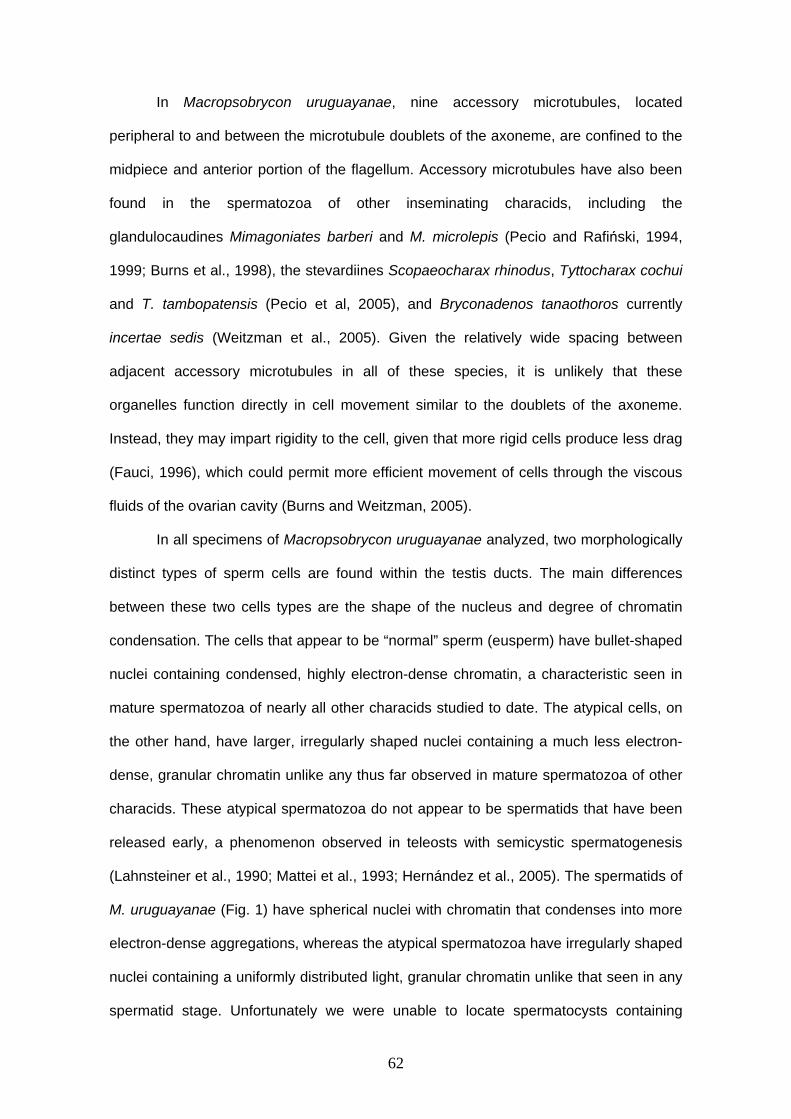
62
In Macropsobrycon uruguayanae, nine accessory microtubules, located
peripheral to and between the microtubule doublets of the axoneme, are confined to the
midpiece and anterior portion of the flagellum. Accessory microtubules have also been
found in the spermatozoa of other inseminating characids, including the
glandulocaudines Mimagoniates barberi and M. microlepis (Pecio and Rafiński, 1994,
1999; Burns et al., 1998), the stevardiines Scopaeocharax rhinodus, Tyttocharax cochui
and T. tambopatensis (Pecio et al, 2005), and Bryconadenos tanaothoros currently
incertae sedis (Weitzman et al., 2005). Given the relatively wide spacing between
adjacent accessory microtubules in all of these species, it is unlikely that these
organelles function directly in cell movement similar to the doublets of the axoneme.
Instead, they may impart rigidity to the cell, given that more rigid cells produce less drag
(Fauci, 1996), which could permit more efficient movement of cells through the viscous
fluids of the ovarian cavity (Burns and Weitzman, 2005).
In all specimens of Macropsobrycon uruguayanae analyzed, two morphologically
distinct types of sperm cells are found within the testis ducts. The main differences
between these two cells types are the shape of the nucleus and degree of chromatin
condensation. The cells that appear to be “normal” sperm (eusperm) have bullet-shaped
nuclei containing condensed, highly electron-dense chromatin, a characteristic seen in
mature spermatozoa of nearly all other characids studied to date. The atypical cells, on
the other hand, have larger, irregularly shaped nuclei containing a much less electron-
dense, granular chromatin unlike any thus far observed in mature spermatozoa of other
characids. These atypical spermatozoa do not appear to be spermatids that have been
released early, a phenomenon observed in teleosts with semicystic spermatogenesis
(Lahnsteiner et al., 1990; Mattei et al., 1993; Hernández et al., 2005). The spermatids of
M. uruguayanae (Fig. 1) have spherical nuclei with chromatin that condenses into more
electron-dense aggregations, whereas the atypical spermatozoa have irregularly shaped
nuclei containing a uniformly distributed light, granular chromatin unlike that seen in any
spermatid stage. Unfortunately we were unable to locate spermatocysts containing
63
developing atypical spermatozoa, so we do not know if they are formed within separate
spermatocysts or if they develop along with eusperm within the same spermatocyst.
Atypical sperm, often referred to as dimorphic sperm or parasperm, are relatively
common in some invertebrate taxa (Jamieson, 1986; Oppliger et al., 1998; Ferraguti et
al., 2002). Within the vertebrates, on the other hand, they have only been reported in
certain cottoid fishes (Hayakawa and Munehara, 2004), unless the production of
deformed sperm in mammals, including humans, can be included in this category (Baker
and Bellis, 1988). The parasperm of cottoid fishes appear to result from an incomplete
second meiotic division (Hayakawa and Munehara, 2004). Depending on the species,
these cells have either two nuclei or a bi-lobed nucleus, and two or no flagella
(Hayakawa et al., 2002a,b; Hayakawa and Munehara, 2004). Because of the abnormal
morphology and diploid genome of the cottoid parasperm, these cells appear to be
incapable of fertilizing eggs. However, experimental evidence indicates that parasperm
may help reduce the lateral dispersion of semen and thus increase the number of
eusperm that can reach an egg mass (Hayakawa et al., 2002b; Hayakawa and
Munehara, 2004), as well as play a role in sperm competition in cottoids (Hayakawa et
al., 2002a). The atypical sperm of Macropsobrycon uruguayanae bear little resemblance
to the parasperm of cottoids. Their possible function in the reproductive biology of this
species remains to be determined.
ACKNOWLEDGMENTS
Amy D. Meisner, formerly of George Washington University, Washington, DC,
USA, Irani Quagio Grassiotto and technicians from Universidade Estadual Paulista,
Botucatu, Brazil for help with transmission electron microscopy and technicians from
Pontifícia Universidade Católica de Porto Alegre, Rio Grande do Sul, Brazil for help with
scanning electron microscopy. This research was supported by Conselho Nacional de
64
Desenvolvimento Científico e Tecnológico (CNPq; Proc. 476821/2003-7; Proc
478002/2006-8) from Brazil.
65
LITERATURE CITED
Baker RR, Bellis MA. 1988. ‘Kamikaze’ sperm in mammals? Anim Beh 36:936-939.
Burns JR, Weitzman SH. 2005. Insemination in ostariophysan fishes. In: Uribe MC,
Grier HJ, editors. Viviparous Fishes. Homestead, FL: New Life Publications. pp 107-
134.
Burns JR, Weitzman SH, Malabarba LR. 1997. Insemination in eight species of
cheirodontine fishes (Teleostei: Characidae: Cheirodontinae). Copeia 1997: 433-
438.
Burns JR, Weitzman SH, Lange KR, Malabarba LR. 1998. Sperm ultrastructure in
characid fishes (Teleostei: Ostariophysi). In: Malabarba LR, Reis RE, Vari RP,
Lucena ZMS, Lucena CAS, editors. Phylogeny and Classification of Neotropical
Fishes. Porto Alegre, Brazil: EDIPUCRS. pp 235-244.
Fauci LJ. 1996. A computational model of the fluid dynamics of undulatory and flagellar
swimming. Amer Zool 36:599-607.
Ferraguti M, Fascio U, Boi S. 2002. Mass production of basal bodies in
paraspermiogenesis of Tubificinae (Annelida, Oligochaeta). Biology of the Cell
94:109-115.
Gardiner DM.1978. Fine structure of the spermatozoon of the viviparous teleost
Cymatogaster aggregata. J Fish Biol 13:435-438.
Ginzburg AS. 1968. Fertilization in Fishes and the Problem of Polyspermy. Moscow:
Academy of Science USSR. 366 pp.
66
Gracenea M, Ferrer JR, González-Moreno O, Trullols M. 1997. Ultrastructural study of
spermatogenesis and the spermatozoon in Postorchigenes gymnesicus (Trematoda,
Lecithodendriidae). J Morphol 234:223-232.
Gusmão-Pompiani GP. 2003. Ultraestrutura da espermiogênese e dos
espermatozóides de peixes da ordem Characiformes, família Characidae
(Teleostei, Ostariophysi): uma abordagem filogenética. Tese de doutorado, Instituto
de Biociências da Universidade Estadual Paulista, São Paulo. 86 pp.
Hayakawa Y, Munehara H. 2004. Ultrastrutural observations of euspermatozoa and
paraspermatozoa in a copulatory cottoid fish Blepsias cirrhosus. J Fish Biol 64:6
1530-1539.
Hayakawa Y, Munehara H, Komaru A. 2002a. Obstructive role of the dimorphic sperm
in a non-copulatory marine sculpin, Hemilepidotus gilberti, to present other males’
eusperm from fertilization. Env Biol Fish 64:419-427.
Hayakawa Y, Akiyama R, Munehara H, Komaru A. 2002b. Dimorphic sperm influence
semen distribution in a non-copulatory sculpin Hemilepidotus gilberti. Env Biol Fish
65:311-317.
Hernández MR, Sábat M, Muñoz M, Casadevall M. 2005. Semicystic spermatogenesis
and reproductive strategy in Ophidion barbatum (Pisces, Ophidiidae). Acta Zool
86:295-300.
Ito S, Karnovsky M J. 1968. Formaldehyde glutaraldehyde fixatives containing trinitro
compounds. J Cell Biol 36:168.
Jamieson BGM. 1986. Some recent studies on the ultrastructure and phylogeny of
annelid and uniramian spermatozoa. Dev Growth Differ 28(s1):25-26.
67
Jamieson BGM. 1991. Fish evolution and systematics: Evidence from spermatozoa.
Cambridge, UK: Cambridge University Press. 319 pp.
Javonillo R, Burns JR, Weitzman SH. 2007. Reproductive morphology of Brittanichthys
axelrodi (Teleostei: Characidae), a miniature inseminating fish from South America. J
Morphol 268:23-32.
Justine J-L. 1995. Spermatozoal ultrastructure and phylogeny in the parasitic
platyhelminthes. In: Jamieson BGM, Ausio J, Justine J-L, editors. Advances in
Spermatozoal Phylogeny and Taxonomy, Vol 166. Paris: Mém Mus Natn Hist Nat. pp
55-86.
Lahnsteiner F, Richtarski U, Patzner RA. 1990. Functions of the testicular gland in two
blenniid fishes, Salaria (= Blennius) pavo and Lipophrys (= Blennius) dalmatinus
(Blenniidae, Teleostei) as revealed by electron microscopy and enzyme
histochemistry. J Fish Biol 37:85-97.
Malabarba LR. 1998. Monophyly of the Cheirodontinae, characters and major clades
(Ostariophysi: Characidae). In: Malabarba LR, Reis RE, Vari RP, Lucena ZMS,
Lucena CAS, editors. Phylogeny and Classification of Neotropical Fishes. Porto
Alegre, Brazil: EDIPUCRS. pp 193-233.
Malabarba LR. 2003. Subfamily Cheirodontinae (Characins, tetra). In: Reis RE,
Kullander SO, Ferraris CJ. Check list of the freshwater fishes of South and Central
America. Porto Alegre, Brazil: EDIPUCRS. 742 pp.
Malabarba LR, Lima FCT, Weitzman SH. 2004. A new species of Kolpotocheirodon
(Teleostei: Characidae: Cheirodontinae: Compsurini) from Bahia, Northeastern
Brazil, with a new diagnosis of the genus. Proc Biol Soc Wash 117:317-329.
68
Mattei X. 1991. Spermatozoon ultrastructure and its systematic implications in fishes.
Can J Zool 69: 3038-3055.
Mattei X, Siau Y, Thiaw OT, Thiam D. 1993. Peculiarities in the organization of testis of
Ophidion sp. (Pisces Teleostei). Evidence for two types of spermatogenesis in
teleost fish. J Fish Biol 43:931-937.
Nelson JS. 2006. Fishes of the World, 4th ed. NY: John Wiley & Sons. 601 pp.
Oppliger A, Hosken DJ, Ribi G. 1998. Snail sperm production characteristics vary with
sperm competition risk. Proc R Soc Lond B 265:1527-1534.
Pecio A, Burns JR, Weitzman SH. 2005. Sperm and spermatozeugma ultrastructure in
the inseminating species Tyttocharax cochui, T. tambopatensis, and Scopaeocharax
rhinodus (Pisces: Teleostei: Characidae: Glandulocaudinae: Xenurobryconini). J.
Morphol 263:216-226.
Pecio A, Rafiński J. 1994. Structure of the testes, spermatozoa and spermatozeugmata
of Mimagoniates barberi Regan, 1907 (Teleostei: Characidae), an internally
fertilizing, oviparous fish. Acta Zool 75:179-185.
Pecio A, Rafiński J. 1999. Spermiogenesis in Mimagoniates barberi (Teleostei:
Ostariophysi: Characidae), an oviparous, internally fertilizing fish. Acta Zool 80: 35-
45.
Weitzman SH, Menezes NA, Evers H-G, Burns JR. 2005. Putative relationships among
inseminating and externally fertilizing characids, with a description of a new genus
and species of Brazilian inseminating fish bearing an anal-fin gland in males
(Characiformes: Characidae). Neotrop Ichthyol 3:329-360.
Yang J, Liu X, Yue G, Adamian M, Bulgakov O, Li T. 2002. Rootletin, a novel coiled-coil
69
protein, is a structural component of the ciliary rootlet. J Cell Biol 159:431-440.
Yang J, Gao J, Adamian M, Wen X-H, Pawlyk B, Zhang L, Sanderson MJ, Zuo J,
Makino CL, Li T. 2005.The ciliary rootlet maintains long-term stability of sensory cilia.
Mol Cell Biol 25:4129-4137.
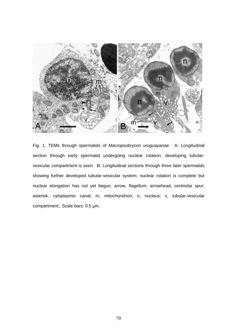
70
Fig. 1. TEMs through spermatids of Macropsobrycon uruguayanae. A: Longitudinal
section through early spermatid undergoing nuclear rotation; developing tubular-
vesicular compartment is seen. B: Longitudinal sections through three later spermatids
showing further developed tubular-vesicular system; nuclear rotation is complete but
nuclear elongation has not yet begun. arrow, flagellum; arrowhead, centriolar spur;
asterisk, cytoplasmic canal; m, mitochondrion; n, nucleus; v, tubular-vesicular
compartment;. Scale bars: 0.5 µm.
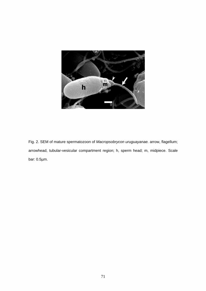
71
Fig. 2. SEM of mature spermatozoon of Macropsobrycon uruguayanae. arrow, flagellum;
arrowhead, tubular-vesicular compartment region; h, sperm head; m, midpiece. Scale
bar: 0.5µm.
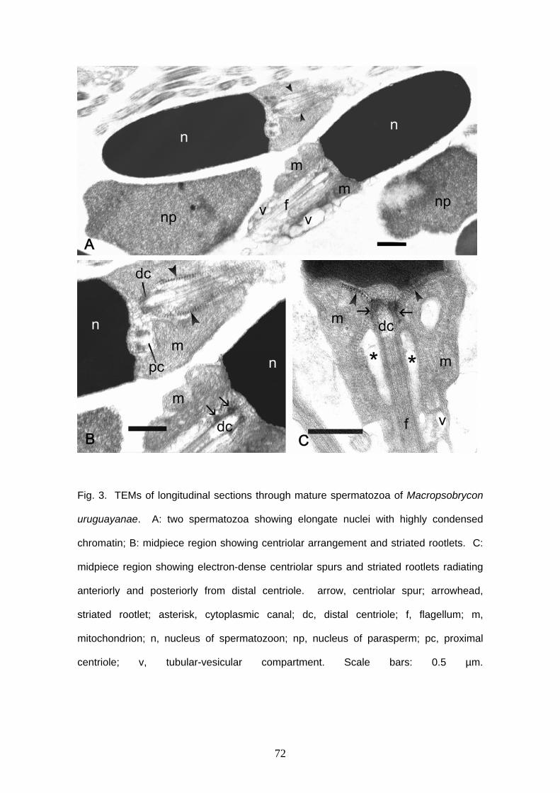
72
Fig. 3. TEMs of longitudinal sections through mature spermatozoa of Macropsobrycon
uruguayanae. A: two spermatozoa showing elongate nuclei with highly condensed
chromatin; B: midpiece region showing centriolar arrangement and striated rootlets. C:
midpiece region showing electron-dense centriolar spurs and striated rootlets radiating
anteriorly and posteriorly from distal centriole. arrow, centriolar spur; arrowhead,
striated rootlet; asterisk, cytoplasmic canal; dc, distal centriole; f, flagellum; m,
mitochondrion; n, nucleus of spermatozoon; np, nucleus of parasperm; pc, proximal
centriole; v, tubular-vesicular compartment. Scale bars: 0.5 µm.
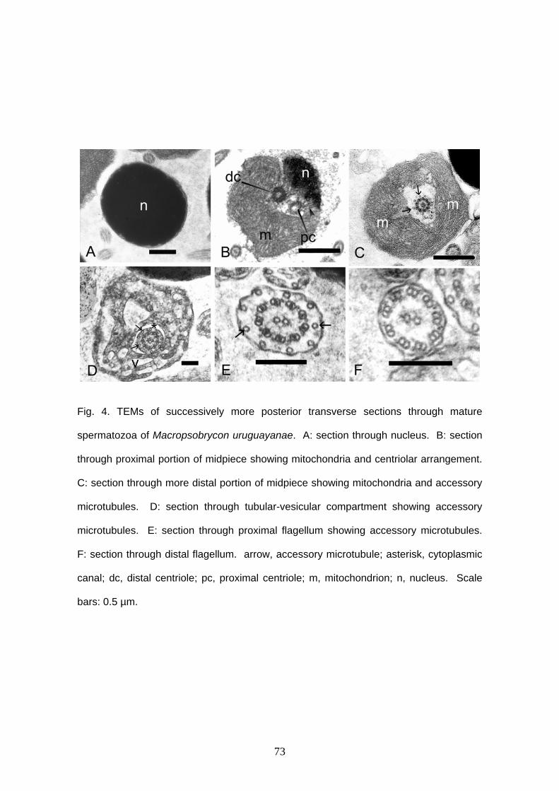
73
Fig. 4. TEMs of successively more posterior transverse sections through mature
spermatozoa of Macropsobrycon uruguayanae. A: section through nucleus. B: section
through proximal portion of midpiece showing mitochondria and centriolar arrangement.
C: section through more distal portion of midpiece showing mitochondria and accessory
microtubules. D: section through tubular-vesicular compartment showing accessory
microtubules. E: section through proximal flagellum showing accessory microtubules.
F: section through distal flagellum. arrow, accessory microtubule; asterisk, cytoplasmic
canal; dc, distal centriole; pc, proximal centriole; m, mitochondrion; n, nucleus. Scale
bars: 0.5 µm.
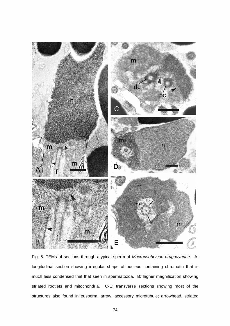
74
Fig. 5. TEMs of sections through atypical sperm of Macropsobrycon uruguayanae. A:
longitudinal section showing irregular shape of nucleus containing chromatin that is
much less condensed that that seen in spermatozoa. B: higher magnification showing
striated rootlets and mitochondria. C-E: transverse sections showing most of the
structures also found in eusperm. arrow, accessory microtubule; arrowhead, striated
75
rootlet; dc, distal centriole; f, flagellum; m, mitochondria; n, nucleus; pc, proximal
centriole. Scale bars: 0.5 µm
76
Capítulo 3:
Gill-derived glands morphology in Cheirodontinae (Teleostei: Characidae)
Author’s names and addresses:
Cristina L. C. de Oliveira 1*, Luiz R. Malabarba 2 and John R. Burns 1, 3
1 Departamento de Zoologia, Universidade Federal do Rio Grande do Sul, Porto Alegre,
Rio Grande do Sul, Brazil, 2 Museu de Ciências e Tecnologia, Pontifícia Universidade Católica de Porto Alegre, Rio
Grande do Sul, Brazil. 3 Department of Biological Sciences, George Washington University, Washington, D.C.
20052 * Correspondence to: Cristina Luísa Conceição de Oliveira. Av. Bento Gonçalves 9500
prédio 43435, sala 104, Departamento de Zoologia, Universidade Federal do Rio
Grande do Sul, Porto Alegre, Rio Grande do Sul, Brazil, CEP 91501-970. E-mail:
Number of text pages:
Number of figures: 7
Number of tables: 1
Abbreviated title (running headline): Gill glands morphology in Cheirodontinae
Send proofs to Cristina Luísa Conceição de Oliveira, Av. Bento Gonçalves 9500 prédio
43435, sala 104, Departamento de Zoologia, Universidade Federal do Rio Grande do
Sul, Porto Alegre, Rio Grande do Sul, Brazil, CEP 91501-970. Phone: (55 51) 3225
8881. E-mail: [email protected]
Keywords: gill gland, gills, filaments, lamellae, Cheirodontinae
77
ABSTRACT
The gill gland of 17 species of Cheirodontinae is described. The gill gland is
located in the anterior region of the gill cavity on either side of the midline of all analyzed
mature males of the Cheirodontinae. The gill gland is small in externally fertilizing
species of the Cheirodontinae, reaching up to 10 gill filaments. In the inseminating
species of the tribe Compsurini, the gland occupies a large extension or almost the
entire gill arch. In some portions of the gland of A. hemigrammus, C. heterura, K.
theloura, M. uruguayanae and S. hastatus, the lamellae are not retained, remaining only
secretor gill gland cells in these regions. Kolpotocheirodon theloura showed noncellular
material within the gill gland chambers and they were turgid. There is no relation
between the presence of a gill gland and insemination since both externally fertilizing
and inseminating characids have or not gill glands. The function of the gill gland is not
known yet, but presence of the gill gland in mature males and absence in females
suggest that the secretion produced may be used to attract the females during the
reproductive period or competition between males. The gill glands of cheirodontines and
other characids are compared.
78
INTRODUCTION
Cheirodontinae is composed by two tribes, Cheirodontini and Compsurini, plus
some genera considered incertae sedis in the subfamily by Malabarba (1998). These
incertae sedis genera were hypothesized to belong to a single tribe, the Odontostilbini,
in the unpublished work of Bührnheim (2006). The Cheirodontini and the incertae sedis
genera include are all externally fertilizing species and Compsurini the inseminating
species (species that transfer sperm of the mature testes for the ovaries of females,
although the actual moment of fertilizing is not known, Burns et al., 1997, 1998; Burns
and Weitzman, 2005).
The gills of teleosts are formed by four gill arches (holobranches) on both sides
of the body. Each gill arch is formed by two rows of filaments (Roberts, 1981),
denominated hemibranches, one more internal and another more external. These
primary filaments are sustained by cartilage and they have fine parallel secondary
lamellae on both sides. The secondary lamellae are constituted by several capillary
inserted by sustentation cells, the pillar cells, and a layer of epithelial cells which cover
the structure. Gaseous exchanges occur in those lamellae, when water flows among
them in a direction, and blood in the opposite direction (Schmidt-Nielsen, 1996). The gill
arch is covered by typical epidermic tissue, thicker in the origin of the primary lamellae
and usually with mucous cells (Roberts, 1981).
Burns and Weitzman (1996) described a gill-derived gland, termed gill gland in
Corynopoma riisei Gill 1858, a stevardiine. The union and functional modification of the
most ventral filaments of the first right and left gill arches form the gill gland. The gill
gland filaments are united externally by stratified epithelial tissue, maintaining
individuality internally. Secretor cells are found between lamellae that compose the
gland. The union between each pair of primary filaments forms chambers, through
where the secretion is led to the exterior (Burns and Weitzman, 1996, Bushmann et al.,
2002). These chemical signals may help during reproduction. The gill gland was found in
79
some Characidae subfamilies, such as Stevardiinae (Burns and Weitzman, 1996;
Bushmann et al., 2002); Cheirodontinae (Oliveira, 2000, 2003 - unpublished data;
Azevedo, 2004 - unpublished data; Bührnheim, 2006 - unpublished data);
Aphyocharacinae (Gonçalves et al., 2005), and some species incertae sedis in
Characidae (Weitzman et al., 2005). The purpose of the present study is to determine
the distribution of gill glands in the Cheirodontinae by histological and scanning electron
microscopy, to describe the structure gill glands and to compare the gland between
inseminating and externally fertilizing species of the Cheirodontinae and other
Characidae.
MATERIAL AND METHODS
At least 1 species of each cheirodontine genera was analyzed, totalizing 17
species, plus 4 comparative species of other characid genera. Species of
Pseudocheirodon Meek and Hildebrand, 1916 and Spintherobolus Eigenmann, 1911
were not analyzed due to the lack of mature specimens. Gills of at least 3 mature males
and 2 mature females of each species were selected. Histological sections were made
in the gonads to confirm degree of sexual maturity. The specimens were obtained from
museum collections; therefore the whole fishes had been fixed in formalin and stored in
70% ethanol. The first and second gill arches in both sides of body/head were removed,
the right arches were used in histology and the left arches were used in Scanning
Electron Microscopy (SEM). The first left gill arches of 5 mature males of Compsura
heterura were fixed in 2.5% glutaraldehyde and 2% paraformaldehyde in 0.1 M
phosphate buffer, pH 7.4 and kept under refrigeration until the start of sample
processing for Transmission Electron Microscopy (TEM) analysis.
Inseminanting species analyzed: Acinocheirodon melanogramma Malabarba and
Weiztman, 1999 (MCP 19142, males: 32.21, 34.97, 32.3 mm SL, females: 33.57, 38.3
80
mm SL); Compsura heterura Eigenmann, 1915 (UFRGS 8978, males: 29.7, 28.1, 29.1
mm SL, females: 28.1, 28.7 mm SL); Kolpotocheirodon theloura Malabarba and
Weitzman, 2000 (MNRJ 18081, males: 25.96, 25.48, 26.30, 26.11, 25.12, 25.03 mm SL,
females: 24.13, 24.84 mm SL); Macropsobrycon uruguayanae Eigenmann, 1915
(UFRGS 8792, males: 35.25, 32.89, 32.77 mm SL, females: 29.44, 38.85 mm SL);
Odontostilbe dialeptura (Fink and Weitzman, 1974) (USNM 209511, males: 32.61,
34.92, 32.17, 35.05 mm SL, females: 32.64, 32.51 mm SL); Saccoderma hastatus
(Eignmann, 1913) (ANSP 139487 males: 29.22, 30.13, 29.14, 29.64, 30.5, 30.02, 31.25,
29.90 mm SL, females: 29.47, 29.51, 29.02 mm SL, CAS 70918 males: 26.25, 25.11
mm SL, females 26.11, MCP 16169 males: 24.44, 24.60, females: 26.05 mm SL).
Externally fertilizing species analyzed: Aphyocheirodon hemigrammus
Eigenmann, 1915 (NRM 17307 male: 29.0 mm SL), Cheirodon ibicuhiensis Eigenmann,
1915 (UFRGS 9042 males: 34.1, 32.4, 31.5 mm SL, UFRGS 9041 females: 38.5, 43,1
mm SL); Cheirodontops geayi Schultz, 1944 (MCNG 14197, males: 30.04, 30.12, 27.71,
29.91, 29.19 mm SL, females: 29.94, 31.31 mm SL); Heterocheirodon jacuiensis
Malabarba and Bertaco, 1999 (MCP 14283, males: 42.09, 40.05, 39.66 mm SL,
females: 43.04, 38.92, 39.62 mm SL), Heterocheirodon yatai (Casciotta, Miquelarena
and Protogino, 1992) MCP 14283, males: 32.88, 32.70, 34.85, 31,57 mm SL, females:
35.25, 34.48, 33.69 mm SL), Nanocheirodon insignis (Steindachner, 1880) (USNM
121518 males: 25.62; 22.53; 21.04; 20.3 mm SL; female: 21.10 mm SL); Odontostilbe
fugitiva Cope, 1870 (MCP 35775 males: 36.0, 33.8 34.27, 35.02, 34.5 mm SL);
Odontostilbe pequira (Steindachner, 1882) (UFRGS 8980, males: 37.2, 38.7, 36.7 mm
SL, females: 41.1, 40.9 mm SL); Prodontocharax melanotus Pearson, 1924 (USNM
326941, male: 38.1 mm SL); Serrapinnus heterodon (Eigenmann, 1915) (UFRGS 8793,
males: 29.7, 27.8, 29.4, 30.8, 29.97, 29.93 mm SL, females: 29.0, 28.7 mm SL) and
Serrapinnus piaba (Lütken, 1875) (UFRGS 8794, males: 28.5, 28.23, 28.42 mm SL,
females: 31.23, 28.97 mm SL).
81
Examined specimens belong to ANSP - Academy of Natural Sciences,
Philadelphia, USA; CAS – California Academy of Sciences, San Francisco, USA; MCP -
Museu de Ciências e Tecnologia, Porto Alegre, Brazil; MCNG – Museo de Ciencias
Naturales, Guanare, Venezuela; MNRJ - Museu Nacional, Rio de Janeiro, Brazil; NRM -
Swedish Museum of Natural History, Stockholm, Sweden; UFRGS - Universidade
Federal do Rio Grande do Sul, Porto Alegre, Brazil; and USNM - National Museum of
Natural History, Washington D.C., USA.
Histology
The first and second right gill arches were decalcified in ethylenediamine
tetracetic acid (EDTA) 10% during 5 days, dehydrated in ethanol series, infiltrated and
embedded in glycol methacrylate. The gills were cut in sagital position with thickness
varying from 3 to 5µm and stained with Hematoxilin-Eosin (HE) and Toluidin Blue. Some
slides were stained with periodic acid of Schiff (PAS).
SEM
The first and second left gill arches were cleaned with ultrasound equipment
during 6 minutes, dehydrated in a graded ethanol series, critical point dried in liquid CO2,
glued on stub with carbon double-stick tape and sputter-coated with carbon and gold.
Images were visualized and photographed in a Philips XL 30 scanning electron
microscopy from Pontifícia Universidade Católica do Rio Grande do Sul, Brazil or in a
Jeol 6060 scanning electron microscopy from Universidade Federal do Rio Grande do
Sul, Brazil.
TEM
At field, the fishes were sacrificed by a cut through the spinal cord. The first left
gill arches of 5 mature males of Compsura heterura were fixed in 2.5% glutaraldehyde
and 2% paraformaldehyde in 0.1 M phosphate buffer, pH 7.4 and kept under

82
refrigeration until the start of sample processing. The gills were post-fixed in 1% osmium
tetroxide in phosphate buffer, dehydrated in acetone series, infiltrated and embedded in
Araldite-Epon (Embed-812). Ultrathin sections were stained with a saturated of uranyl
acetate at 50% alcohol and lead citrate. The sections were viewed in a Philips CM100
transmission electron microscope from Universidade Estadual Paulista- Campus
Botucatu, Brazil.
RESULTS
The gill glands were located in the anterior region of the gill cavity on either side
of the midline (Fig. 1) of all analyzed mature males of Cheirodontinae (Figs. 2, 3). They
were absent in females. The specimens of Spintherobolus broccae are immature. The
development of the gill glands begins with the multiplication of the epithelial cells that
cover the anteriormost portion of the first gill arch, extending then for the following
filaments. Unmodified filaments are located soon after of the gill glands (Figs. 2- 5),
when they do not occupy the whole gill arch. They are covered by stratified epithelial
tissue externally, and internally the gill filaments form chambers. There is no connection
between chambers (Figs. 4, 5). In some sectioning was viewed skeletal muscle
associated with hyaline cartilage of gill filaments (Fig.4f, i). The secretion supposedly
produced leaves freely through the existent space in the distal extremity of the filaments
that are not united. Secretor cells fill out the secondary lamellae that form the gill glands
(Figs. 4, 5).
The pseudostratified epithelial tissue is formed by tall columnar cells. They are
located between secondary lamellae, when these are present, or covering the chambers
formed among the gill filaments. In TEM is possible to see several dark vesicles filling
almost the whole cell (Fig. 6). The nucleus, endoplasmatic reticule and Golgi complex
are in the base of cells.
In spite of gill glands structures are similar among species, some differences
were observed in the gland size and in the degree of gill modification. The gill glands are
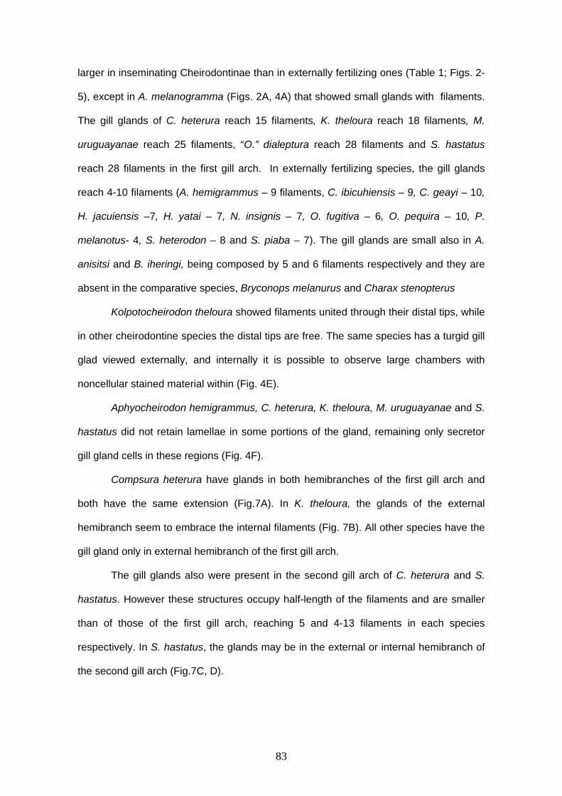
83
larger in inseminating Cheirodontinae than in externally fertilizing ones (Table 1; Figs. 2-
5), except in A. melanogramma (Figs. 2A, 4A) that showed small glands with filaments.
The gill glands of C. heterura reach 15 filaments, K. theloura reach 18 filaments, M.
uruguayanae reach 25 filaments, “O.” dialeptura reach 28 filaments and S. hastatus
reach 28 filaments in the first gill arch. In externally fertilizing species, the gill glands
reach 4-10 filaments (A. hemigrammus – 9 filaments, C. ibicuhiensis – 9, C. geayi – 10,
H. jacuiensis –7, H. yatai – 7, N. insignis – 7, O. fugitiva – 6, O. pequira – 10, P.
melanotus- 4, S. heterodon – 8 and S. piaba – 7). The gill glands are small also in A.
anisitsi and B. iheringi, being composed by 5 and 6 filaments respectively and they are
absent in the comparative species, Bryconops melanurus and Charax stenopterus
Kolpotocheirodon theloura showed filaments united through their distal tips, while
in other cheirodontine species the distal tips are free. The same species has a turgid gill
glad viewed externally, and internally it is possible to observe large chambers with
noncellular stained material within (Fig. 4E).
Aphyocheirodon hemigrammus, C. heterura, K. theloura, M. uruguayanae and S.
hastatus did not retain lamellae in some portions of the gland, remaining only secretor
gill gland cells in these regions (Fig. 4F).
Compsura heterura have glands in both hemibranches of the first gill arch and
both have the same extension (Fig.7A). In K. theloura, the glands of the external
hemibranch seem to embrace the internal filaments (Fig. 7B). All other species have the
gill gland only in external hemibranch of the first gill arch.
The gill glands also were present in the second gill arch of C. heterura and S.
hastatus. However these structures occupy half-length of the filaments and are smaller
than of those of the first gill arch, reaching 5 and 4-13 filaments in each species
respectively. In S. hastatus, the glands may be in the external or internal hemibranch of
the second gill arch (Fig.7C, D).
84
DISCUSSION
The gill gland of all Characidae species in which this organ has been described
possess the same structure. They are formed externally by the growth of
pseudostratified epithelial tissue over and around the most anterior gill filaments of gill
arch, and internally by chambers covered with a columnar or cuboidal epithelium
between secondary lamellae (Burns and Weitzman, 1996; Bushmann et al., 2002). This
similar structural pattern of the gill gland suggests it is homologous among the species
of Characidae.
In Cheirodontinae, the gland was observed in C. ibicuhiensis (Oliveira, 2000 –
unplublished data), C. heterura, O. pequira (Oliveira, 2003 – unplublished data) and in
all mature males of the Cheirodontinae examined by Bührnheim (Acinocheirodon
melanogramma, Amblystilbe alleni, Aphyocheirodon hemigrammus, Cheirodon
ibicuhiensis, C. interruptus, Cheirodontops geayi, Compsura heterura, Kolpotocheirodon
theloura, Macropsobrycon uruguayanae, Odontostilbe fugitiva, O. pequira, O. euspilurus
Fowler 1945, O. paraguayensis Eigenmann & Kennedy 1903, O. ecuadorensis, O.
nareuda, O. parecis, O.microcephala, O. splendida, O. pao, plus 8 new species of
Odontostilbe, Prodontocharax melanotus, Pseudocheirodon terrabae, P. arnoldi,
Saccoderma hastatus, Serrapinnus heterodon, S. microdon, S. micropterus), excepting
in Amblystilbe alleni and Spintherobolus sp. n, whose specimens were immature.
The gill glands were described by Bushmann et al. (2002) in 12 inseminating
species of the Stervardiinae (Acrobrycon Eigenmann & Pearson 1924, Argopleura
Eigenmann 1913, Chrysobrycon Weitzman & Menezes 1998, Corynopoma Gill 1858,
Gephyrocharax Eigenmann 1912, Hysteronotus Eigenmann 1911, Phenacobrycon
Eigenmann 1922, Planaltina Böhlke 1954, Pterobrycon Eigenmann 1913,
Scopaeocharax Weitzman & Fink 1985, Tyttocharax Fowler 1913 and Xenurobrycon
Myers & Miranda Ribeiro 1945), but were absent in the inseminating species of 3
85
stervardiine genera (Diapoma Cope 1894, Pseudocorynopoma Perugia 1891 and
Ptychocharax Weitzman, Fink, Machado-Allison & Royero L. 1994), and in all
inseminating species of the Glandulocaudinae (Bushmann et al., 2002; Burns, personal
communication).
The gill glands were viewed in mature inseminating males of genera considered
incertae sedis in Characidae and belonging to Clade A of Malabarba and Weitzman
(2003), as Attonitus bounites Vari & Ortega 2000, Attonitus irisae Vari & Ortega 2000,
Bryconadenos tanaothoros Weitzman, Menezes, Evers & Burns 2005 (Weitzman et al.,
2005), Creagrutus melasma Vari, Harold & Taphorn 1994 and Monotocheirodon
Eigenmann & Pearson 1924 (Burns, personal communication) and in one undescribed
new genus and species of inseminating characid (Malabarba et al., in prep.).
These glands are also present in mature males with externally fertilizing species
of the Clade A of Malabarba and Weitzman (2003), as observed in the genera
Bryconamericus Eigenmann 1907, Caiapobrycon Malabarba & Vari 2000, Creagrutus
Günther 1864, Cyanocharax Malabarba & Weitzman 2003, Hemibrycon Günther 1864,
Hypobrycon Malabarba & Malabarba 1994, Knodus Eigenmann 1911, Piabina Reinhardt
1867, Rhinobrycon Myers 1944 and Rhinopetitia Géry 1964 (Burns, personal
communication) as well as Phenacogaster franciscoensis Eigenmann 1911, a
Characinae and Aphyocharacidium bolivianum Géry 1973, incertae sedis in Characidae
(Bührnheim, 2006 – unplublished data). In Aphyocharacinae, the gland was found in A.
anisitsi by Gonçalves et al. (2005).
Through of the list of species with gill gland, we can observe that externally
fertilizing and inseminating Characidae have or not gill glands, therefore there is no
relation between the presence of a gill gland and insemination.
Although the gill gland is viewed in many Characidae species, little is know about
number of gill filaments that compose the gill gland and details about its internal
morphology. In spite of the gill glands possess similar structure and to be located in the
same area of the first gill arch, some differences among cheirodontine species and
86
between Cheirodontinae and other Characidae were observed, in the extension of the
gland, the format of the cells, the presence of the gland in both hemibranchs of the first
arch, the presence of the gland in the second gill arch, the turgidity and the presence or
of the secondary lamellae.
The gill gland is small in externally fertilizing species of the Cheirodontinae, being
composed by 10 gill filaments. Nevertheless, the gland occupies a large extension or
almost the entire gill arch in the inseminating species of the tribe Compsurini, such as in
M. uruguayanae, O. dialeptura and S. hastatus. Acinocheirodon melanogramma is the
exception among compsurins and show a small gland. Considering the non basal
position of Acinocheirodon in both phylogenies proposed in Malabarba (1998) and
Oliveira et al. (this volume), the possession of a large gill gland may constitute a
synapomorphy for the Compsurini, and the small gland a reversal in A. melanogramma,
because the specimens analysed had mature testis. Although there is no relation
between the presence or absence of a gill gland and insemination in Characidae, the
increasing size of the gill gland seems to be advantageous for the inseminating
cheirodontines that possess a clearly larger organ than those of the externally fertilized
species of this subfamily.
The presence of the glands in both hemibranches of first arch is a characteristic
showed only by C. heterura. The presence of glands in the second gill arch is a
characteristic shared by C. heterura and S. hastatus. These species formed one natural
group in the analyses of Malabarba (1998) and Bührnheim (2006), and gland
morphology seems to corroborate their hypotheses. Nevertheless, Oliveira et al. (2007-
this volume) presents a hypothesis based on spermatozoa ultrastructure characters that
groups S. hastatus in a clade with A. melanogramma, “O”. mitoptera and “O”. dialeptura,
instead of Compsura. Therefore, additional characters are necessary to solve these
relationships.
The gill glands of most species of Compsurini reach larger sizes than those of
the Stevardiinae. Stervardiinae also show varying degrees of gill gland extension
87
(Bushmann et al., 2002). The glands are formed by 4-13 gill filaments. It is bigger in
Hysteronotus megalostomus Eigenmann 1911 (11 gill filaments) and in Stevardiini
species (= Corynopomini to past authors). In this tribe the gill gland is composed by 7-9
gill filaments in Gephyrocharax melanocheir Eigenmann 1912, by 11-12 gill filaments in
Pterobrycon myrnae Bussing 1974 and by 8-13 gill filaments in Corynopoma riisei
(Bushmann et al., 2002). Other Stevardiinae species analyzed by Bushmann et al.
(2002) had less than 9 gill filaments in the gill gland. Bryconadenos tanaothoros had a
gill gland composed by 5 filaments and unmodified gills were observed at the extreme
left and right of gill glands (Weitzman et al., 2005). In A. bounties, the glands were
formed by 14 gill filaments and in A. irisae by 3 gill filaments, but the glands appeared to
be in the initial stages of formation (Weitzman et al., 2005).
Kolpotocheirodon theloura showed noncellular material within the gill gland
chambers and they were turgid. The state of expansion and secretion of glands varies
greatly (Burns and Weitzman, 1996) and it is related with maturation of testis. The
immature does not have glands, early stage of sexual maturation males have initial
glands and mature males have glands developed (Burns and Weitzman, 1996; Oliveira,
2003; Azevedo, 2004; Gonçalves et al., 2005). Some specimens of C. riisei had gill
glands with regions more expanded and turgid, one specimen had a light yellow viscous
secretion in the enlarged chambers, and a third specimen showed involuted glands
(Burns and Weitzman, 1996). However, none reduction was observed by Oliveira (2003)
in the gill glands of C. heterura after the reproductive period.
In some species occur reduction or disappearance of secondary lamellae, as
observed in C. heterura, K. theloura, M. uruguayanae and S. hastatus. The union of the
filaments, the covering with a stratified epithelium, and the loss of the secondary
lamellae, decrease or impede gaseous exchanges in the portion of the gill glands (Burns
and Weitzman, 1996).
The function of the gill gland is not known yet, but the development of the gill
gland in males occurs along with testis maturation (Gonçalves et al., 2005). The
88
presence of the gill gland in mature males and absence in females suggest that the
secretion produced may be used to attract the females during the reproductive period
(Burns and Weitzman, 1996; Bushmann et al., 2002) or intermale aggression
(Bushmann et al., 2002). The comparatively larger size of the gill gland in inseminating
versus externally fertilized species of the Cheirodontinae demonstrates these secretions
are particularly important in the reproductive success of inseminating cheirodontines.
Bührnheim (2006) viewed that the concavity of the posteriormost branchiostegal
ray forms a hole ventrally on head exactly near the most ventral portion of the gill gland
of the first gill arch of the most of Cheirodontinae. This author suggests that the osseous
modification could be related to the gill gland, functioning as an opening to facilitate
excretion. Posterior studies about gill gland physiology are necessary for understand its
function and working.
ACKNOWLEDGMENTS
We thank the technicians of the Centro de Microscopia Eletrônica of the
Pontifícia Universidade Católica do Rio Grande do Sul, Universidade Federal do Rio
Grande do Sul, Porto Alegre, Brazil and Universidade Estadual Paulista, Botucatu,
Brazil. This work was supported by Conselho Nacional de Desenvolvimento Científico e
Tecnológico (CNPq) proc. CNPq; Proc. 476821/2003-7; Proc. 478002/2006-8) from
Brazil.
89
LITERATURE CITED
Azevedo MA. 2004. Análise comparada de caracteres reprodutivos em três linhagens
de Characidae (Teleostei: Ostariophysi) com inseminação. Tese de doutorado,
Universidade Federal do Rio Grande do Sul, Porto Alegre, Brasil. 238p.
Buhrnheim CM. 2006. Sistemática de Odontostilbe Cope, 1870 com a proposição de
uma nova tribo Odontostilbini e redefinição dos gêneros incertae sedis de
Cheirodontinae (Ostariophysi: Characiformes: Characidae). Tese de doutorado.
Pontifícia Universidade Católica de Porto Alegre, Porto Alegre, Brazil. 315p.
Burns JR, Weitzman SH. 1996. Novel gill-derived gland in the male swordtail characin,
Corynopoma riisei (Teleostei: Characidae: Glandulocaudinae). Copeia 1996 (3):
627-633.
Burns JR, Weitzman SH. 2005. Insemination in ostariophysan fishes. In: Uribe MC,
Grier HJ, editors. Viviparous Fishes. Homestead, FL: New Life Publications. pp 107-
134.
Burns JR, Weitzman SH, Lange KR, Malabarba LR. 1998. Sperm ultrastructure in
characid fishes (Teleostei: Ostariophysi). In: Malabarba LR, Reis RE, Vari RP,
Lucena ZMS, Lucena CAS, editors. Phylogeny and Classification of Neotropical
Fishes. Porto Alegre, Brazil: EDIPUCRS. pp 235-244.
Burns JR, Weitzman SH, Malabarba LR. 1997. Insemination in eight species of
cheirodontine fishes (Teleostei: Characidae: Cheirodontinae). Copeia 1997: 433-
438.
Bushmann PJ, Burns JR, Weitzmann SH. 2002. Gill-derived glands in glandulocaudine
fishes (Teleostei: Characidae: Glandulocaudinae). J. Morphol. 253: 187-195.
90
Gonçalves TK, Azevedo MA, Malabarba LR. 2005. Reproductive biology and
development of sexually dimorphic structures in Aphyocharax anisitsi (Ostariophysi:
Characidae). Neotrop Ichthyol, 3(3):433-438.
Malabarba LR. 1998. Monophyly of the Cheirodontinae, characters and major clades
(Ostariophysi: Characidae). In: Malabarba LR, Reis RE, Vari RP, Lucena ZMS,
Lucena CAS, editors. Phylogeny and Classification of Neotropical Fishes. Porto
Alegre, Brazil: EDIPUCRS. pp 193-233.
Malabarba LR, Weitzman, SH. 2003. Description of a new genus with six new species
from Southern Brazil, Uruguay and Argentina, with a discussion of a putative
characid clade (Teleostei: Characiformes: Characidae). Comun. Mus. Ciênc. Tecnol.
PUCRS, Ser. Zool. 16(1): 67-151.
Oliveira CLC. 2000. Biologia reprodutiva e estudo do desenvolvimento dos caracteres
sexuais secundários de Cheirodon ibicuhiensis Eigenmann, 1915 (Teleostei:
Characidae: Cheirodontinae). Dissertação de Bacharelado, Universidade Federal do
Rio Grande do Sul, Porto Alegre, Brasil. 33p.
Oliveira CLC. 2003. Análise comparada de caracteres reprodutivos e da glândula
branquial de duas espécies de Cheirodontinae (Teleostei: Characidae). Dissertação
de Mestrado, Programa de Pós-Graduação em Biologia Animal. Universidade
Federal do Rio Grande do Sul, Porto Alegre, Brasil. 80p.
Roberts RJ. 1981. Patologia de los peces. Madrid, Ediciones Mundi-Prense, 366p.
Schmidt-Nielsen K. 1996. Fisiologia animal - adaptação e meio ambiente. São Paulo:
Santos, 600p.
Weitzman SH, Menezes NA, Evers H-G, Burns JR. 2005. Putative relationships among
inseminating and externally fertilizing characids, with a description of a new genus

91
and species of Brazilian inseminating fish bearing an anal-fin gland in males
(Characiformes: Characidae). Neotrop Ichthyol 3:329-360.
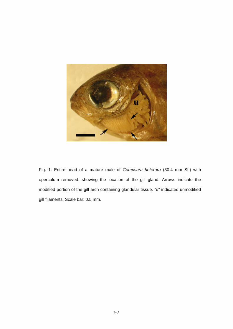
92
Fig. 1. Entire head of a mature male of Compsura heterura (30.4 mm SL) with
operculum removed, showing the location of the gill gland. Arrows indicate the
modified portion of the gill arch containing glandular tissue. “u” indicated unmodified
gill filaments. Scale bar: 0.5 mm.
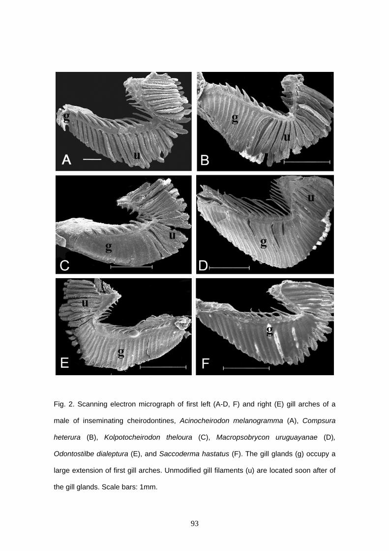
93
Fig. 2. Scanning electron micrograph of first left (A-D, F) and right (E) gill arches of a
male of inseminating cheirodontines, Acinocheirodon melanogramma (A), Compsura
heterura (B), Kolpotocheirodon theloura (C), Macropsobrycon uruguayanae (D),
Odontostilbe dialeptura (E), and Saccoderma hastatus (F). The gill glands (g) occupy a
large extension of first gill arches. Unmodified gill filaments (u) are located soon after of
the gill glands. Scale bars: 1mm.
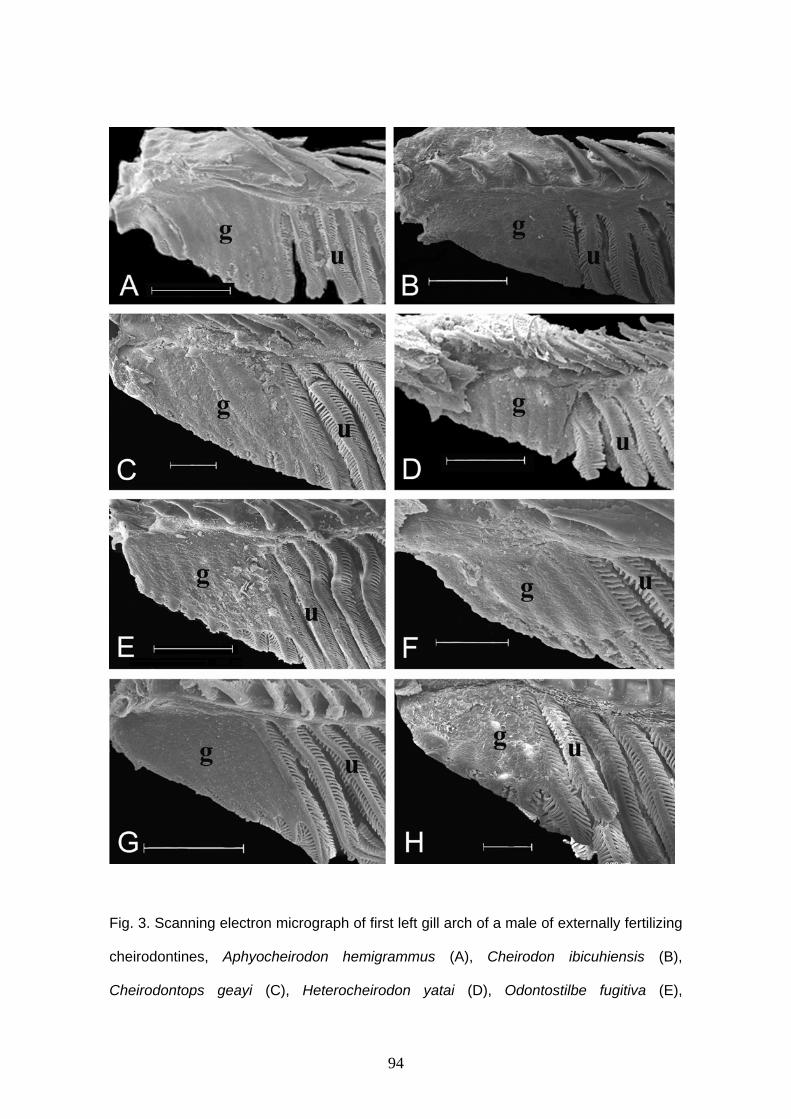
94
Fig. 3. Scanning electron micrograph of first left gill arch of a male of externally fertilizing
cheirodontines, Aphyocheirodon hemigrammus (A), Cheirodon ibicuhiensis (B),
Cheirodontops geayi (C), Heterocheirodon yatai (D), Odontostilbe fugitiva (E),
95
Nanocheirodon insignis (F), Prodontocharax melanotus (G), Serrapinnus heterodon (H).
The gill glands (g) occupy a small extension of first gill arches. Unmodified gill filaments
(u) are located soon after of the gill glands. Scale bars: 200 µm (A, B, C, D, H), 500 µm
(E, G).
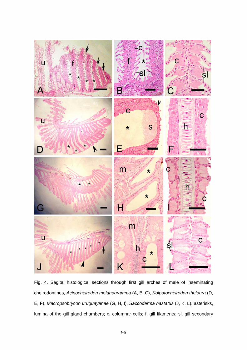
96
Fig. 4. Sagital histological sections through first gill arches of male of inseminating
cheirodontines, Acinocheirodon melanogramma (A, B, C), Kolpotocheirodon theloura (D,
E, F), Macropsobrycon uruguayanae (G, H, I), Saccoderma hastatus (J, K, L). asterisks,
lumina of the gill gland chambers; c, columnar cells; f, gill filaments; sl, gill secondary

97
lamellae; s, secretion; u, unmodified gill filaments; arrow, ventral chamber opening;
arrowhead, gill gland covering; m, skeletal muscle; h, hyaline cartilage of gill filaments.
Scale bar: 150 µm (A, D, G, J), 50 µm (B, E, H, K), 25 µm (C, F, I, L).
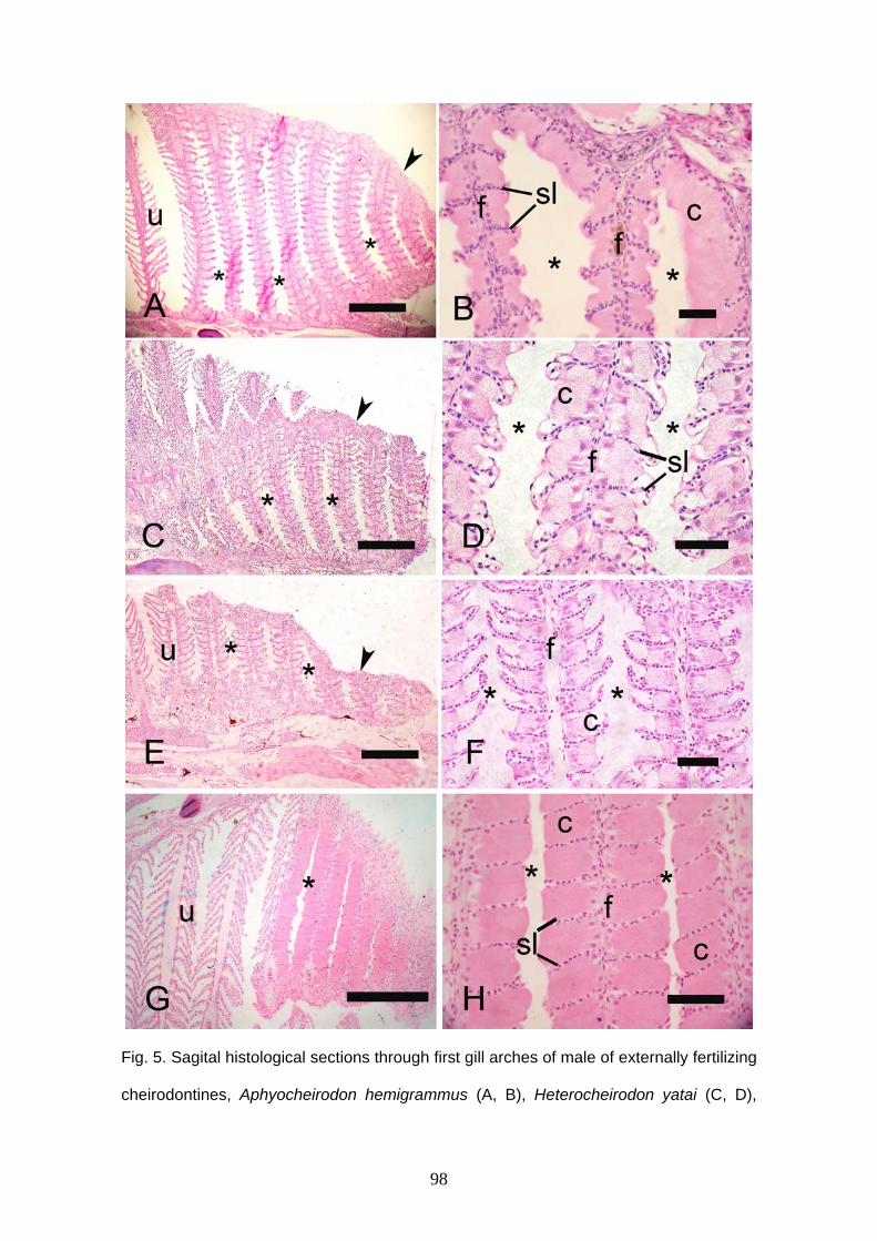
98
Fig. 5. Sagital histological sections through first gill arches of male of externally fertilizing
cheirodontines, Aphyocheirodon hemigrammus (A, B), Heterocheirodon yatai (C, D),
99
Prodontocharax melanotus (E, F), Serrapinnus heterodon (G, H). asterisks, lumina of
the gill gland chambers; f, gill filaments; sl, gill secondary lamellae; u, unmodified gill
filaments; arrowhead, ventral chamber opening; arrow, gill gland covering. Scale bar:
150 µm (A, C, E, G), 25 µm (B, D, F, H).

100
Fig. 6. Transmission electron micrograph of sagital sections through gill glands of
Compsura heterura. Tall columnar cells (c) are found between secondary lamellae (sl).
Several dark vesicles (v) occupy almost whole cell and nuclei (n) are in the base of cells.
h, hyaline cartilage. Scale bar: 8µm (A) 5µm (B).
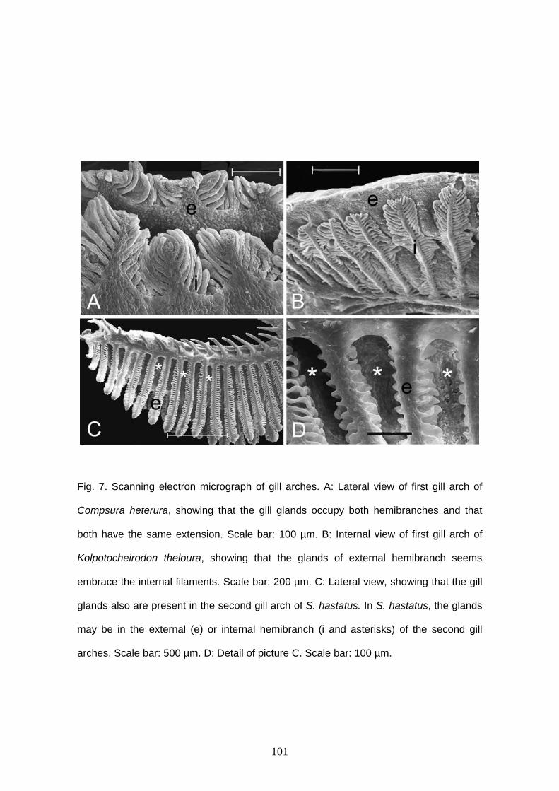
101
Fig. 7. Scanning electron micrograph of gill arches. A: Lateral view of first gill arch of
Compsura heterura, showing that the gill glands occupy both hemibranches and that
both have the same extension. Scale bar: 100 µm. B: Internal view of first gill arch of
Kolpotocheirodon theloura, showing that the glands of external hemibranch seems
embrace the internal filaments. Scale bar: 200 µm. C: Lateral view, showing that the gill
glands also are present in the second gill arch of S. hastatus. In S. hastatus, the glands
may be in the external (e) or internal hemibranch (i and asterisks) of the second gill
arches. Scale bar: 500 µm. D: Detail of picture C. Scale bar: 100 µm.

102
CONCLUSÕES
- O uso de caracteres de ultraestrutura de espermatozóides mostrou-se informativo,
contribuindo para o reconhecimento de Compsurini como um grupo monofilético e
permitindo o estabelecimento relações parciais entre as espécies da tribo.
- Os espermatozóides de espécies de fertilização externa mostram ter estruturas
conservativas enquanto que os espermatozóides de espécies inseminadoras apresentam
mudanças estruturais, principalmente no núcleo espermático.
- Compsurinis apresentam três formas de espermatozóides: aquasperma, forma de
projétil e alongado.
- O aquasperma de Cheirodontini e Odontostilbini difere de outras espécies de caracídeos
de fertilização externa por apresentar vesículas numerosas e pequenas na região basal
da peça intermediária.
- Kolpotocheirodon theloura apresenta um aquasperma derivado que difere das espécies
de Cheirodontini e Odontostilbini por apresentar peça intermediária mais alongada que se
estreita progressivamente no sentido distal, e vesículas mais largas, com formato
irregular e em menor número. Cheirodontini e Odontostilbini possuem um aquasperma
com peças intermediárias mais curtas e que terminam abruptamente, e vesículas
menores, com formato regular e em maior número.
- Compsura heterura, M. uruguayanae, A. melanogramma, S. hastatus, “Odontostilbe”
mitoptera and “O.” dialeptura formam um grupo monofilético sustentado por três
caracteres, forma do núcleo, relação comprimento e largura do núcleo e posição do
complexo centriolar em relação à fossa nuclear.
- Acinocheirodon melanogramma, “O”. dialeptura, “O”. mitoptera and S. hastatus
103
constituem um grupo monofilético de Compsurini sustentado por quatro caracteres, forma
do núcleo, a ausência de rotação nuclear, a posição dos centríolos, e a posição das
mitocôndrias.
- A rotação nuclear ocorre em todos Cheirodontini e Odontostilbini analisados e em C.
heterura, K. theloura e M. uruguayanae, estando ausente no clado formado por
Acinocheirodon melanogramma, “O”. dialeptura, “O”. mitoptera and S. hastatus .
- Macropsobrycon uruguayanae possui espermatozóides normais e espermatozóides
atípicos denominados paraespermatozóides, encontrados no lúmen testicular.
- Os espermatozóides de M. uruguayanae apresentam duas autapomorfias, como
microtúbulos acessórios ao redor do axonema e estrias centriolares denominadas
rootlets.
- A glândula branquial possui a mesma estrutura em todos os caracídeos analisados até o
momento, diferindo em sua extensão, presença ou não no segundo arco branquial e
presença ou não nas duas hemibranquias.
- A glândula branquial é foi encontrada em todos machos maduros, estando ausente em
fêmeas.
- Queirodontíneos de fertilização externa possuem glândula branquial menor ocupando
no máximo 10 filamentos branquiais e as espécies inseminadoras apresentam uma
extensa glândula (até 28 filamentos branquiais), com exceção de Acinocheiron
melanogramma.
- As glândulas branquiais de compsurinis alcançam maiores extensões do que as
glândulas branquiais de Stervardiinae.
- A presença da glândula branquial nas duas hemibranquias é uma característica de
104
somente C. heterura e a presença da glândula no segundo arco branquial é uma
característica dividida por C. heterura and S. hastatus.
- A glândula branquial está presente tanto em espécies inseminadoras quanto em
espécies de fertilização externa, não existindo relação entre a presença de glândula
branquial e inseminação.
- Em C. heterura, K. theloura, M. uruguayanae e S. hastatus observou-se à redução ou
desaparecimento das lamelas secundárias.
- A função da glândula não é conhecida, mas devido a sua presença em somente machos
maduros e em maturação, esta estrutura poderia ser usada na produção e liberação de
secreção para a atração de fêmeas durante o período reprodutivo. Estudos posteriores
sobre a fisiologia da glândula são necessários para conhecer a função e funcionamento
da glândula.
105
Anexo 1 Journal Morphology For Authors
For additional tools visit Author Resources - an enhanced suite of online tools for Wiley InterScience journal authors, featuring Article Tracking, E-mail Publication Alerts and Customized Research Tools.
• Copyright Transfer Agreement • Permission Request Form • The National Institutes of Health Public Access Initiative
Instructions to Authors Disk Submission Instructions Wiley's Journal Styles and EndNote Reprints of this Guide for Authors, published periodically and in the first issue of each year, are available on request to the editor, Dr. Frederick W. Harrison, 191 Wildwood Drive, Sylva, North Carolina, 28779, USA; Fax: 828-293-7029; Phone: 828-293-5566; E-mail: [email protected], or the publisher, Wiley-Liss, Inc., a division of John Wiley & Sons, 111 River Street, Hoboken, NJ 07030.
Manuscripts
General instructions. The manuscript should conform to Journal of Morphology style with respect to use of capital and lower case letters in headings and should be submitted exactly as it is to appear in print. It should consist of the following subdivisions, each prepared as a unit on separate sheets. Components of the text (Introduction through Acknowledgments) constitute one unit and should follow one another consequently, utilizing all available space.
• Title page (p. 1) • Abstract (p. 2) • Text o Introduction (p. 3) o Materials and Methods o Results o Discussion o Acknowledgments • Literature Cited • Footnotes • Tables • Figure/legends
The manuscript, including Literature Cited and other sections, should be typed double-spaced on bond or heavy-bodied paper 8 1/2" x 11" (22 cm x 28 cm) with a 1" (2.5 cm) margin on one side. Number the manuscript pages consecutively beginning with the title page. Submit the original manuscript and original prints of all illustrations, as well as two review copies. Manuscripts submitted in final, revised post-review form should be accompanied by a disk and the appropriate disk identification form, along with hard copy of text and illustrations.
- Do not divide words at the end of lines (hyphenate); if they are unfamiliar to the printer, they may be incorrectly hyphenated.
- Do not justify right margin of text whether or not your word processor permits you to do this.

106
o Corrections to the manuscript should be typed or printed legibly in ink.
- Do not begin sentences with abbreviations.
- The word "Figure" is not abbreviated in the text, except when appearing in parentheses: (Fig. 2); (Figs. 4-6).
- The spelling of nontechnical terms should be that recommended in the current Webster's International Dictionary.
- Italics and boldface should be indicated accordingly.
- Always spell out numbers when they stand as the first word in a sentence; do not follow such numbers with abbreviations. Numbers indicating time, weight, and measurements are to be in Arabic numerals when followed by abbreviations (e.g., 2 mm; 1 sec; 3 ml).
Title page. The title page should contain:
- Author's name (or names)
- Institution and department from which the chapter emanated, with city, state, and zip code
- Number of text pages, figures, graphs, and charts, each on a separate line
- Abbreviated title (running headline) not to exceed 48 characters and spaces
- Name, address, telephone number, fax number, and E-mail address of the person to whom the proof is to be sent
- Any special instructions regarding joint publication with other articles, return of artwork, and color plates.
Key words. Key words should be included and should not exceed 85 characters and spaces.
Abstract. The abstract should be used to disclose findings rather than aims. Indicate techniques in passing. When published, it will precede the introductory section of the text. The abstract should be written in complete sentences; it should be intelligible without reference to the rest of the chapter. Do not repeat information in title. Text introduction. A generic name should be spelled out the first time but abbreviated the second time it appears in a paragraph. Example, Danus plectopus the first time and D. plectopus the second. Materials and Methods. Repeat the name of the organism (this is the only place in the paper where the author of the Latin name may be appropriately included; if so spell it out rather than, for instance, listing an "L.") Include the source of the material, sex, weight and, if appropriate, conditions of laboratory acclimation, and one or two introductory sentences. Unless it is a very obvious form, indicate how, by whom and where the material was collected and identified. Then follow this by a terse description of the techniques. Even if some of these are published elsewhere, include enough information so that the remaining material can be placed into context. Do not force the reader to return to the library to make sense out of what is reported here. Always remember that this section should provide enough information to let the observation and experiments be repeated. Results. Group results under appropriate subheadings. Primary subheadings should be italicized and centered, secondary headings marginalized, and tertiary headings italicized and indented in accordance with Journal of Morphology style. Describe the observations but do not discuss them. Present tense is preferred, although past tense is acceptable. Use either one or the other.
107
Discussion. Briefly review those aspects of the results that appear significant and perhaps permit conclusions regarding past work by others. Organize the review of past work in terms of topics and organisms rather than as a mere historical treatment of previous studies. Do not make the authors of the papers the subject of the sentences. Organize ideas so that each particular issue is discussed only once. The most important points of the discussion should be placed into the first and last paragraphs.
Literature Cited.
Wiley's Journal Styles Are Now in EndNote EndNote is a software product that we recommend to our journal authors to help simplify and streamline the research process. Using EndNote's bibliographic management tools, you can search bibliographic databases, build and organize your reference collection, and then instantly output your bibliography in any Wiley journal style. Download Reference Style for this Journal: If you already use EndNote, you can download the reference style for this journal. How to Order: To learn more about EndNote, or to purchase your own copy, click here. Technical Support: If you need assistance using EndNote, contact [email protected], or visit www.endnote.com/support.
The list of Literature Cited should be double-spaced. In the text, references should be cited by author's surname followed by year of publication: . . . do not cite items as "submitted" or "in preparation." . . . studies by Whittier (1992) reveal . . . . . . studies by Trueb and Hanken (1992) reveal . . . . . . studies by Wittmann et al. (1993) reveal . . . . . . an earlier report (Whittier, 1992) . . . . . . earlier reports (Quinn and Baumel, 1993; Lindholm and Bass, 1993) . . . Unpublished data should be referred to as personal communication. An example would be... studies by Mary Packard (personal communication)...In this case mention the full address in the acknowledgments, not in the literature. When references are made to more than one paper by the author published in the same year, they should be designated in the text as (Condon et al., 1990a,b) and in the literature list as follows: Condon K, Silberstein L, Blau HM, Thompson WJ. 1990a. Development of muscle fiber types in the prenatal rat hindlimb. Dev Biol 138:256–274. Condon K, Silberstein L, Blau HM, Thompson WJ. 1990b. Differentiation of fiber types in aneural musculature of the prenatal rat hindlimb. Dev Biol 138:275–295. The literature list must be arranged alphabetically by author's surname in the following style: Author's name (or names), year of publication, complete title, volume, and inclusive pages as follows: Journal article King VM, Armstrong DM, Apps R, Trott JR. 1998. Numerical aspects of pontine, lateral reticular, and inferior olivary projections to two paravermal cortical zones of the cat cerebellum. J Comp Neurol 390:537-551. Book Voet D, Voet JG. 1990. Biochemistry. New York: John Wiley & Sons. 1223 p. Book chapter Gilmor ML, Rouse ST, Heilman CJ, Nash NR, Levey AI. 1998. Receptor fusion proteins and analysis. In: Ariano MA, editor. Receptor localization. New York: Wiley-Liss. p 75-90. References to papers by two authors follow those by the senior author. If there is more than one paper with a second author, these are listed alphabetically. References by three or more authors
108
follow those by the senior author (including two-author references) and are arranged chronologically independent of the order of the second and third authors. Abbreviations of journal titles should follow those used in Index Medicus. Non-English titles should be in the original language unless this uses a different alphabet. Please follow appropriate spelling and capitalization. Include accents and umlauts. References in the text to papers published before 2001 should not be abbreviated: (1784), (1889), (1900), (2000). In the Literature Cited section the year must never be abbreviated. Footnotes. Footnotes to the text should be limited as much as possible and must be numbered consecutively. The corresponding reference numbers must be clearly indicated in the text. Additional references to the identical footnotes are to be numbered with the next following consecutive number, for example: 3 See footnote 2, page . . . Footnotes to a table should be typed directly beneath the table and numbered 1,2,3, etc. They should not be numbered in sequences with the footnotes in the text. Tables. All tables must be cited in the text. Since tabular material is expensive to reproduce, it should be simple and uncomplicated, with as few vertical and horizontal rules as possible. Indicate in the margin where the tables are to appear in the text. Table titles should be complete but brief. Information other than that defining the data should be presented in footnotes. Figures. All figures must be cited in the text. Photographs or drawings mounted together as a group may be given separate figure numbers, preferably in the lower lefthand corner. If group-mounted illustrations are closely related, however, it is preferable to assign them a single figure number and letter the individual prints as A, B, C (a, b, c), etc. in the lower left corner. The code to abbreviations for each figure should appear in the figure caption and may be summed at its end. The abbreviations should be ordered alphabetically, followed by numerical codes. List the codes consecutively, with each code entry followed by a comma and then the full term and a semicolon or terminal period rather than setting them in a double column with each entry on a separate line. Do not provide a separate list of abbreviations for inclusion at the end of the article. However, you may appropriately give an abbreviation in parentheses after the first mention of a term in the text. Whenever possible, figures should be integrated into the text. Group figures to fit a single page along with their appropriate legends. Reference to relevant text passages can often reduce the length of legends and avoid redundancy. Metric system. The metric system should be used for all measurements, weight, etc. Temperatures should be expressed in degrees Celsius (centigrade). Metric abbreviations, as listed below, should be expressed in lower-case without periods.
Length Volume
km kilometer km3 cubic
m meter kilometer
cm centimeter m3 cubic
mm millimeter meter
µm micrometer cm3 cubic
(micron) centimeter
nm nanometer mm3 cubic
pm picometer millimeter
Å Angstrom unit µm3 cubic
109
(10 Å = 1 nm) micrometer
nm3 cubic
nanometer
Area kl kiloliter
km2 square l liter, always
kilometer spell out
m2 square ml milliliter
meter µl microliter
cm2 square nl nanoliter
centimeter pl picoliter
mm2 square kg kilogram
millimeter gm gram
µm2 square mg milligram
micrometer µg microgram
mm2 square ng nanogram
nanometer pg picogram
Symbols. When preceded by a digit, the following symbols are to be used: % for percent; ° for degrees.
ILLUSTRATIONS
To achieve greatest fidelity and rendition of detail, it is preferable that the printer work directly from original drawings or high-quality photographic prints (but not photocopies made on an office duplicating machine)or disks. All illustrations must be submitted in complete and finished form with adequate labeling. To achieve optimum halftone quality, photographic prints submitted for reproduction must be of adequate contrast and if multiple prints are included in a single figure, they should be of uniform tone. For information on electronic submission of illustrations, figures, and drawings, please see section below. Figures and Legends. Original illustrations, and three sets of good-contrast photographic copies for review purposes should be submitted with the manuscript. Number figures in consecutive series with Arabic numerals, and key them into the text. The reverse side of each figure should have the author's name, figure number, top side of illustration, reduction requested, and "Review Copy" or "Original" indicated. It is best, whenever possible, to plan for a one-to-one duplication. The maximum printed figure dimensions are 41 picas wide by 58 picas deep (8.5 cm wide by 24.5 cm deep) for single-column placement. Illustrations cannot be reduced less than 20% of their submitted size and must be less than 11" x 14" (28 x 36 cm). Black-and-white prints. Prints should be on white, nonmatte paper.
110
Reduction to printed size. The author should indicate clearly on each illustration the reduction desired, bearing the following in mind:
• Lettering and labels must be readable after reduction. When reduced, the minimum height of a capital letter should not be less than 2.5 mm for a photomicrograph and 1 mm for a graph or chart.
• Do not write directly on glossy prints or use devices like paper clips which can damage the print. Submit a descriptive legend for each illustration. Abbreviations used on figures should be defined in the legend and must match exactly those used in the text.
Cover Illustrations. Authors may submit color figures for consideration as cover illustrations. These figures must be enclosed with the submitted manuscript, preferably sized to 8" x 10" (21 x 26 cm). Color Prints. Authors are encouraged to submit color illustrations when the color conveys essential scientific information. Color illustration is available at no cost to the author only after consultation between author and Editor. Additional pages of color reproduction will be subsidized by the publisher, reducing author costs to $500 per page. All color figures will be reproduced in full color in the online edition of the journal at no cost to authors. Authors are requested to pay the cost of reproducing color figures in print. Authors are encouraged to submit color illustrations that highlight the text and convey essential scientific information. For best reproduction, bright, clear colors should be used. Dark colors against a dark background do not reproduce well; please place your color images against a white background wherever possible. Please contact Farah Alladin at [email protected] for further information. Proofs and Reprints. Upon acceptance of a manuscript for publication in Journal of Morphology, the author will be asked to sign a "Copyright Transfer Agreement" which transfers copyright to the Publisher. No published material may be reproduced or published elsewhere without written consent of the Publisher and the author. All statements (or omissions) in published manuscripts are the responsibility of the authors who will be asked to review a single set of page and illustration proofs. Reprints may be purchased at prices quoted on the reprint order form which accompanies the proofs. As far as possible, the publisher will adhere to the author's suggested reduction. However, discretionary adjustments may have to be made. Line drawings. Figures should be drawn with black ink on medium-weight white paper or light-weight artboard. To reduce weight and postal charges, photographic prints may be submitted in lieu of original drawings. The artwork should be sharp and black to achieve maximum contrast. Use stippling and hatching techniques to achieve tonal quality. Avoid the use of shading (pencil, wash, or airbrush) for tonal effect unless the drawing is to be reproduced as a halftone with its attendant gray-tint background. If original graphs are submitted, they should be drawn on blue-ruled paper; colors other than blue will reproduce. Line art may be submitted on disk. Please see the accompanying "Disk Submission Instructions form" for details. Mounting figures. Photomicrographs and illustrations should be mounted as follows:
• Figures should be trimmed straight on all sides and "squared." • Figures should be mounted on strong bristol board of about 15 points (0.4 mm) thickness
with at least a 1" (2.5 cm) margin surrounding the figure or grouping of figures. • Figures should be attached to bristol board using appropriate dry mounting materials, or
a cement or glue that is white or colorless when set. • When two or more figures are assembled, they should be mounted close together and
separated by no more than 1/8" (3 mm). • Illustrations grouped to form a single figure should be of similar density to tone to prevent
loss of detail.
Lettering and labels. Illustrations should be lettered and numbered with printed paste-on or transfer labels.
111
• Labels should be large enough to allow for suitable reduction and sturdy enough to withstand mailing and handling in the production process.
• For protection, it is recommended that labeling be sprayed with clear adhesive to prevent it from becoming scratched or being torn off.
• Labeling should be done directly on the drawing or photographic print, never on an overlay.
• All labeling should be placed at least 1/4" (6 mm) in from the edges of the illustration. • To achieve adequate contrast between the label or letter and its background, place white
labels over dark backgrounds and black labels over light backgrounds, or shadow the labels with an appropriately light or dark highlight.
Numbering. Figures, including charts and graphs, must be numbered consecutively. General illustration instructions. Original illustrations and two review copies should be submitted with the manuscript. Copies may be photographs of the originals or very high quality photocopies
• If the original drawings are too large for shipment, photographic prints should be submitted.
• The reverse side of each illustration should indicate: Author's name; Figure number; Top side of illustration; Reduction requested.
• Do not fasten illustrations with paper clips, staples, etc., because fastener may mark the surface of the illustration.
• Illustrations should be shipped flat and protected by heavy cardboard.
Special reproduction problems. Color illustration is available, at the discretion of the Editor, without cost to the author. Prior to submission of color illustrations, the author must contact the Editor. Authors are encouraged to submit an 8 x 10 inch format color illustration suitable for use as the cover of the journal. An author wishing to submit a color illustration for the journal cover should also contact the Editor.
ELECTRONIC SUBMISSION OF ARTICLES
Storage medium. 3-1/2" high-density disk in IBM MS-DOS, Windows, or MacIntosh format. Software. Microsoft Word 6.0 is preferred, although manuscripts prepared with any other microcomputer word processor are acceptable. Do not use desktop publishing software such as Adobe PageMaker© or Quark XPress©. If you prepared your manuscript with one of these programs, export the text to a word processing format. Please make sure your word processing program's "fast save" feature is turned off. Format. Refrain from complex formatting; the Publisher will style your manuscript according to the Journal design specifications. File names. Submit the text and tables of each manuscript as a single file. Name each file with your last name (up to eight letters). Text files should be given the three-letter extension that identifies the file format. MacIntosh users should maintain the MS-DOS "eight dot three" file-naming convention. Illustrations. Submit as separate files from text files, on separate diskette or cartridges. If feasible, full color files should be submitted on separate disks from other image files, and all print reproduction requires files for full color images to be in a CMYK color space. 3-1/2" high-density diskettes, CD, Iomega Zip, and 5 1/4" 44- or 88-MB SyQuest© cartridges can be submitted. At authors' request, cartridges and diskettes will be returned after publication. All illustration files should be in TIFF or EPS (with preview) formats. Do not submit native application formats. Journal quality reproduction will require greyscale and color files at resolutions yielding approximately 300 ppi. Bitmapped line art should be submitted at resolutions yielding 600-1200 ppi. These resolutions refer to the output size of the file; if you anticipate that your images will be enlarged or reduced, resolutions should be adjusted accordingly. Illustration files should be given the 2- or 3-letter extension that identifies the file format used (i.e., .tif, .eps).
112
Labels. Label all disks with your name, the file name, and the word processing program and version used. Paper copy. The disk must be accompanied by hard copy printout. If the disk and paper copy differ, the paper copy will be used for typesetting and this may delay publication of the article. Once the paper has been accepted, firm quotes will be supplied by the publisher, and the author will have the opportunity to approve both costs and proofs prior to printing. Color prints, transparencies, zip disks, and CD-ROMs are acceptable for reproduction. The frame of the transparency should be marked to indicate the area that can safely be cropped to arrive at the critical image that is to appear in the final printing.
MISCELLANEOUS
The editor and publisher reserve the right to return to the author for revision manuscripts and illustrations that are not in proper finished form. Upon acceptance of an article for publication, the author will be asked to sign a copyright transfer agreement, transferring rights to the publisher, who reserves copyright. It is the current policy for the publisher to underwrite all normal black and white tabular and illustration costs. However, because of the very high cost of color work, color illustration is available at no cost to the author only after consultation between author and Editor. Proofs. A single set of page and illustration proofs will be sent to the author. All corrections should be marked clearly directly on page proofs. Reprints. Reprints may be purchased at prices quoted on the reprint order form. Reprint orders should be returned with the proofs. It is important to order initially a sufficient quantity of reprints, because the price is substantially higher if they are ordered after the paper has been published. ???Production Questions??? Michele Azzaretto Phone: 201-748-6492
Disk Submission Instructions
Please return your final, revised manuscript on disk as well as hard copy. The hard copy must match the disk. The Journal strongly encourages authors to deliver the final, revised version of their accepted manuscripts (text, tables, and, if possible, illustrations) on disk. Given the near-universal use of computer word-processing for manuscript preparation, we anticipate that providing a disk will be convenient for you, and it carries the added advantages of maintaining the integrity of your keystrokes and expediting typesetting. Please return the disk submission slip below with your manuscript and labeled disk(s).
Guidelines for Electronic Submission
Text Storage medium. 3-1/2" high-density disk in IBM MS-DOS, Windows, or Macintosh format. Software and format. Microsoft Word 6.0 is preferred, although manuscripts prepared with any other microcomputer word processor, including TeX and LaTeX, are acceptable. Refrain from complex formatting; the Publisher will style your manuscript according to the Journal design specifications. Do not use desktop publishing software such as Aldus PageMaker or Quark XPress. If you prepared your manuscript with one of these programs, export the text to a word processing format. Please make sure your word processing program's "fast save" feature is turned off. Please do not deliver files that contain hidden text: for example, do not use your word processor's automated features to create footnotes or reference lists. File names. Submit the text and tables of each manuscript as a single file. Name each file with your last name (up to eight letters). Text files should be given the three-letter extension that identifies the file format. Macintosh users should maintain the MS-DOS "eight dot three" file-naming convention. Labels. Label all disks with your name, the file name, and the word processing program and version used.
113
Illustrations All print reproduction requires files for full color images to be in a CMYK color space. If possible, ICC or ColorSync profiles of your output device should accompany all digital image submissions. Storage medium. Submit as separate files from text files, on separate disks or cartridges. If feasible, full color files should be submitted on separate disks from other image files. 3-1/2" high-density disks, CD, Iomega Zip, and 5 1/4" 44- or 88-MB SyQuest cartridges can be submitted. At authors' request, cartridges and disks will be returned after publication. Software and format. All illustration files should be in TIFF or EPS (with preview) formats. Do not submit native application formats. Resolution. Journal quality reproduction will require greyscale and color files at resolutions yielding approximately 300 ppi. Bitmapped line art should be submitted at resolutions yielding 600-1200 ppi. These resolutions refer to the output size of the file; if you anticipate that your images will be enlarged or reduced, resolutions should be adjusted accordingly. File names. Illustration files should be given the 2- or 3-letter extension that identifies the file format used (i.e., .tif, .eps). Labels. Label all disks and cartridges with your name, the file names, formats, and compression schemes (if any) used. Hard copy output must accompany all files.