Cosmopolitan, Sep 2011
3
Directional Movement of Dendritic Macromolecules on Gradient Surfaces Theresa Chang, † Dorota I. Rozkiewicz, ‡ Bart Jan Ravoo, ‡ E. W. Meijer,* ,† and David N. Reinhoudt* ,‡ Laboratory of Macromolecular and Organic Chemistry, EindhoVen UniVersity of Technology, PO Box 513, 5600 MB EindhoVen, The Netherlands, and Laboratory of Supramolecular Chemistry and Technology, MESA + Institute for Nanotechnology, UniVersity of Twente, PO Box 217, 7500 AE Enschede, The Netherlands Received January 2, 2007; Revised Manuscript Received March 7, 2007 ABSTRACT A gradient-driven methodology has been developed to manipulate the movement of dendritic macromolecules. Poly(propyleneim ine) dendrimers, labeled with rhodamine B, are attached to glass substrates via multiple imine bonds. The dendrimers are able to move on the surface by the hydrolysis and re-formation of these imine bonds. In the absence of an external stimulus, this random movement results in a two-dimensional diffus ion on the substrate. We are able to bias the movement of these nanoparti cles by means of an aldehyde gradient on the glass substrat e. Chemotaxis, 1 the response of cells to chemical gradients by directed movement, is a universal phenomenon that is vital to the survival of both microorg anis ms and mult icel lula r organisms. For example, bacteria are able to sense changes in concentrations of certain chemicals and move toward or away from this chemical by means of altering its tumbling frequency. 2 Likewise, multicellular organisms are able to fight off bacterial infections using white blood cells (neu- trophils) that are able to sense chemicals left by bacteria to fin d and des tro y the invadi ng micro org ani sms. Hence, directional sensing and response play a central role in health and diseases. 3 Fundamental ly, the same prin ciple s shoul d apply for particles of any size that are able to sense and res pond to a gra die nt, alt hough the mec han ism may be completely different. 4 In this Letter, we present the directed movement of dendritic macromolecules on glass substrates. Dendrimers have been used in a variety of applications; in this case it is the multivalent, globular nature and their ability to deform their shape that is essential for gradient sensing. For our purposes, the dendrimers need to be attached to the substrate, yet have some freedom to move about on the surface, to “sense” and respond to their environment. This freedom requires that their attachment to the surfaces be reversible, making use of the dynamic covalent chemistry, 5 to allow the dendrimer to move across the substrate. An amine-terminated dendrimer will attach to a surface containing aldehyde groups by imine condensation. One or mor e of these imine bon ds can hyd rol yze , 6 giv ing the dendrimer additional freedom to move about and attach itself via imine formation with another aldehyde on the surface. The net result is random motion on the surface, resembling a random walk. In the presence of a gradient, it is more likely for the dendrimer to move in one direction (with the gradient) as it is statistically more likely to form imine bonds where more aldehyde groups are present. The number of imine bonds formed between the two will dep end on the ava il abi lit y and pro ximity of ami ne and aldehyde groups. On a fifth generation dendrimer there are 64 amine groups, some (approximately 8) of which are used to attach rhodamine labels, so it is possible to follow by fluorescence. Although dendrimers do deform on surfaces, flattening to maximize favorable interactions, not all of the amine groups are accessible at the same time for attachment to the surface; hence when the dendrimer moves around, tho se fre e ami ne gro ups are fre e to react wit h ald ehy de groups on the substrate. Reactive aldehyde substrates were prepared by reacting glas s slide s with trimethox ysily lalky lald ehyde . Supplier: Fluorochem UK. The aldehyde gradients are achieved by a modified reverse dip-coating procedure, 7 whereas the control substrates were prepared by full immersion of the substrate for a specified amount of time. The dendrimers are labeled with rhodami ne B by reac ting the fift h gener atio n poly - propy lenei mine dendrimer with 8 equiv of rhoda mine B isothiocyanate. The resulting labeled dendrimers were applied onto the subst rate by micr ocont act prin ting. 8 The print ed substrates 9 were fully immersed 10 in water overnight and then analyzed using confocal microscopy. * Co rr espo nd in g auth o rs . E- ma il : e .w . meij er @t ue . nl and [email protected]. † Eindhoven University of Technology. ‡ University of Twente. NANO LETTERS 2007 Vol. 7, No. 4 978-980 10.1 021/n l0700 05v CCC : $37. 00 © 2007 A meric an Chem ical S ociet y Published on Web 03/21/2007
Transcript of Cosmopolitan, Sep 2011
8/3/2019 Cosmopolitan, Sep 2011
http://slidepdf.com/reader/full/cosmopolitan-sep-2011 1/3
Directional Movement of DendriticMacromolecules on Gradient Surfaces
Theresa Chang,† Dorota I. Rozkiewicz,‡ Bart Jan Ravoo,‡ E. W. Meijer,*,† and
David N. Reinhoudt*,‡
Laboratory of Macromolecular and Organic Chemistry, EindhoVen UniVersity of
Technology, PO Box 513, 5600 MB EindhoVen, The Netherlands, and Laboratory of
Supramolecular Chemistry and Technology, MESA+ Institute for Nanotechnology,
UniVersity of Twente, PO Box 217, 7500 AE Enschede, The Netherlands
Received January 2, 2007; Revised Manuscript Received March 7, 2007
ABSTRACT
A gradient-driven methodology has been developed to manipulate the movement of dendritic macromolecules. Poly(propyleneimine) dendrimers,
labeled with rhodamine B, are attached to glass substrates via multiple imine bonds. The dendrimers are able to move on the surface by thehydrolysis and re-formation of these imine bonds. In the absence of an external stimulus, this random movement results in a two-dimensional
diffusion on the substrate. We are able to bias the movement of these nanoparticles by means of an aldehyde gradient on the glass substrate.
Chemotaxis,1 the response of cells to chemical gradients by
directed movement, is a universal phenomenon that is vital
to the survival of both microorganisms and multicellular
organisms. For example, bacteria are able to sense changes
in concentrations of certain chemicals and move toward or
away from this chemical by means of altering its tumbling
frequency.2 Likewise, multicellular organisms are able to
fight off bacterial infections using white blood cells (neu-
trophils) that are able to sense chemicals left by bacteria tofind and destroy the invading microorganisms. Hence,
directional sensing and response play a central role in health
and diseases.3 Fundamentally, the same principles should
apply for particles of any size that are able to sense and
respond to a gradient, although the mechanism may be
completely different.4 In this Letter, we present the directed
movement of dendritic macromolecules on glass substrates.
Dendrimers have been used in a variety of applications;
in this case it is the multivalent, globular nature and their
ability to deform their shape that is essential for gradient
sensing. For our purposes, the dendrimers need to be attached
to the substrate, yet have some freedom to move about on
the surface, to “sense” and respond to their environment.
This freedom requires that their attachment to the surfaces
be reversible, making use of the dynamic covalent chemistry,5
to allow the dendrimer to move across the substrate.
An amine-terminated dendrimer will attach to a surface
containing aldehyde groups by imine condensation. One or
more of these imine bonds can hydrolyze,6 giving the
dendrimer additional freedom to move about and attach itself
via imine formation with another aldehyde on the surface.
The net result is random motion on the surface, resembling
a random walk. In the presence of a gradient, it is more likely
for the dendrimer to move in one direction (with the gradient)
as it is statistically more likely to form imine bonds where
more aldehyde groups are present.
The number of imine bonds formed between the two willdepend on the availability and proximity of amine and
aldehyde groups. On a fifth generation dendrimer there are
64 amine groups, some (approximately 8) of which are used
to attach rhodamine labels, so it is possible to follow by
fluorescence. Although dendrimers do deform on surfaces,
flattening to maximize favorable interactions, not all of the
amine groups are accessible at the same time for attachment
to the surface; hence when the dendrimer moves around,
those free amine groups are free to react with aldehyde
groups on the substrate.
Reactive aldehyde substrates were prepared by reacting
glass slides with trimethoxysilylalkylaldehyde. Supplier:Fluorochem UK. The aldehyde gradients are achieved by a
modified reverse dip-coating procedure,7 whereas the control
substrates were prepared by full immersion of the substrate
for a specified amount of time. The dendrimers are labeled
with rhodamine B by reacting the fifth generation poly-
propyleneimine dendrimer with 8 equiv of rhodamine B
isothiocyanate. The resulting labeled dendrimers were applied
onto the substrate by microcontact printing.8 The printed
substrates9 were fully immersed10 in water overnight and then
analyzed using confocal microscopy.
* Corresponding authors. E-mail: [email protected] [email protected].
† Eindhoven University of Technology.‡ University of Twente.
NANO
LETTERS
2007Vol. 7, No. 4
978-980
10.1021/nl070005v CCC: $37.00 © 2007 American Chemical SocietyPublished on Web 03/21/2007
8/3/2019 Cosmopolitan, Sep 2011
http://slidepdf.com/reader/full/cosmopolitan-sep-2011 2/3
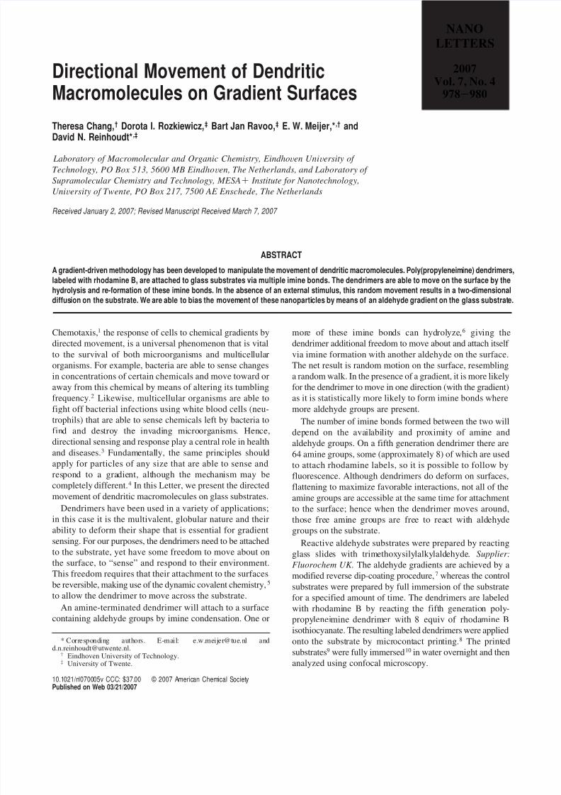
The confocal microscope images of the top and bottom
of a rectangular pattern of dendrimers printed on a glass
substrate before and after overnight immersion in water are
shown in Figure 2.Before immersion, the image is sharp as can be observed
by the edges of the pattern as well as defects in the stamp.
This can be seen clearly in the fluorescence intensity graphs,
where sharp slopes can be seen at the edges of the stamp.
After immersion, the patterns are blurred as a consequence
of diffusion of the dendrimers. In the absence of a gradient,
we see that the edges are no longer clear, there is migration
of dendrimer away from where they were originallly
stamped, and the amount of migration is similar on all edges.
The fluorescence intensity is lower after immersion which
can be attributed to loss of dendrimers either that were
attached to other dendrimers or that were weakly bound.
Dendrimers on the gradient substrate, however, appear to
move with the gradient. On the top of the stamp, the edge
remains sharp, whereas at the bottom we see a large amount
of fluorescence past the edge of the stamp.
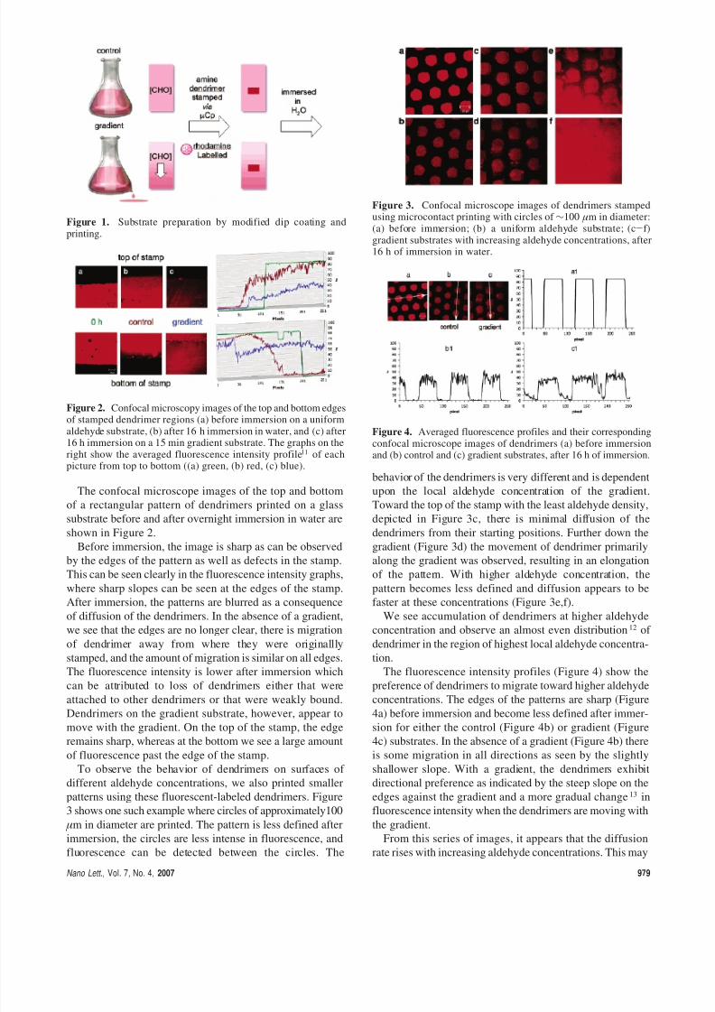
To observe the behavior of dendrimers on surfaces of
different aldehyde concentrations, we also printed smaller
patterns using these fluorescent-labeled dendrimers. Figure
3 shows one such example where circles of approximately100
µm in diameter are printed. The pattern is less defined after
immersion, the circles are less intense in fluorescence, and
fluorescence can be detected between the circles. The
behavior of the dendrimers is very different and is dependent
upon the local aldehyde concentration of the gradient.
Toward the top of the stamp with the least aldehyde density,
depicted in Figure 3c, there is minimal diffusion of the
dendrimers from their starting positions. Further down thegradient (Figure 3d) the movement of dendrimer primarily
along the gradient was observed, resulting in an elongation
of the pattern. With higher aldehyde concentration, the
pattern becomes less defined and diffusion appears to be
faster at these concentrations (Figure 3e,f).
We see accumulation of dendrimers at higher aldehyde
concentration and observe an almost even distribution12 of
dendrimer in the region of highest local aldehyde concentra-
tion.
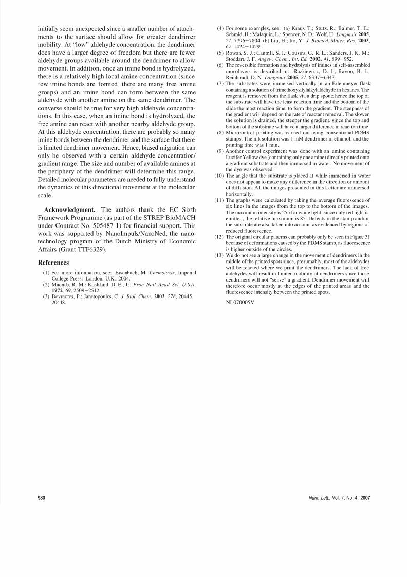
The fluorescence intensity profiles (Figure 4) show the
preference of dendrimers to migrate toward higher aldehyde
concentrations. The edges of the patterns are sharp (Figure
4a) before immersion and become less defined after immer-
sion for either the control (Figure 4b) or gradient (Figure
4c) substrates. In the absence of a gradient (Figure 4b) there
is some migration in all directions as seen by the slightly
shallower slope. With a gradient, the dendrimers exhibit
directional preference as indicated by the steep slope on the
edges against the gradient and a more gradual change13 in
fluorescence intensity when the dendrimers are moving with
the gradient.
From this series of images, it appears that the diffusion
rate rises with increasing aldehyde concentrations. This may
Figure 1. Substrate preparation by modified dip coating andprinting.
Figure 2. Confocal microscopy images of the top and bottom edgesof stamped dendrimer regions (a) before immersion on a uniformaldehyde substrate, (b) after 16 h immersion in water, and (c) after16 h immersion on a 15 min gradient substrate. The graphs on theright show the averaged fluorescence intensity profile11 of eachpicture from top to bottom ((a) green, (b) red, (c) blue).
Figure 3. Confocal microscope images of dendrimers stampedusing microcontact printing with circles of ∼100 µm in diameter:(a) before immersion; (b) a uniform aldehyde substrate; (c-f)gradient substrates with increasing aldehyde concentrations, after16 h of immersion in water.
Figure 4. Averaged fluorescence profiles and their correspondingconfocal microscope images of dendrimers (a) before immersionand (b) control and (c) gradient substrates, after 16 h of immersion.
Nano Lett., Vol. 7, No. 4, 2007 979
8/3/2019 Cosmopolitan, Sep 2011
http://slidepdf.com/reader/full/cosmopolitan-sep-2011 3/3
initially seem unexpected since a smaller number of attach-
ments to the surface should allow for greater dendrimer
mobility. At “low” aldehyde concentration, the dendrimer
does have a larger degree of freedom but there are fewer
aldehyde groups available around the dendrimer to allow
movement. In addition, once an imine bond is hydrolyzed,
there is a relatively high local amine concentration (since
few imine bonds are formed, there are many free amine
groups) and an imine bond can form between the same
aldehyde with another amine on the same dendrimer. Theconverse should be true for very high aldehyde concentra-
tions. In this case, when an imine bond is hydrolyzed, the
free amine can react with another nearby aldehyde group.
At this aldehyde concentration, there are probably so many
imine bonds between the dendrimer and the surface that there
is limited dendrimer movement. Hence, biased migration can
only be observed with a certain aldehyde concentration/
gradient range. The size and number of available amines at
the periphery of the dendrimer will determine this range.
Detailed molecular parameters are needed to fully understand
the dynamics of this directional movement at the molecular
scale.
Acknowledgment. The authors thank the EC Sixth
Framework Programme (as part of the STREP BioMACH
under Contract No. 505487-1) for financial support. This
work was supported by NanoImpuls/NanoNed, the nano-
technology program of the Dutch Ministry of Economic
Affairs (Grant TTF6329).
References
(1) For more information, see: Eisenbach, M. Chemotaxis; ImperialCollege Press: London, U.K., 2004.
(2) Macnab, R. M.; Koshland, D. E., Jr. Proc. Natl. Acad. Sci. U.S.A.1972, 69, 2509-2512.
(3) Devreotes, P.; Janetopoulos, C. J. Biol. Chem. 2003, 278, 20445-
20448.
(4) For some examples, see: (a) Kraus, T.; Stutz, R.; Balmer, T. E.;
Schmid, H.; Malaquin, L.; Spencer, N. D.; Wolf, H. Langmuir 2005,
21, 7796-7804. (b) Liu, H.; Ito, Y. J. Biomed. Mater. Res. 2003,
67 , 1424-1429.
(5) Rowan, S. J.; Cantrill, S. J.; Cousins, G. R. L.; Sanders, J. K. M.;
Stoddart, J. F. Angew. Chem., Int. Ed. 2002, 41, 899-952.
(6) The reversible formation and hydrolysis of imines in self-assembled
monolayers is described in: Rozkiewicz, D. I.; Ravoo, B. J.;
Reinhoudt, D. N. Langmuir 2005, 21, 6337-6343.
(7) The substrates were immersed vertically in an Erlenmeyer flask
containing a solution of trimethoxysilylalkylaldehyde in hexanes. The
reagent is removed from the flask via a drip spout; hence the top of
the substrate will have the least reaction time and the bottom of theslide the most reaction time, to form the gradient. The steepness of
the gradient will depend on the rate of reactant removal. The slower
the solution is drained, the steeper the gradient, since the top and
bottom of the substrate will have a larger difference in reaction time.
(8) Microcontact printing was carried out using conventional PDMS
stamps. The ink solution was 1 mM dendrimer in ethanol, and the
printing time was 1 min.
(9) Another control experiment was done with an amine containing
Lucifer Yellow dye (containing only one amine) directly printed onto
a gradient substrate and then immersed in water. No movement of
the dye was observed.
(10) The angle that the substrate is placed at while immersed in water
does not appear to make any difference in the direction or amount
of diffusion. All the images presented in this Letter are immersed
horizontally.
(11) The graphs were calculated by taking the average fluorescence of six lines in the images from the top to the bottom of the images.
The maximum intensity is 255 for white light; since only red light is
emitted, the relative maximum is 85. Defects in the stamp and/or
the substrate are also taken into account as evidenced by regions of
reduced fluorescence.
(12) The original circular patterns can probably only be seen in Figure 3f
because of deformations caused by the PDMS stamp, as fluorescence
is higher outside of the circles.
(13) We do not see a large change in the movement of dendrimers in the
middle of the printed spots since, presumably, most of the aldehydes
will be reacted where we print the dendrimers. The lack of free
aldehydes will result in limited mobility of dendrimers since those
dendrimers will not “sense” a gradient. Dendrimer movement will
therefore occur mostly at the edges of the printed areas and the
fluorescence intensity between the printed spots.
NL070005V
980 Nano Lett., Vol. 7, No. 4, 2007