Congosaurus bequaerti, a Paleocene (Crocodyliformes...
18
bulletin de l'institut royal des sciences naturelles de belgique bulletin van het koninklijk belgisch instituut voor natuurwetenschappen sciences de la terre, 74: 129-146, 2004 aardwetenschappen, 74: 129-146, 2004 Congosaurus bequaerti, a Paleocene Dyrosaurid (Crocodyliformes; Mesoeucrocodylia) from Landana (Angola) by Stéphane JOUVE & Daniela SCHWARZ Jouve, S. & Schwarz, D., 2004. - Congosaurus bequaerti, a Paleo¬ cene Dyrosaurid (Crocodyliformes; Mesoeucrocodylia) from Landana (Angola). Bulletin de l'Institut royal des Sciences naturelles de Bel¬ gique, Sciences de la Terre 74: 129-146, 4 pis., 3 figs., 4 tables, Bruxelles-Brussel, March 31, 2004. - ISSN 0374-6291. Abstract A complete redescription of the material of Congosaurus bequaerti from the Cabinda enclave (Angola), first described in 1914 by Dollo, enables a taxonomie revision of this species. The genus Congosaurus was considered by Buffetaut (1976, 1980) as a junior synonym of the genus Hyposaurus. Buffetaut (1976, 1980) used the dorsoventral flattening of the mandible to distinguish Hyposaurus from the genus Rhabdognathus. A scatter plot comparing width/height ratio of the mandible according to the tooth position, supports that the two genera can clearly be separated by this method. On the other hand the values obtained for Congosaurus bequaerti are closer to Rhabdognathus than to Hyposaurus, underlining a clear différence between Congosaurus and Hyposaurus. The anatomical characteristics furnish more argu¬ ments to differentiate these two genera. In C. bequaerti, the lachrymal is extremely anteriorly extended and the posterior maxillary and den- tary teeth are labio-lingually compressed, contrary to the condition observed in Hyposaurus or Rhabdognathus. In Hyposaurus, the ex- ternal mandibular fenestra is large and the teeth exhibit ventrolingually "twisted" anterior carinae, contrary to the extremely reduced fenestra and the linear anterior carinae of Congosaurus bequaerti. The snout proportion of C. bequaerti (about 65 % of the total skull length) is about the same than in Hyposaurus (66 %) and far from Rhabdog¬ nathus (73 %). Ail these characters suggest that Congosaurus bequaerti should be distinguished from Hyposaurus and Rhabdognathus, and then, the original Dollo's genus should be retained for this species. Kev-words: Congosaurus, Dyrosauridae, Crocodyliformes, taxonomy, Paleocene, Angola. Résumé Une description complète du matériel de Congosaurus bequaerti de l'enclave de Cabinda (Angola), décrit pour la première fois en 1914 par Dollo, permet une révision taxonomique de cette espèce. Le genre Congosaurus fut considéré par Buffetaut (1976, 1980) comme un synonyme plus récent du genre Hyposaurus. Buffetaut (1976, 1980) utilisa l'aplatissement dorso-ventral plus important de la mandibule pour distinguer Hyposaurus du genre Rhabdognathus. Un graphique, comparant le rapport largeur/hauteur de la mandibule en fonction de la position sur celle ci, prouve que ces deux genres peuvent clairement être distingués par cette méthode. Par contre, les valeurs obtenues pour Congosaurus bequaerti sont plus proches de celles de Rhabdognathus que d'Hyposaurus, soulignant une nette différence entre Congosaurus et Hyposaurus. Les caractéristiques anatomiques fournissent d'autres arguments pour différencier ces deux genres. Chez Congosaurus be¬ quaerti, le lacrymal est extrêmement étendu antérieurement et les dents maxillaires et dentaires postérieures sont comprimées labio-linguale- ment contrairement à ce que l'on a observé chez Hyposaurus ou Rhabdognathus. Chez Hyposaurus, la fenêtre mandibulaire externe est large et les dents montrent une carène antérieure ventro-linguale- ment déviée, contrairement à la fenêtre très réduite et aux carènes droites de Congosaurus bequaerti. Les proportions du museau de C. bequaerti (environ 65 % de la longueur total du crâne) sont à peu près les mêmes que celles d'Hyposaurus (66 %) et très éloignées de Rhabdognathus (73 %). Tous ces caractères suggèrent que Congosaurus bequaerti peut être distingué à'Hyposaurus et de Rhabdognathus, et donc le genre original décrit par Dollo doit être conservé pour cette espèce. Mots-clefs: Congosaurus, Dyrosauridae, Crocodyliformes, taxonomie, Paléocène, Angola. Introduction In 1913, the skeletal remains of a fossil crocodyliform, discovered by J. Bequaert (Bequaert, 1923) during a botanical expédition in Landana (Cabinda enclave, Ango¬ la) (Fig. 1), were brought to the Musée d'Afrique Centrale in Tervuren, Belgium. These comprised the nearly com¬ plete skeleton of a dyrosaurid crocodylomorph. They were described preliminarily and named Congosaurus bequaerti by Dollo (1914). Though Dollo (1914) promised a more complete description of the individual for a later date, he was never able to finish this work. Therefore, in 1948 the director of the fonner "Musée du Congo belge" in Ter- vuren invited W. E. Swinton from London to re-examine the Congosaurus skeleton. Swinton (1950) prepared an extended description of the remains of this dyrosaur spe¬ cies. The taxonomie status of Congosaurus bequaerti was questioned by Arambourg (1952) and Antunes (1964); they regarded Congosaurus as a junior subjective synonym of Dyrosaurus. Buffetaut (1976, 1980) revised the mate¬ rial once again and, in contrast to the former authors, argued that the Cabinda dyrosaur belongs to the genus Hyposaurus. This remained until recently the taxonomical status for the Cabinda dyrosaur. Studies of dyrosaur material from North Africa (Jouve, pers. obs.) indicate that the Cabinda dyrosaur can be distinguished from the North American and North Afri-
Transcript of Congosaurus bequaerti, a Paleocene (Crocodyliformes...
bulletin de l'institut royal des sciences naturelles de belgiquebulletin van het koninklijk belgisch instituut voor natuurwetenschappen
sciences de la terre, 74: 129-146, 2004aardwetenschappen, 74: 129-146, 2004
Congosaurus bequaerti, a Paleocene Dyrosaurid(Crocodyliformes; Mesoeucrocodylia) from Landana (Angola)
by Stéphane JOUVE & Daniela SCHWARZ
Jouve, S. & Schwarz, D., 2004. - Congosaurus bequaerti, a Paleo¬cene Dyrosaurid (Crocodyliformes; Mesoeucrocodylia) from Landana(Angola). Bulletin de l'Institut royal des Sciences naturelles de Bel¬gique, Sciences de la Terre 74: 129-146, 4 pis., 3 figs., 4 tables,Bruxelles-Brussel, March 31, 2004. - ISSN 0374-6291.
Abstract
A complete redescription of the material of Congosaurus bequaertifrom the Cabinda enclave (Angola), first described in 1914 by Dollo,enables a taxonomie revision of this species. The genus Congosauruswas considered by Buffetaut (1976, 1980) as a junior synonym of thegenus Hyposaurus. Buffetaut (1976, 1980) used the dorsoventralflattening of the mandible to distinguish Hyposaurus from the genusRhabdognathus. A scatter plot comparing width/height ratio of themandible according to the tooth position, supports that the two generacan clearly be separated by this method. On the other hand the valuesobtained for Congosaurus bequaerti are closer to Rhabdognathus thanto Hyposaurus, underlining a clear différence between Congosaurusand Hyposaurus. The anatomical characteristics furnish more argu¬ments to differentiate these two genera. In C. bequaerti, the lachrymalis extremely anteriorly extended and the posterior maxillary and den-tary teeth are labio-lingually compressed, contrary to the conditionobserved in Hyposaurus or Rhabdognathus. In Hyposaurus, the ex-ternal mandibular fenestra is large and the teeth exhibit ventrolingually"twisted" anterior carinae, contrary to the extremely reduced fenestraand the linear anterior carinae of Congosaurus bequaerti. The snoutproportion of C. bequaerti (about 65 % of the total skull length) isabout the same than in Hyposaurus (66 %) and far from Rhabdog¬nathus (73 %).
Ail these characters suggest that Congosaurus bequaerti should bedistinguished from Hyposaurus and Rhabdognathus, and then, theoriginal Dollo's genus should be retained for this species.
Kev-words: Congosaurus, Dyrosauridae, Crocodyliformes, taxonomy,Paleocene, Angola.
Résumé
Une description complète du matériel de Congosaurus bequaerti del'enclave de Cabinda (Angola), décrit pour la première fois en 1914 parDollo, permet une révision taxonomique de cette espèce. Le genreCongosaurus fut considéré par Buffetaut (1976, 1980) comme unsynonyme plus récent du genre Hyposaurus. Buffetaut (1976, 1980)utilisa l'aplatissement dorso-ventral plus important de la mandibulepour distinguer Hyposaurus du genre Rhabdognathus. Un graphique,comparant le rapport largeur/hauteur de la mandibule en fonction de laposition sur celle ci, prouve que ces deux genres peuvent clairementêtre distingués par cette méthode. Par contre, les valeurs obtenues pourCongosaurus bequaerti sont plus proches de celles de Rhabdognathusque d'Hyposaurus, soulignant une nette différence entre Congosaurus
et Hyposaurus. Les caractéristiques anatomiques fournissent d'autresarguments pour différencier ces deux genres. Chez Congosaurus be¬quaerti, le lacrymal est extrêmement étendu antérieurement et les dentsmaxillaires et dentaires postérieures sont comprimées labio-linguale-ment contrairement à ce que l'on a observé chez Hyposaurus ouRhabdognathus. Chez Hyposaurus, la fenêtre mandibulaire externeest large et les dents montrent une carène antérieure ventro-linguale-ment déviée, contrairement à la fenêtre très réduite et aux carènesdroites de Congosaurus bequaerti. Les proportions du museau deC. bequaerti (environ 65 % de la longueur total du crâne) sont à peuprès les mêmes que celles d'Hyposaurus (66 %) et très éloignées deRhabdognathus (73 %).
Tous ces caractères suggèrent que Congosaurus bequaerti peut êtredistingué à'Hyposaurus et de Rhabdognathus, et donc le genre originaldécrit par Dollo doit être conservé pour cette espèce.
Mots-clefs: Congosaurus, Dyrosauridae, Crocodyliformes, taxonomie,Paléocène, Angola.
Introduction
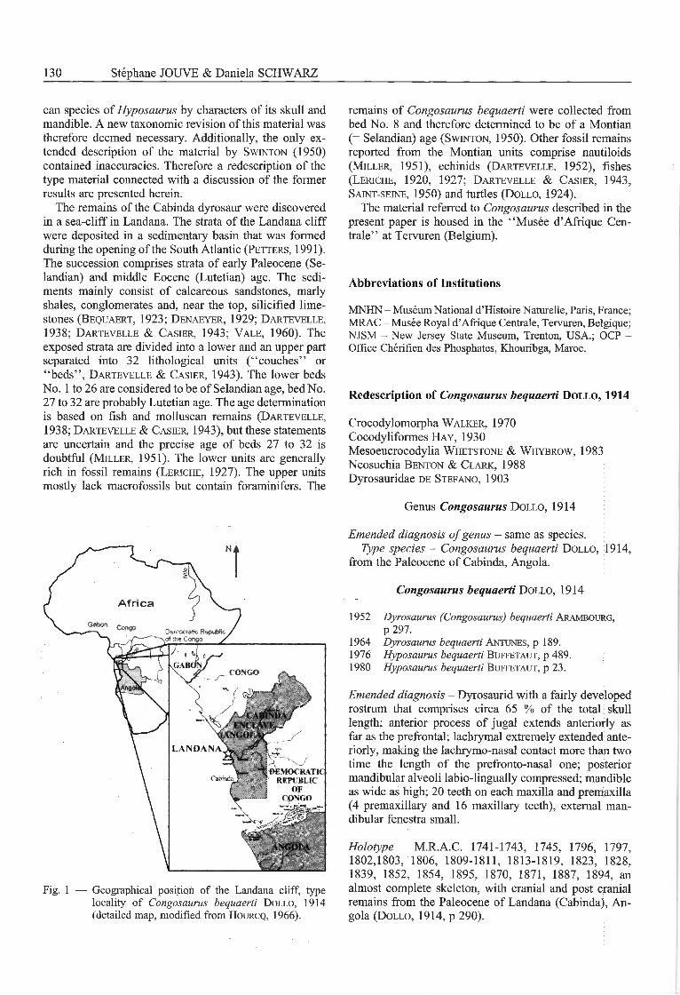
In 1913, the skeletal remains of a fossil crocodyliform,discovered by J. Bequaert (Bequaert, 1923) during abotanical expédition in Landana (Cabinda enclave, Ango¬la) (Fig. 1), were brought to the Musée d'Afrique Centralein Tervuren, Belgium. These comprised the nearly com¬plete skeleton of a dyrosaurid crocodylomorph. They weredescribed preliminarily and named Congosaurus bequaertiby Dollo (1914). Though Dollo (1914) promised a morecomplete description of the individual for a later date, hewas never able to finish this work. Therefore, in 1948 thedirector of the fonner "Musée du Congo belge" in Ter-vuren invited W. E. Swinton from London to re-examinethe Congosaurus skeleton. Swinton (1950) prepared anextended description of the remains of this dyrosaur spe¬cies. The taxonomie status of Congosaurus bequaerti was
questioned by Arambourg (1952) and Antunes (1964);they regarded Congosaurus as a junior subjective synonymof Dyrosaurus. Buffetaut (1976, 1980) revised the mate¬rial once again and, in contrast to the former authors,argued that the Cabinda dyrosaur belongs to the genusHyposaurus. This remained until recently the taxonomicalstatus for the Cabinda dyrosaur.
Studies of dyrosaur material from North Africa (Jouve,pers. obs.) indicate that the Cabinda dyrosaur can bedistinguished from the North American and North Afri-
130 Stéphane JOUVE & Daniela SCHWARZ
can species of Hyposaurus by characters of its skull andmandible. A new taxonomie revision of this material was
therefore deemed necessary. Additionally, the only ex-tended description of the material by Swinton (1950)contained inaccuracies. Therefore a redescription of thetype material connected with a discussion of the formerresults are presented herein.
The remains of the Cabinda dyrosaur were discoveredin a sea-cliff in Landana. The strata of the Landana cliffwere deposited in a sedimentary basin that was formedduring the opening of the South Atlantic (Petters, 1991).The succession comprises strata of early Paleocene (Se¬landian) and middle Eocene (Lutetian) âge. The sédi¬ments mainly consist of calcareous sandstones, marlyshales, conglomérâtes and, near the top, silicified lime-stones (Bequaert, 1923; Denaeyer, 1929; Dartevelle,1938; Dartevelle & Casier, 1943; Vale, 1960). Theexposed strata are divided into a lower and an upper partseparated into 32 lithological units ("couches" or"beds", Dartevelle & Casier, 1943). The lower bedsNo. 1 to 26 are considered to be of Selandian age, bed No.27 to 32 are probably Lutetian age. The age déterminationis based on fish and molluscan remains (Dartevelle,1938; Dartevelle & Casier, 1943), but these statementsare uncertain and the précisé age of beds 27 to 32 isdoubtful (Miller, 1951). The lower units are generallyrich in fossil remains (Leriche, 1927). The upper unitsmostly lack macrofossils but contain foraminifers. The
remains of Congosaurus bequaerti were collected frombed No. 8 and therefore determined to be of a Montian
(= Selandian) age (Swinton, 1950). Other fossil remainsreported from the Montian units comprise nautiloids(Miller, 1951), echinids (Dartevelle, 1952), fishes(Leriche, 1920, 1927; Dartevelle & Casier, 1943,Saint-seine, 1950) and turtles (Dollo, 1924).
The material referred to Congosaurus described in thepresent paper is housed in the "Musée d'Afrique Cen¬trale" at Tervuren (Belgium).
Abbreviations of Institutions
MNHN - Muséum National d'Histoire Naturelle, Paris, France;MRAC - Musée Royal d'Afrique Centrale, Tervuren, Belgique;NJSM - New Jersey State Museum, Trenton, USA.; OCP -
Office Chérifien des Phosphates, Khouribga, Maroc.
Redescription of Congosaurus bequaerti Dollo, 1914
Crocodylomorpha Walker, 1970Cocodyliformes Hay, 1930Mesoeucrocodylia Whetstone & Whybrow, 1983Neosuchia Benton & Clark, 1988Dyrosauridae de Stefano, 1903
Genus Congosaurus Dollo, 1914
Emended diagnosis ofgenus - same as species.Type species - Congosaurus bequaerti Dollo, 1914,
from the Paleocene of Cabinda, Angola.
Congosaurus bequaerti Dollo, 1914
1952 Dvrosaurus (Congosaurus) bequaerti Arambourg,p 297.
1964 Dvrosaurus bequaerti Antunes, p 189.1976 Hyposaurus bequaerti Buffetaut, p 489.1980 Hyposaurus bequaerti Buffetaut, p 23.
Emended diagnosis - Dyrosaurid with a fairly developedrostrum that comprises circa 65 % of the total skulllength; anterior process of jugal extends anteriorly asfar as the prefrontal; lachrymal extremely extended ante¬riorly, making the lachrymo-nasal contact more than twotime the length of the prefronto-nasal one; posteriormandibular alveoli labio-lingually compressed; mandibleas wide as high; 20 teeth on each maxilla and premaxilla(4 premaxillary and 16 maxillary teeth), external man¬dibular fenestra small.
Holotvpe - M.R.A.C. 1741-1743, 1745, 1796, 1797,1802,1803, 1806, 1809-1811, 1813-1819, 1823, 1828,1839, 1852, 1854, 1895, 1870, 1871, 1887, 1894, analmost complete skeleton, with cranial and post cranialremains from the Paleocene of Landana (Cabinda), An¬gola (Dollo, 1914, p 290).
Democratie Repubfctof the Congo /
congo
/ ENCIAVE,AM.OISr
landana
Soèmocrat1crfm blic
of
Fig. 1 — Geographical position of the Landana cliff, typelocality of Congosaurus bequaerti Dollo, 1914(detailed map, modified from Hourcq, 1966).
Congosaurus bequaerti, a Paleocene Dyrosaurid 131
Préservation - The skull is broken posterior to the ante-rior margin of the orbit, and the braincase and skull deckare lost. The sutures of the skull have been traced bySwinton (1950) with black ink, but those from the ante-rior wall of the suborbital cavity require reinterpretation(Pl. 1, Fig. 4, 5).
The mandible is almost complete and only the anteriorpart comprising the first six teeth is missing. The twoposterior rami are dorsoventrally compressed and dis-torted.
The remains of the axial skeleton comprise the axisand its odontoid process, the 3rd to 9th cervical vertebrae,the 2nd prothoracic vertebra, the lst, 4th to 8th and 10ththoracic vertebrae, the last presacral vertebra representingone lumbar vertebra as well as the 2nd to 4,h, 6th, 9th to18th and three terminal caudal vertebrae (probably the30th, 31st and 35th). The précisé positions of the caudalvertebrae were determined by a comparison of theircentrum lengths. From the thoracic région one and fromthe caudal région four isolated neural spines are present.Remains of ribs belong to the left 2nd and the right and left3rd to 7th cervical ribs as well as to 5th thoracic ribs and 5thhaemal arches are present. The appendicular skeleton isrepresented by the right scapula, the right and left cor-acoid, the right humérus, the proximal and distal part ofthe right ulna, the proximal part of the right radius, a rightradiale, four right metacarpals, the left ilium, the right andleft femur, the left tibia, the proximal part of the leftfibula, a proximal and a distal fragment of two metatar-sals, six phalanges as well as three ungual phalanges, andseveral dorsal and ventral osteoderms.
The préservation of the remains ranges from the well-preserved and complete vertebrae, ribs and bones of theappendicular skeleton to the very fragmentary, badlydamaged remains. Most of the postcranial elements sufferfrom mediolateral compression and distortion.
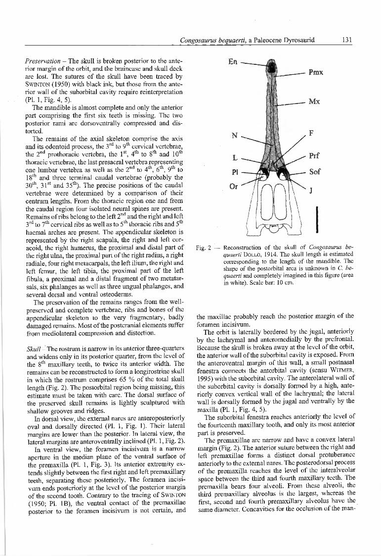
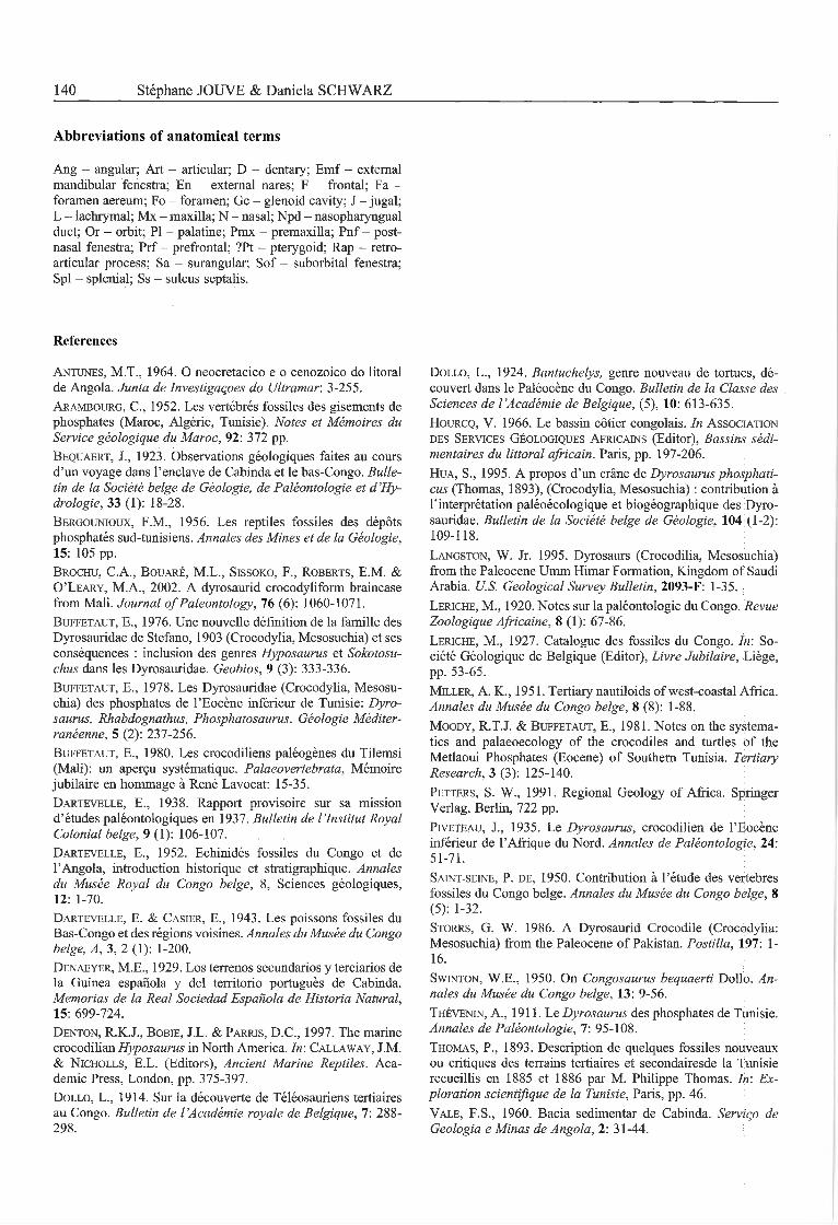
Skull - The rostrum is narrow in its anterior three-quartersand widens only in its posterior quarter, from the level ofthe 8th maxillary teeth, to twice its anterior width. Theremains can be reconstructed to form a longirostrine skullin which the rostrum comprises 65 % of the total skulllength (Fig. 2). The postorbital région being missing, thisestimate must be taken with care. The dorsal surface ofthe preserved skull remains is lightly sculptured withshallow grooves and ridges.
In dorsal view, the external nares are anteroposteriorlyoval and dorsally directed (Pl. 1, Fig. 1). Their latéralmargins are lower than the posterior. In latéral view, thelatéral margins are anteroventrally inclined (Pl. 1, Fig. 2).
In ventral view, the foramen incisivum is a narrowaperture in the médian plane of the ventral surface ofthe premaxilla (Pl. 1, Fig. 3). Its anterior extremity ex-tends slightly between the first right and left premaxillaryteeth, separating these posteriorly. The foramen incisi¬vum ends posteriorly at the level of the posterior marginof the second tooth. Contrary to the tracing of Swinton(1950; Pl. 1B), the ventral contact of the premaxillaeposterior to the foramen incisivum is not certain, and
Pmx
Mx
Fig. 2 — Reconstruction of the skull of Congosaurus be¬quaerti Dollo, 1914. The skull length is estimatedcorresponding to the length of the mandible. Theshape of the postorbital area is unknown in C. be¬quaerti and completely imagined in this figure (areain white). Scale bar: 10 cm.
the maxillae probably reach the posterior margin of theforamen incisivum.
The orbit is laterally bordered by the jugal, anteriorlyby the lachrymal and anteromedially by the prefrontal.Because the skull is broken away at the level of the orbit,the anterior wall of the suborbital cavity is exposed. Fromthe anteroventral margin of this wall, a small postnasalfenestra connects the antorbital cavity (sensu Witmer,1995) with the suborbital cavity. The anterolateral wall ofthe suborbital cavity is dorsally formed by a high, ante¬riorly convex vertical wall of the lachrymal; the latéralwall is dorsally formed by the jugal and ventrally by themaxilla (Pl. 1, Fig. 4, 5).
The suborbital fenestra reaches anteriorly the level ofthe fourteenth maxillary tooth, and only its most anteriorpart is preserved.
The premaxillae are narrow and have a convex latéralmargin (Fig. 2). The anterior suture between the right andleft premaxillae forms a distinct dorsal protubéranceanteriorly to the external nares. The posterodorsal processof the premaxilla reaches the level of the interalveolarspace between the third and fourth maxillary teeth. Thepremaxilla bears four alveoli. From these alveoli, thethird premaxillary alveolus is the largest, whereas thefirst, second and fourth premaxillary alveolus have thesame diameter. Concavities for the occlusion of the man-
132 Stéphane JOUVE & Daniela SCHWARZ
dibular teeth separate the premaxillary teeth, formingshallow latéral notches, slightly dorsally visible. Thesesulci create an irregular outline to the premaxillae indorsal view. The alveolar border projects ventrally, belowthe interalveolar space, so that the latéral margin of thepremaxilla is very wavy in latéral view. A deep occlusalnotch for the first dentary tooth separates the first andsecond premaxillary alveoli. The first right and left pre¬maxillary alveoli are positioned very close to each other.
The right and left maxillae are dorsally separated bythe nasal bone over ail their length (Fig. 2). The latéralmargin of the maxilla is relatively linear posteriorlyand only in its posterior sixth curves laterally. Severalforaminae are present on the medial margin of themaxilla, located just posteriorly to the postnasal fenestra(Pl. 1, Figs. 4, 5). These openings are probably neurovas-cular foraminae, of which the passage of the maxillarynerve (n. alveolaris dorsalis caudalis). The maxilla pos-sesses 16 separated alveoli. The right and left maxillaeventrally contact each other until the level of the 12thmaxillary alveolus. From this level, the maxillae are
medially separated by the palatines and diverge poster-olaterally. As on the premaxilla, the alveolar borderprojects ventrally, below the interalveolar space, so thatthe latéral margin of the maxilla is strongly wavy inlatéral view. The maxillary alveoli are relatively small,and the first to sixth alveoli are circular in outline whereasthe more posterior alveoli are labiolingually compressedand have an anteroposterior oval outline. The interalveo¬lar space between the first to eighth maxillary alveoli ismore than twice as long as the alveoli. From the eighthmaxillary alveolus the interalveolar space continuouslydecreases in length so that the interalveolar space be¬tween the most posterior maxillary teeth is less long thanthe alveoli. The width between the right and the left rowof alveoli is anteriorly twice the width of a maxillaryalveolus and constant from the first to the sixth maxillaryalveoli. This space increases progressively posteriorlyfrom the seventh.
The nasal is a single bone that passes deeply betweenthe posterodorsal processes of the premaxillae (Fig. 2). Itreaches anteriorly the level of the interalveolar spacebetween the first and second maxillary alveoli, and islargely excluded from the external nares. The width of thenasal is constant from its posteriormost contact with thepremaxillae to the level of the seventh maxillary alveolus,posterior to which the nasal expands to twice its anteriorwidth. The nasal is posteriorly separated by a médianprojection of the frontal that reaches the level of the 12thmaxillary alveolus.
Only the anteriorly directed tip of the prefrontal ispreserved (Fig. 2). This tip wedges between the laterallypositioned lacrimal and the medially positioned frontal,exceeds anteriorly the level of the anterior margin of theorbits on a very short length. The dorsal three-quarters ofthe prefrontal pillais are formed by the prefrontal whereasthe basis of each pillar is formed by the palatine (Pl. 1,Fig. 4, 5). Medially, the right and the left prefrontal pillarsare separated from each other by a narrow and high slit-
like opening that is interpreted as a passage for the nasalseptum and the olfactory tract. The frontal forms thedorsal margin of this opening. From the frontal, a smallmedial process descends and probably separates the rightand left olfactory nerves. A medial process on eachprefrontal pillar ascends dorsally and stops at the samelevel as the descending process of the frontal. This pro¬cess does not seem to be formed by the prefrontal, butmaybe by the frontal or an ethmoidal ossification, be-cause a long vertically oriented suture separates thisprocess from the prefrontal pillar. The ventromedial mar¬gin of the prefrontal pillar (sulcus septalis) seems not tobe formed by the palatine, but by another bone, perhapsthe pterygoid. The prefrontal sends a small latéralhooked-shaped process from the prefrontal pillar withinthe postnasal fenestra, a little bit dorsal to the prefrontal-palatine suture.
The lachrymal is large and wide and nearly triangularin dorsal view (Fig. 2). Its contact with the nasal is verybroad anteriorly and exceeds the prefrontal-nasal contactfor more than twice of its length. The lachrymal forms thesemi-circular anterior margin of the orbit.
The jugal constitutes the latéral margin of the orbit(Pl. 1, Fig. 4, 5). Its anterior process ends anterior to theprefrontal at the level of the 12th maxillary alveolus.
Mandible - The mandible is extremely elongated andnarrow in its anterior preserved half. The mandibularsymphysis posteriorly reaches the level of the sixteenthdentary alveolus (varying according to size and âge indyrosaurids (Jouve, pers. obs.)). The external mandibularfenestra is reduced to a small opening, surrounded poster-odorsally by the surangular, anterodorsally and anteriorlyby the dentary and ventrally by the angular (Pl. 2, Fig. 1,2). The retroarticular process is elongated and curvesposterodorsal ly. As a resuit of the strongly concave med¬ial horizontal wing of the articular, the retroarticularprocess is medially deeply concave (Pl. 2, Fig. 3, 4), witha 'L'-shaped cross section at mid length of the retro¬articular process.
The dentary extends for three-quarters of the totallength of the mandible. In latéral view, the dentary endsposteriorly with a high triangular process that forms theanterior and dorsal margin of the external mandibularfenestra (Pl. 2, Fig. 1, 2).
The splenials extend widely anteriorly and reach ante¬riorly the level of the interalveolar space between theninth and the tenth dentary alveolus (varying according tothe size and âge in dyrosaurids (Jouve, pers. obs.)). Thus,they meet along a symphysis. In anterior direction, bothsplenials tapers to a sharp wedge between both dentaries.Posteromedially, the splenials end at about mid lengthbetween the last dentary tooth and the glenoid fossa,anterior to the external mandibular fenestra (Pl. 2,Figs. 3, 4).
The angular extends along the posterior fourth of themandible. Its ventral margin is convex. In ventral view,the angular ends anteriorly with a ventral process thatreaches the level of the posteriormost dentary tooth.
Congosaurus bequaerti, a Paleocene Dyrosaurid 133
Posteriorly, the angular constitutes the ventral margin ofthe retroarticular process and half of its height in latéralview (Pl. 2, Figs. 1, 2). It extends to the posterior end ofthe retroarticular process.
The surangular reaches anteriorly the posteriormostdentary alveolus with a thin dorsomedial process. Theanterodorsal margin of the surangular bears two occlusalpits for the two posteriormost maxillary teeth. The sur¬angular fonns the latéral half of the glenoid fossa, and inthe retroarticular process its suture with the articularcurves laterally. The surangular is posteriorly long, nar-row and faces dorsolaterally on the retroarticular process.Latéral to the glenoid fossa, it bears a laterally orientedlongitudinal rim. The posterior part of the rim bears alarge foramen aereum (Pl. 2, Figs. 5, 6).
The medial half of the glenoid fossa is formed by thearticular. The articular bears an anteromedial process thatgives the glenoid fossa a posteriorly concave anteriormargin (Pl. 2, Figs. 5, 6). This process continues in anarrow lamina that reaches anteroventrally toward themedioventral margin of the mandible. The articular formsail of the medial part of the retroarticular process. Itsmedial wing is in ventral position posterior from the joint,at the same level as the ventral margin of the retroarti¬cular process. This medial wing forms an 'L'-shapedtransverse cross-section (Pl. 2, Figs. 3, 4).
No trace of the coronoid is preserved.
Dentition - The teeth are homodont, conical and slender.In labial view, the teeth are slightly curved posteriorlydistally and end with an acute apex (Pl. 1, Fig. 6). Theflrst to eleventh dentary teeth have a cylindrical crosssection. The posterior dentary and maxillary teeth arelateromedially compressed in cross-section. The rootsof the teeth are not much wider than the tooth crowns.
The teeth are divided into asymmetrical labial and lingualsurfaces by well-developed mesial and distal carinae. Thelingual surface of the tooth crowns have well-developedstriae that run straight from the base to the dorsal third ofthe crowns (Pl. 1, Fig. 7). Striae are present on labial andlingual surfaces of the teeth (Pl. 1, Figs. 6, 7).
The size of the teeth decreases from front to back. Theposterior teeth are more robust than the anterior and theirlingual striations are very less marked or absent.
Axial skeleton - Ail preserved vertébral centra are weaklyamphicoelous. The odontoid process is wedge-shaped inlatéral view and tapers from dorsal to ventral to one thirdof its dorsal length. A well-developed diapophysis isanterodorsally situated at the latéral surface of the odon¬toid process. The parapophysis lies with its anterior halfat the latéral surface of the odontoid process and with itsposterior half at the latéral surface of the axis vertébralcentrum (Pl. 3, Fig. 1). The axis vertébral centrum ishourglass-shaped in ventral view. A knob-like hypapo-physis is positioned centrally in the posterior fourth of theventral surface (Langston, 1995). From the hypapophy-sis, a médian and longitudinally oriented crest runs to theanterior margin of the ventral surface. The neurocentral
suture is in its anterior two-thirds slightly dorsally convexbut curves ventrally in its posterior third. The dorsalmargin of the neural arch inclines in a straight line fromanterior to posterior for approximately 25 degrees andforms the neural spine in its caudal third.
The anterior and the posterior surface of the vertébralcentra of the third to ninth cervical slightly convergedorsally (Pl. 3, Fig. 2-6). The anterior third of the ventralsurface of the vertébral centra is strongly rugose.Whereas the third cervical vertebra bears a hatchet-shaped hypapophysis in the anterior third of the ventralsurface of the centrum, at the fourth and fifth cervical thehypapophysis is replaced by a rugosity (Langston 1995).The hypapophysis of the sixth cervical forms a low, sharpcrest in the posterior third of the ventral surface of thecentrum. At the seventh and eighth cervical, the hypapo-physes are low crests that continue from the anterior halfof the ventral surface of the centrum posteriorly (PI. 3,Fig. 4, 5). The hypapophysis of the ninth cervical projectsventrally from the anterior half of the vertebral centrum(PI. 3, Fig. 6, Storrs, 1986). The anterior margin of thehypapophysis is weakly convex and rugose, and theposterior margin is linear. The ventral margin of thehypapophysis is convex.
In latéral view, the parapophysis is displaced from aposition anteroventrally at the latéral surface of the ver¬tebral centrum to a position caudomedially at the latéralcentrum surface between the third and the eighth cervical(Pl. 3, Fig. 1-5). The parapophysis of the ninth cervicaloverlaps with its dorsal fourth the neurocentral suture.The ventral margin of the diapopysis of the third andfourth cervical is formed by the vertebral centrum. Fromthe fifth cervical on the diapophysis lies directly dorsallyto the neurocentral suture and the parapophysis.
The preserved part of the neural spine of the thirdcervical is positioned in the posterior half of the neuralarch (PI. 3, Fig. 1). The neural spine is rod-like andcaudodorsally directed. The neural spine of the sixthcervical is straight and dorsally directed. The neuralspines of the seventh and eighth cervical are slightlydorsocaudally directed and possess a weakly convexanterior and a weakly concave posterior margin, and aconvex, slightly broadened dorsal margin. The height ofthe neural spines increases from the third to the ninthcervical vertebra (Table 1).
In latéral view, the anterior and posterior surface of thedorsal vertebral centra diverge dorsally. The preservedlast presacral possesses a rugose posterior surface fromwhich the left and right latéral fourth are bent caudolat-erally. The second prothoracic bears a large hypapophysissimilar in outline to the hypapophysis of the ninth cervi¬cal (Pl. 3, Fig. 7). The last preserved hypapophysis is atthe first thoracic vertebra and it is positioned posterome-dially at the ventral surface of the vertebral centrum andpossesses a strongly convex ventral margin. The latéralsurfaces of the thoracics without hypapophyses convergestrongly in ventral direction so that the ventral surface isreduced to a rounded crest.
The parapophysis of the second prothoracic overlaps
134 Stéphane JOUVE & Daniela SCHWARZ
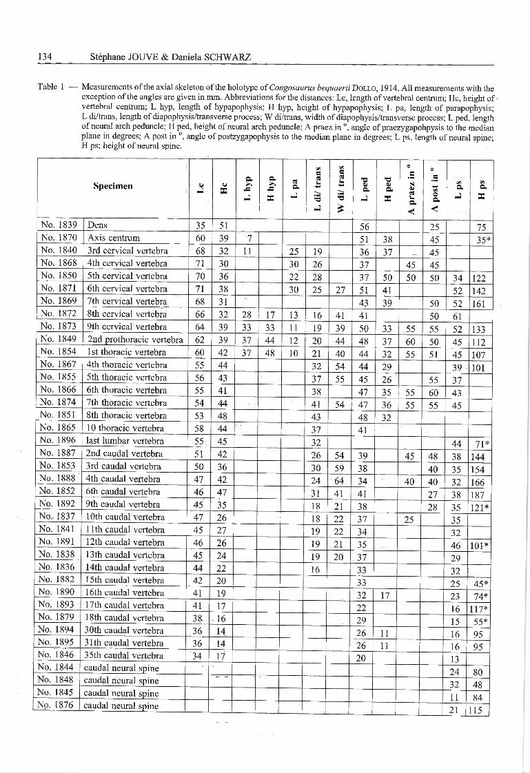
Table 1 — Measurements of the axial skeleton of the holotype of Congosaurus bequaerti Dollo, 1914. Ail measurements with theexception of the angles are given in mm. Abbreviations for the distances: Le, length of vertébral centrum; Hc, height ofvertébral centrum; L hyp, length of hypapophysis; H hyp, height of hypapophysis; L pa, length of parapophysis;L di/trans, length of diapophysis/transverse process; W di/trans, width of diapophysis/transverse process; L ped, lengthof neural arch peduncle; H ped, height of neural arch peduncle; A praez in °, angle of praezygapohpysis to the médianplane in degrees; A post in °, angle of postzygapophysis to the médian plane in degrees; L ps, length of neural spine;H ps; height of neural spine.
Specimen o-
o
X
a.
J=
J
a
s:
X
esa.
J
Ldi/trans Wdi/trans ■oaa.
■aVa
X
Apraezin° Apostin0 </îa
-J
t/3a
X
No. 1839 Dens 35 51 56 25 75No. 1870 Axis centrum 60 39 7 51 38 45 35*No. 1840 3rd cervical vertebra 68 32 11 25 19 36 37 45No. 1868 4th cervical vertebra 71 30 30 26 37 45 45No. 1850 5th cervical vertebra 70 36 22 28 37 50 50 50 34 122No. 1871 6th cervical vertebra 71 38 30 25 27 51 41 52 142No. 1869 7th cervical vertebra 68 31 43 39 50 52 161No. 1872 8th cervical vertebra 66 32 28 17 13 16 41 41 50 61No. 1873 9th cervical vertebra 64 39 33 33 11 19 39 50 33 55 55 52 133No. 1849 2nd prothoracic vertebra 62 39 37 44 12 20 44 48 37 60 50 45 112No. 1854 1 st thoracic vertebra 60 42 37 48 10 21 40 44 32 55 51 45 107No. 1867 4th thoracic vertebra 55 44 32 54 44 29 39 101No. 1855 5th thoracic vertebra 56 43 37 55 45 26 55 37No. 1866 6th thoracic vertebra 55 41 38 47 35 55 60 43No. 1874 7th thoracic vertebra 54 44 41 54 47 36 55 55 45No. 1851 8th thoracic vertebra 53 48 43 48 32No. 1865 10 thoracic vertebra 58 44 37 41No. 1896 last lumbar vertebra 55 45 32 44 71*No. 1887 2nd caudal vertebra 51 42 26 54 39 45 48 38 144No. 1853 3rd caudal vertebra 50 36 30 59 38 40 35 154No. 1888 4th caudal vertebra 47 42 24 64 34 40 40 32 166No. 1852 6th caudal vertebra 46 47 31 41 41 27 38 187No. 1892 9th caudal vertebra 45 35 18 21 38 28 35 121*No. 1837 lOth caudal vertebra 47 26 18 22 37 25 35No. 1841 11 th caudal vertebra 45 27 19 22 34 32No. 1891 12th caudal vertebra 46 26 19 21 35 46 101*No. 1838 13th caudal vertebra 45 24 19 20 37 29No. 1836 14th caudal vertebra 44 22 16 33 32No. 1882 15th caudal vertebra 42 20 33 25 45*No. 1890 16th caudal vertebra 41 19 32 17 23 74*No. 1893 17th caudal vertebra 41 17 22 16 117*No. 1879 18th caudal vertebra 38 16 29 15 55*No. 1894 30th caudal vertebra 36 14 26 11 16 95No. 1895 31 th caudal vertebra 36 14 26 11 16 95No. 1846 35th caudal vertebra 34 17 20 13No. 1844 caudal neural spine 24 80No. 1848 caudal neural spine 32 48No. 1845 caudal neural spine 11 84No. 1876 caudal neural spine 21 115
Congosaurus bequaerti, a Paleocene Dyrosaurid 135
with its dorsal third the neurocentral suture, whereas atthe first thoracic, the parapophysis lies completely at theneural arch. From at least the fourth thoracic, parapophy¬sis and diapophysis are united in a transverse process.Remarkably, the dorsals have a large articular surface forthe elastic ligament that forms a deep groove in theventral two thirds of the anterior and posterior marginof the neural spines (Pl. 3, Fig. 10).
The neural spines of the dorsals are slightly posterodor-sally bent (Langston, 1995) and decrease in height fromthe 2nd prothoracic to the 2nd thoracic (Pl. 3, Fig. 9, Table1). The anterior margin of the spine is slightly convex andthe posterior margin is slightly concave and broadened.The strongly broadened dorsal margin of the neural spinesof the second prothoracic and the first thoracic are slightlyconvex. The anterior margin of the neural spines of thefourth an fifth thoracic curves modestly to the broadeneddorsal margin and the dorsal margin inclines in height ffornanterior to posterior for 20 degrees.
At the third caudal, the ventral surface of the vertebralcentrum bears a posterior articular surface for the haemalarch. From the fourth caudal in the anterior and posteriorfourth of the ventral surface, articular surfaces for thehaemal arches are developed. Between two latéral andlongitudinal ridges, the ventral surface of the vertebralcentrum is concave (Storrs 1986).
The second to fourth caudal possess caudal ribs that arepositioned directly dorsal from the neurocentral suture atthe neural arch. The caudal ribs are in dorsal view sword-shaped with a convex anterior and a strongly concaveposterior margin. A transverse process is preserved at thesixth to seventeenth caudal. The transverse process liesmedially at the latéral surface of the neural arch at thesixth caudal, and it is displaced into the caudal half in themore terminal caudal vertebrae. The preserved caudalswithout transverse process exhibit a rounded crest in themedial third at the latéral surface of the neural arch. Thepre- and postzygapophysis is completely reduced and arod-like process from the twelfth caudal. The neuralspines of the second to sixth caudal are dorsally directed(PI. 3, Fig. 11) whereas those of the terminal caudals arerod-like with a wide dorsal part and caudodorsally direc¬ted (PI. 3, Fig. 13, Langston, 1995). The neural spinesincrease in height until the sixth caudal (Table 1, Pl. 3Fig. 11-13).
The second cervical rib (Pl. 4, Fig. 1,2; Table 2) is aslong as the odontoid process plus axis. The capitulotu-bercular incision is v-shaped. The third to seventh cervi¬cal ribs (Pl. 4, Fig. 3-6) are one fifth longer than thevertebral centrum (Table 2). From the third to the seventhcervical rib, the part of the costal body posterior to thetuberculum decreases in length. The ventral margin of therib is slightly convex medially and the latéral surface iscentrally depressed. At the fifth to seventh cervical ribs,the medial surface of the costal body is only slightlyconcave.
The costal body of the eighth cervical rib (PI. 4, Fig. 7)is oriented in the vertical plane. The preserved thoracicribs are bow-shaped with a strongly convex anterior and a
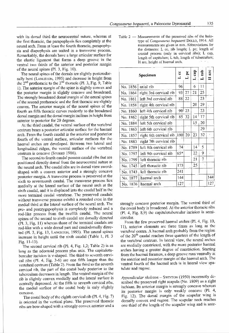
Table 2 — Measurements of the preserved ribs of the holo-type of Congosaurus bequaerti Dollo, 1914. Allmeasurements are given in mm. Abbreviations forthe distances: L cc, rib length; L pc; length ofcranial process (only in cervical ribs); L cap,length of capitulum; L tub, length of tuberculum;H arc, height of haemal arch.
u a -O O
Specimen C-> o. s a
-1 -l J X
No. 1856 axial rib 96 6 11
No. 1864 right 3rd cervical rib 93 27 21 25
No. 1861 left 3rd cervical rib 88 22 23
No. 1858 right 4th cervical rib 20 29
No. 1860 left 4th cervical rib 90 23 23
No. 1862 right 5th cervical rib 85 32 14 17
No. 1884 left 5th cervical rib 15 20
No. 1863 left 6th cervical rib 29
No. 1857 right 6th cervical rib 100 20 23 32
No. 1883 right 7th cervical ribNo. 1789 left 8th cervical rib 74 14 5
No. 1797 left 9th cervical rib 65* 22 9
No. 1799 left thoracic rib 21 7
No. 1745 left thoracic rib 173 25
No. 1743 left thoracic rib 241
No. 1877 haemal arch 144 144
No. 1876 haemal arch 127 127
strongly concave posterior margin. The ventral third ofthe costal body is broadened. At the anterior thoracic ribs(PI. 4, Fig. 8,9) the capitulotubercular incision is semi-circular.
From the few preserved haemal arches (Pl. 4, Fig. 10,11), anterior elements are three times as long as thevertebral centra. A haemal arch probably from the régionof the 20th caudal reaches three quarters of the length ofthe vertebral centrum. In latéral view, the neural archesare medially constricted, with the more posterior haemalarches having a greater degree of constriction. Startingfrom the haemal foramen, a deep groove runs ventrally atthe anterior and posterior margin of the haemal arch. Theventral fourth of the haemal arch is in latéral view spa¬tul ate and rugose.
Appendicular skeleton - Swinton (1950) incorrectly de-scribed the preserved right scapula (No. 1809) as a rightischium. lts anterior margin is strongly concave whereasits posterior margin is only weakly concave (PI. 4,Fig. 12). The dorsal margin of the scapular wing isdorsally convex and rugose. The scapular neck reachesone third of the length of the scapular wing and is ante-
136 Stéphane JOUVE & Daniela SCHWARZ
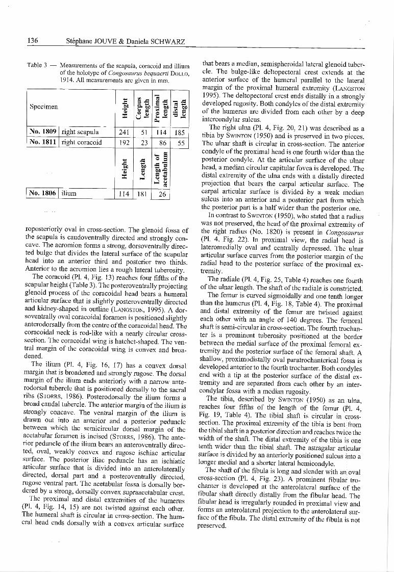
Table 3 — Measurements of the scapula, coracoid and illiumof the holotype of Congosaurus bequaerti Dollo,1914. Ail measurements are given in mm.
Specimen Height Corpus length Proximal length distal lengthNo. 1809 right scapula 241 51 114 185
No. 1811 right coracoid 192 23 86 55
-=ÖD
.s
SX
Lengthof acetabulum0J
X0i-
No. 1806 ilium 114 181 26
roposteriorly oval in cross-section. The glenoid fossa ofthe scapula is caudoventrally directed and strongly con¬cave. The acromion forms a strong, dorsoventrally direc¬ted bulge that divides the latéral surface of the scapularhead into an anterior third and posterior two thirds.Anterior to the acromion lies a rough latéral tuberosity.
The coracoid (Pl. 4, Fig. 13) reaches four fifths of thescapular height (Table 3). The posteroventrally projectingglenoid process of the coracoidal head bears a humeraiarticular surface that is slightly posteroventrally directedand kidney-shaped in outline (Langston, 1995). A dor¬soventrally oval coracoidal foramen is positioned slightlyanterodorsally from the centre of the coracoidal head. Thecoracoidal neck is rod-like with a nearly circular cross-section. The coracoidal wing is hatchet-shaped. The ven¬tral margin of the coracoidal wing is convex and broa-dened.
The ilium (Pl. 4, Fig. 16, 17) has a convex dorsalmargin that is broadened and strongly rugose. The dorsalmargin of the ilium ends anteriorly with a narrow ante-rodorsal tubercle that is positioned dorsally to the sacralribs (Storrs, 1986). Posterodorsally the ilium forms abroad caudal tubercle. The anterior margin of the ilium isstrongly concave. The ventral margin of the ilium isdrawn out into an anterior and a posterior pedunclebetween which the semicircular dorsal margin of theacetabular foramen is incised (Storrs, 1986). The ante¬rior peduncle of the ilium bears an anteroventrally direc¬ted, oval, weakly convex and rugose ischiac articularsurface. The posterior iliac peduncle has an ischiaticarticular surface that is divided into an anterolaterallydirected, dorsal part and a posteroventrally directed,rugose ventral part. The acetabular fossa is dorsally bor-dered by a strong, dorsally convex supraacetabular crest.
The proximal and distal extremities of the humérus(Pl. 4, Fig. 14, 15) are not twisted against each other.The humerai shaft is circular in cross-section. The hum¬erai head ends dorsally with a convex articular surface
that bears a médian, semispheroidal latéral glenoid tuber¬cle. The bulge-like deltopectoral crest extends at theanterior surface of the humerai parallel to the latéralmargin of the proximal humerai extremity (Langston1995). The deltopectoral crest ends distally in a stronglydeveloped rugosity. Both condyles of the distal extremityof the humérus are divided from each other by a deepintercondylar sulcus.
The right ulna (Pl. 4, Fig. 20, 21) was described as atibia by Swinton (1950) and is preserved in two pièces.The ulnar shaft is circular in cross-section. The anteriorcondyle of the proximal head is one fourth wider than theposterior condyle. At the articular surface of the ulnarhead, a médian circular capitular fovea is developed. Thedistal extremity of the ulna ends with a distally directedprojection that bears the carpal articular surface. Thecarpal articular surface is divided by a weak médiansulcus into an anterior and a posterior part from whichthe posterior part is a half wider than the posterior one.
In contrast to Swinton (1950), who stated that a radiuswas not preserved, the head of the proximal extremity ofthe right radius (No. 1820) is present in Congosaurus(Pl. 4, Fig. 22). In proximal view, the radial head islateromedially oval and centrally depressed. The ulnararticular surface curves from the posterior margin of theradial head to the posterior surface of the proximal ex¬tremity.
The radiale (Pl. 4, Fig. 25, Table 4) reaches one fourthof the ulnar length. The shaft of the radiale is constricted.
The fémur is curved sigmoidally and one tenth longerthan the humérus (Pl. 4, Fig. 18, Table 4). The proximaland distal extremity of the fémur are twisted againsteach other with an angle of 140 degrees. The fémoralshaft is semi-circular in cross-section. The fourth trochan-ter is a prominent tuberosity positioned at the borderbetween the medial surface of the proximal fémoral ex¬tremity and the posterior surface of the fémoral shaft. Ashallow, proximodistally oval paratrochanterical fossa isdeveloped anterior to the fourth trochanter. Both condylesend with a tip at the posterior surface of the distal ex¬
tremity and are separated from each other by an inter¬condylar fossa with a médian rugosity.
The tibia, described by Swinton (1950) as an ulna,reaches four fifths of the length of the fémur (Pl. 4,Fig. 19, Table 4). The tibial shaft is circular in cross-section. The proximal extremity of the tibia is bent fromthe tibial shaft in a posterior direction and reaches twice thewidth of the shaft. The distal extremity of the tibia is onetenth wider than the tibial shaft. The astragalar articularsurface is divided by an anteriorly positioned sulcus into alonger medial and a shorter latéral hemicondyle.
The shaft of the fibula is long and slender with an ovalcross-section (Pl. 4, Fig. 23). A prominent fibular tro¬chanter is developed at the anterolateral surface of thefibular shaft directly distally from the fibular head. Thefibular head is irregularly rounded in proximal view andforms an anterolateral projection to the anterolateral sur¬face of the fibula. The distal extremity of the fibula is notpreserved.
Congosaurus bequaerti, a Paleocene Dyrosaurid 137
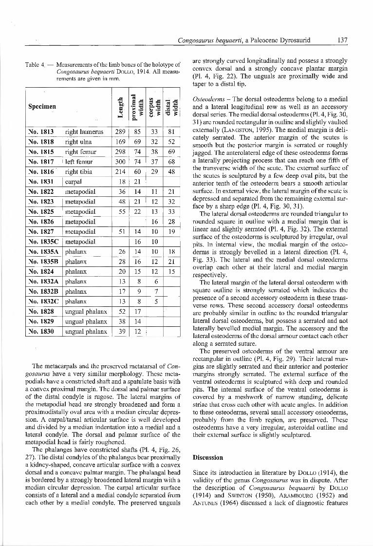
Table 4 — Measurements of the limb bones of the holotype ofCongosaurus bequaerti Dollo, 1914. Ail measu¬rements are given in mm.
Specimen Length proximal width corpus width distal widthNo. 1813 right humérus 289 85 33 81
No. 1818 right ulna 169 69 32 52
No. 1815 right femur 298 74 38 69
No. 1817 left femur 300 74 37 68
No. 1816 right tibia 214 60 29 48
No. 1831 carpal 18 21
No. 1822 metapodial 36 14 11 21
No. 1823 metapodial 48 21 12 32
No. 1825 metapodial 55 22 13 33
No. 1826 metapodial 16 28
No. 1827 metapodial 51 14 10 19
No. 1835C metapodial 16 10
No. 1835A phalanx 26 14 10 18
No. 1835B phalanx 28 16 12 21
No. 1824 phalanx 20 15 12 15
No. 1832A phalanx 13 8 6
No. 1832B phalanx 17 9 7
No. 1832C phalanx 13 8 5
No. 1828 ungual phalanx 52 17
No. 1829 ungual phalanx 38 14
No. 1830 ungual phalanx 39 12
The metacarpals and the preserved metatarsal of Con¬gosaurus have a very similar morphology. These meta-podials have a constricted shaft and a spatulate basis witha convex proximal margin. The dorsal and palmar surfaceof the distal condyle is rugose. The latéral margins ofthe metapodial head are strongly broadened and form aproximodistally oval area with a médian circular dépres¬sion. A carpal/tarsal articular surface is well developedand divided by a médian indentation into a medial and alatéral condyle. The dorsal and palmar surface of themetapodial head is fairly roughened.
The phalanges have constricted shafts (Pl. 4, Fig. 26,27). The distal condyles of the phalanges bear proximallya kidney-shaped, concave articular surface with a convexdorsal and a concave palmar margin. The phalangal headis bordered by a strongly broadened latéral margin with amédian circular dépression. The carpal articular surfaceconsists of a latéral and a medial condyle separated fromeach other by a medial condyle. The preserved unguals
are strongly curved longitudinally and possess a stronglyconvex dorsal and a strongly concave plantar margin(Pl. 4, Fig. 22). The unguals are proximally wide andtaper to a distal tip.
Osteoderms - The dorsal osteoderms belong to a medialand a latéral longitudinal row as well as an accessorydorsal series. The medial dorsal osteoderms (Pl. 4, Fig. 30,31 ) are rounded rectangular in outline and siightly vaultedexternally (Langston, 1995). The medial margin is deli-cately serrated. The anterior margin of the scutes issmooth but the posterior margin is serrated or roughlyjagged. The anterolateral edge of these osteoderms formsa laterally projecting process that can reach one fïfth ofthe transverse width of the scute. The external surface ofthe scutes is sculptured by a few deep oval pits, but theanterior tenth of the osteoderm bears a smooth articularsurface. In extemal view, the latéral margin of the scute isdepressed and separated from the remaining external sur¬face by a sharp edge (PI. 4, Fig. 30, 31).
The latéral dorsal osteodeims are rounded triangular torounded square in outline with a medial margin that islinear and slightly serrated (PI. 4, Fig. 32). The externalsurface of the osteoderms is sculptured by irregular, ovalpits. In internai view, the medial margin of the osteo¬derms is strongly bevelled in a latéral direction (Pl. 4,Fig. 33). The latéral and the medial dorsal osteodermsoverlap each other at their latéral and medial marginrespectively.
The latéral margin of the latéral dorsal osteoderm withsquare outline is strongly serrated which indicates thepresence of a second accessory osteoderm in these trans¬verse rows. These second accessory dorsal osteodermsare probably similar in outline to the rounded triangularlatéral dorsal osteodeims, but possess a serrated and notlaterally bevelled medial margin. The accessory and thelatéral osteoderms of the dorsal armour contact each other
along a serrated suture.The preserved osteoderms of the ventral armour are
rectangular in outline (Pl. 4, Fig. 29). Their latéral mar¬gins are slightly serrated and their anterior and posteriormargins strongly serrated. The external surface of theventral osteoderms is sculptured with deep and roundedpits. The internai surface of the ventral osteoderms iscovered by a meshwork of narrow standing, delicatestriae that cross each other with acute angles. In additionto these osteoderms, several small accessory osteoderms,probably from the limb région, are preserved. Theseosteoderms have a very irregular, asteroidal outline andtheir external surface is slightly sculptured.
Discussion
Since its introduction in literature by Dollo (1914), thevalidity of the genus Congosaurus was in dispute. Afterthe description of Congosaurus bequaerti by Dollo(1914) and Swinton (1950), Arambourg (1952) andAntunes (1964) discussed a lack of diagnostic features
138 Stéphane JOUVE & Daniela SCHWARZ
to distinguish between Congosaurus and Dyrosaurus. Asa resuit, the genus Congosaurus subsumed into synonymyby both authors. In a compilation about the distributionof dyrosaurid fossils in Africa, Buffetaut (1976) men-tioned the genus Congosaurus as a synonym to Hypo-saurus. These assignments were discussed again byBuffetaut (1980), who revised Congosaurus bequaertiand reassigned it to the genus Hyposaurus on the basisof mandibular characters. A new fïnd of the Paleocenefrom Tilemsi in Mali (MNHN TGE 4032) was assignedto the species "Hyposaurus" (Congosaurus) bequaerti(Buffetaut, 1980).
The genus Hyposaurus was erected by Owen in 1849and comprises North American as well as North Africanspecies. In northern Africa, Hyposaurus was describedtogether with several other genera in the Paleocene ofMorocco, with H. paueidens (Arambourg, 1952), and inthe Paleocene of the Sokoto Province in Nigeria, with H.wilsoni and nopscai (Swinton, 1950). The numerousgenera from this late layer were revised by Buffetaut(1980), with new material from the Tilemsi Valley inMali, and only two were regarded as valid. In NorthAmerica only one species, Hyposaurus rogersii, is recog-
nised (Denton et al., 1997), but Rhabdognathus is prob-ably present too (Jouve, pers. obs.). These two genera,Hyposaurus and Rhabdognathus can be easily distin-guished from each other by their rostrum length, whichrepresents 73 % of the skull length in Rhabdognathus(Jouve, pers. obs.) and only 66 % in Hyposaurus rogersii(Jouve, pers. obs.). Unfortunately, both Hyposaurus andRhabdognathus were insufficiently diagnosed by Buffe¬taut (1980), who in his emended diagnosis gave someimprécise and blurred characters for the genus Hypo¬saurus, as "a cross dentary section variable, generallywider than high, oval to circular...". For Rhabdognathus,Buffetaut (1980) described "a long and narrow snoutand the mandibular symphysis higher than wide" asdiagnostic characters.
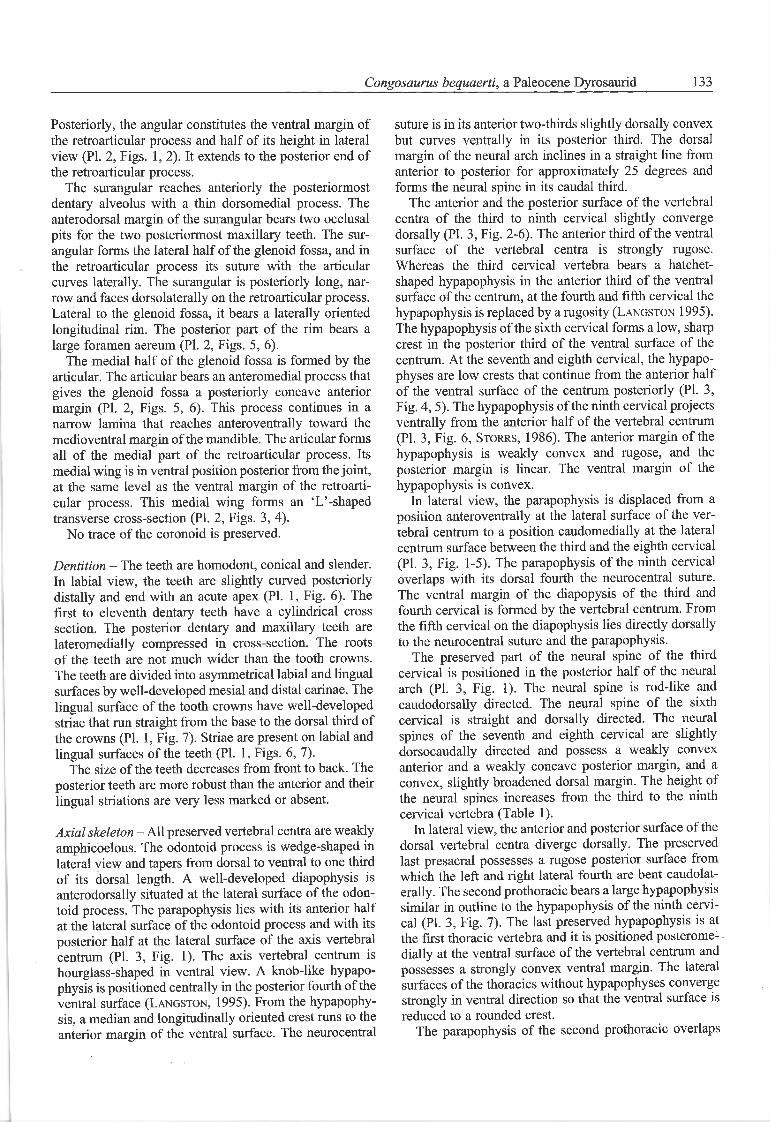
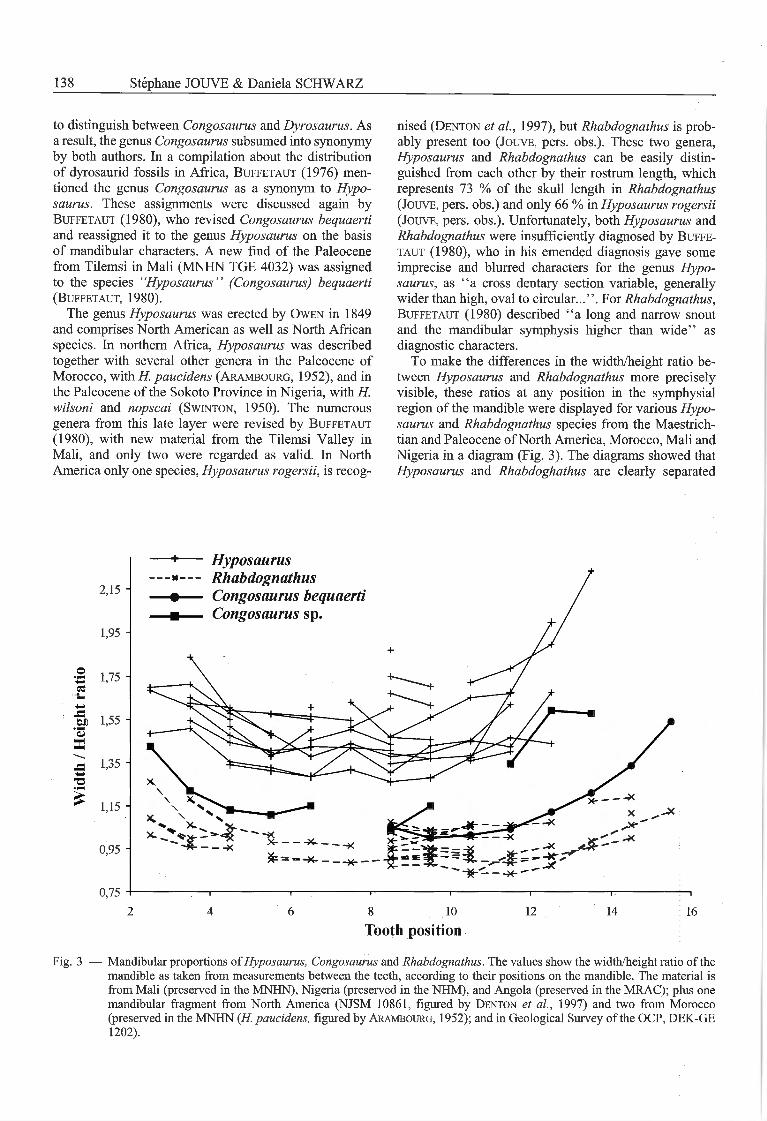
To make the différences in the width/height ratio be¬tween Hyposaurus and Rhabdognathus more preciselyvisible, these ratios at any position in the symphysialrégion of the mandible were displayed for various Hypo¬saurus and Rhabdognathus species from the Maestrich-tian and Paleocene ofNorth America, Morocco, Mali andNigeria in a diagram (Fig. 3). The diagrams showed thatHyposaurus and Rhabdoghathus are clearly separated
2,15
1,95
•2 1,75 -
M 1,55 -'3a
1,35■O
* 1,15 H
0,95 -
0,75
Hyposaurus-n— Rhabdognathus-m Congosaurus bequaerti
Congosaurus sp.
10 12 14 16
Tooth position
Fig. 3 — Mandibular proportions ofHyposaurus, Congosaurus and Rhabdognathus. The values show the width/height ratio of themandible as taken from measurements between the teeth, according to their positions on the mandible. The material isfrom Mali (preserved in the MNHN), Nigeria (preserved in the NHM), and Angola (preserved in the MRAC); plus onemandibular fragment from North America (NJSM 10861, figured by Denton et al, 1997) and two from Morocco(preserved in the MNHN (H. paueidens, figured by Arambourg, 1952); and in Geological Survey of the OCP, DEK-GE1202).
Congosaurus bequaerti, a Paleocene Dyrosaurid 139
from each other by their différences in the mandibularcross section: Rhabdognathus has a width-height ratiothat is below 1.10 between the fourth and the thir-teenth dentary alveolus, the mandibular symphysisrégion is therefore roughly as high as it is wide. Incontrast, in Hyposaurus the mandibular symphysiswidth-height ratio exceeds a value of 1.25 - the mandi¬bular symphysis région in this genus therefore is clearlywider than high.
Surprisingly, the holotype of C. bequaerti from Cabin-da displays the same values as Rhabdognathus (Fig. 3).Also the width-height ratio of the mandibular symphysisof the Malian specimen of "Hyposaurus" bequaerti(MNHN TGE 4032) seems to be problematic. In thisspecimen, the mandibular width between the 4th and the10th tooth is about equal to the height as it occurs inRhabdognathus, whereas the mandible in this région ismore flattened in H. wilsoni, H. nopscai, H. rogersii andthe H. paucidens and H. sp. from Morocco (ratio superiorto 1.25). In the posterior part of the mandible, the Malian"H. " bequaerti fits the values of Hyposaurus. However,the flattening of the posterior part of the mandible is in theholotype of C. bequaerti less important than in the Malianspecimen and fits the values of Rhabdognathus.
In Hyposaurus rogersii, nopscai and wilsoni ail dentaryalveoli are circular in outline as it is the case in ailother dyrosaurids, whereas the posterior dentary alveoliare laterally compressed in C. bequaerti. Moreover, inH. rogersii, some teeth bring a particular shape of theiranterior carinae which is ventrolingually "twisted"(Denton et al., 1997; Jouve, pers.obs.). This character-istic is not observed on ail the teeth of each specimen ofthe North American Hyposaurus, but it is very frequent inthe Maastrichtian specimens, and, contrary to Denton etal. (1997), have also been observed in some of the rarePaleocene forms (Jouve, pers.obs.). In the Malian sédi¬ments, the teeth are extreme ly rare, and only known forone specimen (TGE 4373), exhibiting also this "twisted"carinae. In Hyposaurus from Morocco, the same condi¬tion is observed (Jouve, pers. obs.). This "twisted" car¬inae is present in ail Hyposaurus where the teeth areknown, but is not developed at any teeth of Congosaurusbequaerti.
Furthermore, in C. bequaerti, the anterior process ofthe jugal extends anteriorly to reach at least the anteriorend of the prefrontal. The prefrontal, in contrast toC. bequaerti, extends anterior to the anterior extent ofthe jugal in other known dyrosaurids, such as Hyposaurusrogersii (Jouve, pers. obs.), Dyrosaurus phosphaticus(Thévenin, 1911; Pivetau, 1935; Bergounioux, 1956;Moody & Buffetaut, 1981; FIua, 1995), Phosphato-saurus gavialoides (Bergounioux, 1956; Buffetaut,1978; Moody & Buffetaut, 1981) and Rhabdognathussp. (Brochu et al., 2002). Likewise, the lachrymal inC. bequaerti is very long anteriorly and its contact withthe nasal is more than twice the prefrontal-nasal suture, aproportion much more significant than observed in ailother dyrosaurids. In the type species of Hyposaurus,(H. rogersii), which is the best known, the external man¬
dibular fenestra forms a wide opening, contrary to thevery modest one in the C. bequaerti mandible. C. be¬quaerti has at least 20 teeth in the premaxilla and maxilla,whereas H. rogersii has only 15 teeth in the upper jaw.
To summarize, C. bequaerti can be distinguished fromail other valid Hyposaurus species (Hyposaurus nopcsaiand wilsoni from Mali and Nigeria, H. paucidens andsp. from Morocco, and from the type species, H. rogersiifrom North America), by a mandibular symphysis as wideas high, straight mesial and distal carinae, a stronglyreduced external mandibular fenestra, a jugal extendinganteriorly past the prefrontal and a lacrimo-nasal suturethat is at least twice as long as the prefronto-nasal suture.Conversely, Hyposaurus can be diagnosed by the prob-ably diagnostic characters of a mandibular symphysiswider than high and "twisted" anterior tooth carinae(in species where the teeth are known). but this will bethe matter of a different paper (Jouve, in prep. ).
C. bequaerti has a similar rostrum length (65 % of thetotal skull length), as H. rogersii (66 %), in contrast toRhabdognathus rarus (73 %). Even if the snout proportionis the same, the tooth number differs between C. bequaer¬ti, which has 17 teeth anterior to the orbits, and H.rogersii, with only 13 teeth anterior to the orbits, andR. rarus having 25 teeth. Congosaurus bequaerti differsfrom Hyposaurus by its higher mandible, and from Rhab¬dognathus by its shorter snout.
Contrary to Arambourg (1952) and Antunes (1964),C. bequaerti is also not a member of the genus Dyro¬saurus, since its rostrum is much shorter (17 preorbitalteeth; rostrum: 65 % of the total skull length) than thesnout of Dyrosaurus phosphaticus (Thomas, 1893) (23preobital teeth, and snout proportion at about 70 %(Jouve, pers. obs.)).
It is therefore reasonable to retain Dollo's originalgenus Congosaurus for this species. The Malian speci¬men (TGE 4032) possesses a rostrum length and ananterior mandibular height like Congosaurus bequaertiand therefore may belong to the same genus. Flowever,the flattening of the posterior part of the mandible makesit doubtful, if this individual belongs to the same speciesas Congosaurus bequaerti. This différence could be alsoan effect of an individual variation, but since no otherspecimen is known for comparison, it seems preferable torefer this specimen only as Congosaurus sp.
Acknowledgments
The authors thank Drs. Chris Brochu and Hans Larsson for reviewingthis paper, and Nathalie Bardet (Paris) for help and advice. The authorsalso thank Drs. D. Baudet (Tervuren), P. Godefroit and A. Dhondt(Brussels), Sandra Chapman and Angela Milner (London), E. Gaffney,M. Norell, Ivy Rutzky and C. Mehling (New York City), MaureenO'Leary and R. Hill (Stony Brook), L. Murray and D. Brinkman (NewHaven), and D. Parris (Trenton, New Jersey) for their welcome andaccess to collections. Support was provided by the European Commu-nity - Access to Research Infrastructure action of the Improving Hu-man Potential Programme (with the ABC-Resource of the IRScNB andthe SYS-Resource of the NHM). This work also benefited from thesupport of the AMNH with the "Collection study grant", and the"Fondation des Treilles".
140 Stéphane JOUVE & Daniela SCHWARZ
Abbreviations of anatomical terms
Ang - angular; Art - articular; D - dentary; Emf - externalmandibular fenestra; En - external nares; F - frontal; Fa -
foramen aereum; Fo - foramen; Gc - glenoid cavity; J - jugal;L - lachrymal; Mx - maxilla; N - nasal; Npd - nasopharyngualduct; Or - orbit; PI - palatine; Pmx - premaxilla; Pnf - post¬nasal fenestra; Prf - prefrontal; ?Pt - pterygoid; Rap - retro-articular process; Sa - surangular; Sof - suborbital fenestra;Spl - splenial; Ss - sulcus septalis.
References
Antunes, M.T., 1964. O neocretacico e o cenozoico do litoralde Angola. Junta de Investigaqoes do Ultramar. 3-255.Arambourg, C., 1952. Les vertébrés fossiles des gisements dephosphates (Maroc, Algérie, Tunisie). Notes et Mémoires duSen'ice géologique du Maroc, 92: 372 pp.
Bequaert, J., 1923. Observations géologiques faites au coursd'un voyage dans l'enclave de Cabinda et le bas-Congo. Bulle¬tin de la Société belge de Géologie, de Paléontologie et d'Hy¬drologie, 33 (1): 18-28.Bergounioux, F.M., 1956. Les reptiles fossiles des dépôtsphosphatés sud-tunisiens. Annales des Mines et de la Géologie,15: 105 pp.
Brochu, C.A., Bouaré, M.L., Sissoko, F., Roberts, E.M. &O'Leary, M.A., 2002. A dyrosaurid crocodyliform braincasefrom Mali. Journal ofPaleontology, 76 (6): 1060-1071.Buefetaut, E., 1976. Une nouvelle définition de la famille desDyrosauridae de Stefano, 1903 (Crocodylia, Mesosuchia) et sesconséquences : inclusion des genres Hyposaurus et Sokotosu-chus dans les Dyrosauridae. Geobios, 9 (3): 333-336.Buffetaut, E., 1978. Les Dyrosauridae (Crocodylia, Mesosu¬chia) des phosphates de l'Eocène inférieur de Tunisie: Dyro-saurus, Rhabdognathus, Phosphatosaurus. Géologie Méditer¬ranéenne, 5 (2): 237-256.Buffetaut, E., 1980. Les crocodiliens paléogènes du Tilemsi(Mali): un aperçu systématique. Palaeovertebrata, Mémoirejubilaire en hommage à René Lavocat: 15-35.Dartevelle, E., 1938. Rapport provisoire sur sa missiond'études paléontologiques en 1937. Bulletin de l'Institut RoyalColonial belge, 9 (1): 106-107.Dartevelle, E., 1952. Echinidés fossiles du Congo et del'Angola, introduction historique et stratigraphique. Annalesdu Musée Roval du Congo belge, 8, Sciences géologiques,12: 1-70.
Dartevelle, E. & Casier, E., 1943. Les poissons fossiles duBas-Congo et des régions voisines. Annales du Musée du Congobelge, A, 3, 2 (1): 1-200.Denaeyer, M.E., 1929. Los terrenos secundarios y terciarios dela Guinea espanola y del territorio portuguès de Cabinda.Memorias de la Real Sociedad Espanola de Historia Natural,15: 699-724.
Denton, R.K.J., Bobie, J.L. & Parris, D.C., 1997. The marinecrocodilian Hyposaurus in North America. In: Callaway, J.M.& Nicholls, E.L. (Editors), Ancient Marine Reptiles. Aca¬demie Press, London, pp. 375-397.Dollo, L., 1914. Sur la découverte de Téléosauriens tertiairesau Congo. Bulletin de l'Académie royale de Belgique, 7: 288-298.
Dollo, L., 1924. Bantuchelys, genre nouveau de tortues, dé¬couvert dans le Paléocène du Congo. Bulletin de la Classe desSciences de l'Académie de Belgique, (5), 10: 613-635.Hourcq, V. 1966. Le bassin côtier congolais. In Associationdes Services Géologiques Africains (Editor), Bassins sédi-mentaires du littoral africain. Paris, pp. 197-206.Hua, S., 1995. A propos d'un crâne de Dyrosaurus phosphati-cus (Thomas, 1893), (Crocodylia, Mesosuchia) : contribution àl'interprétation paléoécologique et biogéographique des Dyro¬sauridae. Bulletin de la Société belge de Géologie, 104 (1-2):109-118.
Langston, W. Jr. 1995. Dyrosaurs (Crocodilia, Mesosuchia)from the Paleocene Umm Himar Formation, Kingdom of SaudiArabia. U.S. Geological Survey Bulletin, 2093-F: 1-35.Leriche, M., 1920. Notes sur la paléontologie du Congo. RevueZoologique Africaine, 8 (1): 67-86.Leriche, M., 1927. Catalogue des fossiles du Congo. In: So¬ciété Géologique de Belgique (Editor), Livre Jubilaire, Liège,pp. 53-65.Miller, A. K., 1951. Tertiary nautiloids of west-coastal Africa.Annales du Musée du Congo belge, 8 (8): 1-88.Moody, R.T.J. & Buffetaut, E., 1981. Notes on the systema-tics and palaeoecology of the crocodiles and turtles of theMetlaoui Phosphates (Eocene) of Southern Tunisia. TertiatyResearch, 3 (3): 125-140.Petters, S. W., 1991. Régional Geology of Africa. SpringerVerlag, Berlin, 722 pp.
Piveteau, J., 1935. Le Dyrosaurus, crocodilien de l'Eocèneinférieur de l'Afrique du Nord. Annales de Paléontologie, 24:51-71.
Saint-seine, P. de, 1950. Contribution à l'étude des vertebresfossiles du Congo belge. Annales du Musée du Congo belge, 8(5): 1-32.Storrs, G. W. 1986. A Dyrosaurid Crocodile (Crocodylia:Mesosuchia) from the Paleocene of Pakistan. Postilla, 197: 1-16.
Swinton, W.E., 1950. On Congosaurus bequaerti Dollo. An¬nales du Musée du Congo belge, 13: 9-56.Thévenin, A., 1911. Le Dyrosaurus des phosphates de Tunisie.Annales de Paléontologie, 7: 95-108.Thomas, P., 1893. Description de quelques fossiles nouveauxou critiques des terrains tertiaires et secondairesde la Tunisierecueillis en 1885 et 1886 par M. Philippe Thomas. In: Ex¬ploration scientifique de la Tunisie, Paris, pp. 46.Vale, F.S., 1960. Bacia sedimentar de Cabinda. Sen'iço deGeologia e Minas de Angola, 2: 31-44.
Congosaurus bequaerti, a Paleocene Dyrosaurid 141
Witmer, L.M., 1995. Homology on facial structures in extantArchosaurs (Birds and Crocodilians), with special référencé toparanasal pneumaticity and nasal conchae. Journal ofMorphol-ogy, 225 (3): 269-327.
Stéphane JouveUMR 8569 du CNRS
Département Histoire de la Terre, Muséum National d'HistoireNaturelle de Paris8 rue Buffon, F-75005 ParisFrance.E-mail: [email protected]
Daniela SchwarzNaturhistorisches Museum Basel
Augustinergasse 2, CH-4001 BaselSwitzerland.Email: [email protected]
Typescript submitted May 22, 2003Revised typescript received 29.12. 2003
Explanation of plates
Plate 1
Congosaurus bequaerti Dollo, 1914
Fig. 1, 2, 3 — holotype, MRAC 1741c, anterior extremity of the snout in dorsal, left latéral and ventral view.Figs. 4, 5 — holotype, MRAC 1741c, posterior view of the skull, anterior wall of suborbital cavity.Figs. 6, 7 — holotype, MRAC 1744, lingual and labial view of two mandibular teeth.
Scale bars represent 5 cm.
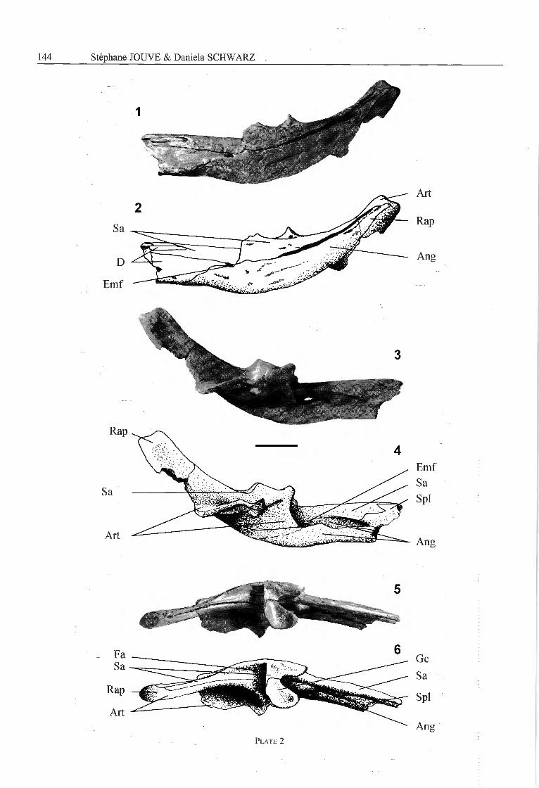
Plate 2
Congosaurus bequaerti Dollo, 1914, holotype, MRAC 1801
Fig. 1,2 — latéral view of the posterior part of the mandible.Fig. 3,4 — medial view of the posterior part of the mandible.Fig. 5,6 — dorsal view of the posterior part of the mandible.
Scale bar represents 5 cm.
Plate 3
Congosaurus bequaerti Dollo, 1914
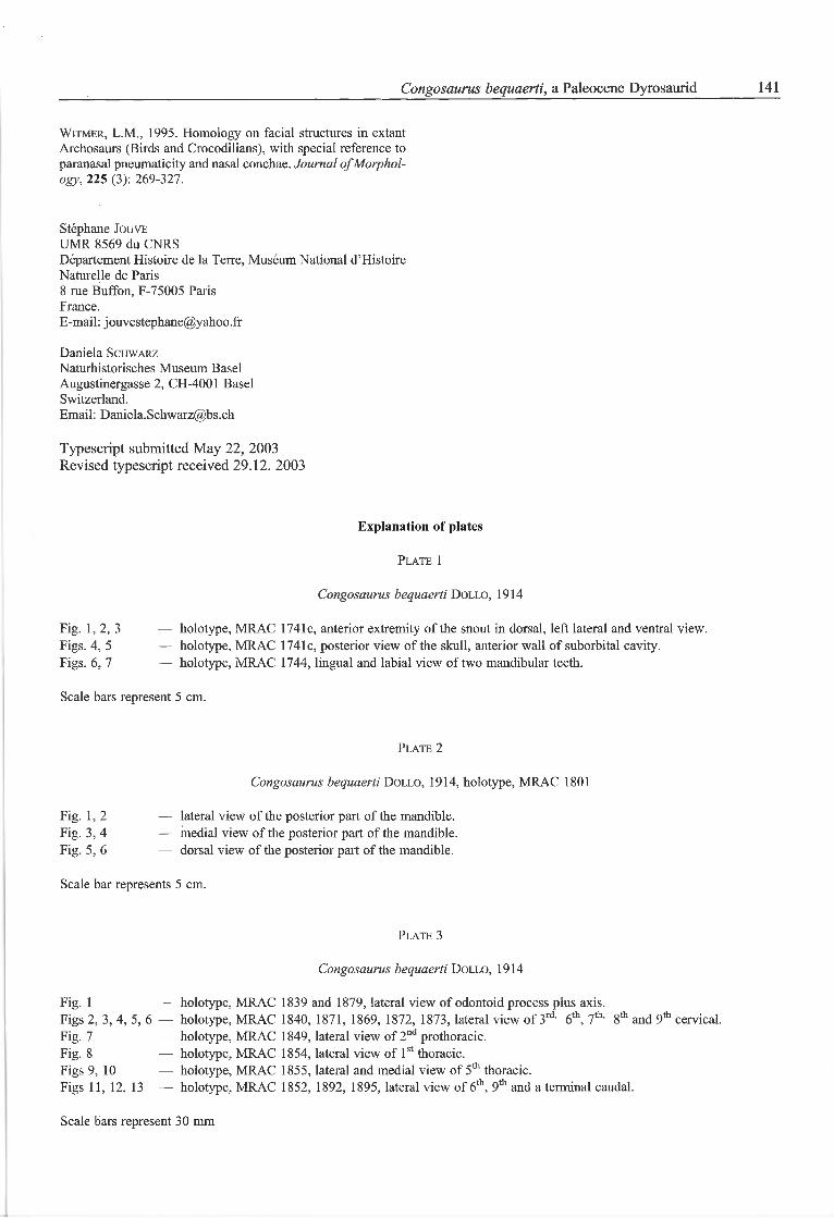
Fig. 1 — holotype, MRAC 1839 and 1879, latéral view of odontoid process plus axis.Figs 2, 3, 4, 5, 6 — holotype, MRAC 1840, 1871, 1869, 1872, 1873, latéral view of 3rd' 6th, 7,h' 8th and 9,h cervical.Fig. 7 — holotype, MRAC 1849, latéral view of 2nd prothoracic.Fig. 8 — holotype, MRAC 1854, latéral view of lst thoracic.Figs 9, 10 — holotype, MRAC 1855, latéral and medial view of 5th thoracic.Figs 11, 12, 13 — holotype, MRAC 1852, 1892, 1895, latéral view of 6th, 9th and a terminal caudal.
Scale bars represent 30 mm
142 Stéphane JOUVE & Daniela SCHWARZ
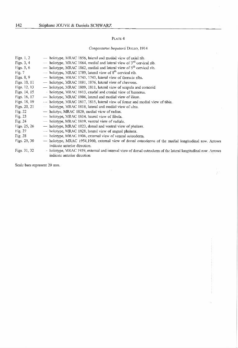
Plate 4
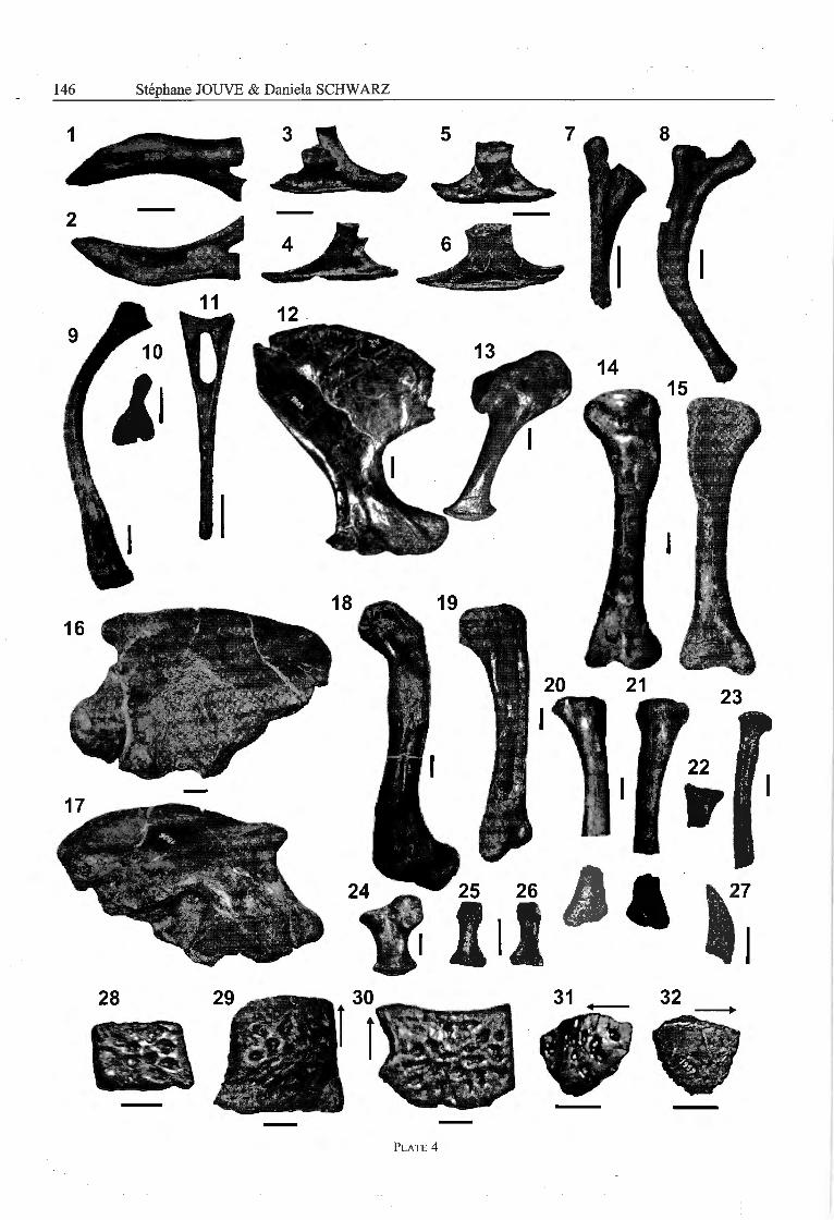
Congosaurus bequaerti Dollo, 1914
Figs. 1,2 — holotype, MRAC 1856, latéral and medial view of axial rib.Figs. 3, 4 — holotype, MRAC 1864, medial and latéral view of 3rd cervical rib.Figs. 5, 6 — holotype, MRAC 1862, medial and latéral view of 5th cervical rib.Fig. 7 — holotype, MRAC 1789, latéral view of 8th cervical rib.Figs. 8, 9 — holotype, MRAC 1745, 1743, latéral view of thoracic ribs.Figs. 10, 11 — holotype, MRAC 1881, 1876, latéral view of chevrons.Figs. 12, 13 — holotype, MRAC 1809, 1811, latéral view of scapula and coracoidFigs. 14, 15 — holotype, MRAC 1813, caudal and cranial view of humérus.Figs. 16, 17 — holotype, MRAC 1806, latéral and medial view of ilium.Figs. 18, 19 — holotype, MRAC 1817, 1815, latéral view of fémur and medial view of tibia.Figs. 20, 21 — holotype, MRAC 1818, latéral and medial view of ulna.Fig. 22 — holotye, MRAC 1820, medial view of radius.Fig. 23 — holotype, MRAC 1814, latéral view of fibula.Fig. 24 — holotype, MRAC 1819, ventral view of radiale.Figs. 25, 26 — holotype, MRAC 1823, dorsal and ventral view of phalanx.Fig. 27 — holotype, MRAC 1828, latéral view of ungual phalanx.Fig. 28 — holotype, MRAC 1906, external view of ventral osteoderm.Figs. 29, 30 — holotype, MRAC 1954,1960, external view of dorsal osteoderms of the medial longitudinal row. Arrows
indicate anterior direction.
Figs. 31, 32 — holotype, MRAC 1959, external and internai view of dorsal osteoderm of the latéral longitudinal row. Arrowsindicate anterior direction
Scale bars represent 20 mm.
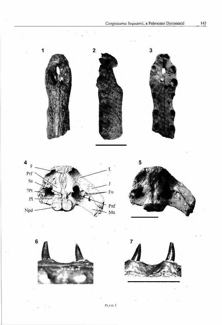
Plate 1
144 Stéphane JOUVE & Daniela SCHWARZ
Plate 2
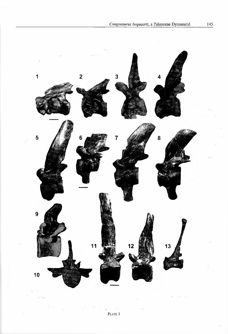
Plate 3
146 Stéphane JOUVE & Daniela SCHWARZ
Plate 4