21-d-24-c - LiK YOLU 7 6.1 9.0 61 aa 61 61 8.0 620 g 6233 ...
Cell, Vol. 121, 607–620, May 20, 2005, Copyright ©2005 by Elsevier Inc. DOI 10.1016/j.cell.2005.03.012
The v-ATPase V0 Subunit a1 Is Requiredfor a Late Step in Synaptic VesicleExocytosis in Drosophila
P. Robin Hiesinger,1 Amir Fayyazuddin,2
Sunil Q. Mehta,3 Tanja Rosenmund,2
Karen L. Schulze,1 R. Grace Zhai,1
Patrik Verstreken,2 Yu Cao,2 Yi Zhou,2
Jeannette Kunz,4 and Hugo J. Bellen1,2,3,5,*1Howard Hughes Medical Institute2Department of Molecular and Human Genetics3Program in Developmental Biology4Department of Molecular Physiology and Biophysics5Department of NeuroscienceBaylor College of MedicineHouston, Texas 77030
Summary
The V0 complex forms the proteolipid pore of anATPase that acidifies vesicles. In addition, an inde-pendent function in membrane fusion has been pro-posed largely based on yeast vacuolar fusion experi-ments. We have isolated mutations in the largest V0
component vha100-1 in flies in an unbiased geneticscreen for synaptic malfunction. The protein is onlyrequired in neurons, colocalizes with markers for syn-aptic vesicles as well as active zones, and interactswith t-SNAREs. Loss of vha100-1 leads to vesicle ac-cumulation in synaptic terminals, suggesting a deficitin release. The amplitude of spontaneous releaseevents and release with hypertonic stimulation indi-cate normal levels of neurotransmitter loading, yetmutant embryos display severe defects in evokedsynaptic transmission and FM1-43 uptake. Our datasuggest that Vha100-1 functions downstream of SNAREsin synaptic vesicle fusion.
Introduction
Membrane fusion lies at the heart of numerous cell bio-logical processes, most notably vesicle trafficking eventsunderlying cellular function (Jahn, 2004; Jahn et al.,2003; Mayer, 2002). The basic fusion machinery in virtu-ally all membrane fusion assays analyzed to date relieson SNAREs (soluble NSF-attachment protein receptors)(Jahn, 2004; Südhof, 2004). The commonly acceptedmodel for a general membrane fusion machinery de-notes that membranes are forced into close proximityby the action of SNAREs. In support of this idea,SNAREs have been shown to be sufficient to drive fu-sion in numerous assays (Hu et al., 2003; Weber et al.,1998). In contrast to this “proximity model” of mem-brane fusion, the proteolipid pore-forming V0 complexhas been shown to be required for membrane fusiondownstream of SNARE action in a yeast vacuolar fusionassay (Bayer et al., 2003; Peters et al., 2001). Although“pore models” for membrane fusion have been sug-gested repeatedly in the past, it has been difficult togather compelling data to verify the presence of a pro-
*Correspondence: [email protected]
teolipid pore during membrane fusion (Jahn, 2004;Jena, 2004; Lindau and Almers, 1995; Mayer, 2002; Mo-rel, 2003; Weimer and Jorgensen, 2003).
One of the most intensively studied membrane fusionevents is the exocytosis of synaptic vesicles that re-lease neurotransmitter at chemical synapses. Neuro-transmitter release is tightly regulated to ensure correctinformation exchange between neurons and other cells(Südhof, 2004). Consequently, synaptic vesicle fusionis regulated by a complex machinery in which SNAREproteins play a prominent role (Chen and Scheller, 2001;Jahn et al., 2003). Hence, loss of synaptic SNAREsgenerally causes severe defects in vesicle exocytosis(Deitcher et al., 1998; Jahn, 2004; Richmond and Broa-die, 2002; Schulze et al., 1995). The prevalent modelstates that the SNARE complex stabilizes readily re-leasable vesicles at the membrane at basal Ca2+ con-centrations. This complex triggers membrane fusion atelevated Ca2+ levels (Jahn et al., 2003; Sørensen, 2004).Hence, priming for fusion and the actual fusion step areboth thought to be directly linked to SNARE function.
The existence of a proteinaceous pore capable of re-leasing neurotransmitter has first been proposed forvesicles isolated from electroplaques of the electric or-gan of Torpedo marmorata. It was found to consist ofthe 16 kDa subunit of the v-ATPase V0 complex andwas shown to induce quantal release when expressedin a specific cell line (Falk-Vairant et al., 1996; Israël etal., 1986; Morel, 2003). It should be noted that thesestudies did not address the role of V0 proteins at thesynapse in vivo and they were met with skepticism, asit is difficult to reconcile that a complex event such assynaptic vesicle exocytosis could be mimicked bytransfecting a single protein in cells (Falk-Vairant et al.,1996). More recently, the Torpedo V0 subunit a1 (theortholog of the fly protein discussed here) was shownto localize specifically to nerve terminals and interactwith SNAREs (Morel et al., 2003). However, a directrequirement of V0 proteolipids for membrane fusion hasonly been demonstrated for a yeast vacuolar fusion as-say (see below; Peters et al., 2001).
The vesicular or vacuolar ATPase is a multisubunitprotein complex consisting of a V1 and V0 subcomplex(Nelson, 2003; Nishi and Forgac, 2002). The V1 sectorcomprises a protein complex that hydrolyzes ATP, pro-viding the energy to pump protons through the proteo-lipid pore formed by the V0 sector. The v-ATPases areimportant proton pumps that acidify a wide variety ofintracellular and some extracellular compartments (Nel-son, 2003; Nishi and Forgac, 2002). In neurons, loadingof synaptic vesicles with neurotransmitter requires thev-ATPase-dependent acidification of synaptic vesicles(Amara and Kuhar, 1993). For the largest V0 subunit,four genes (a1–a4) have been identified in human,mouse, C. elegans, and Drosophila. In C. elegans, thefour genes display highly tissue-specific distributionswith subunit a1 (unc-32) exhibiting predominantly neu-ronal expression (Oka et al., 2001; Pujol et al., 2001).Similarly, the two subunit a1 proteins in yeast, Vph1pand Stv1p, exhibit strong compartmental specificity,
Cell608
with Vph1p being the vacuolar version (Kawasaki-Nishi obet al., 2001; Perzov et al., 2002). VPH1-deficient vacu-
oles do not fuse in the vacuolar fusion assay (Bayer acet al., 2003). The defect was placed downstream of
SNARE function and is independent of the decreased goacidification of the vacuoles. Hence, VPH1 provided a
means to untangle the role of the v-ATPase V0 complex epin acidification from vacuolar fusion in yeast (Bayer et
al., 2003; Peters et al., 2001; Thorngren et al., 2004). atHowever, the relevance of this work for fusion of synap-
tic vesicles in neurons and other fusion events in yeast thas not been documented.
We have isolated mutations in a neuron-specific puVPH1 homolog of the v-ATPase V0 complex in a forward
genetic screen in Drosophila. Mutations in vha100-1 hw(v100), the fly subunit a1, allow a functional analysis of
the role of the V0 complex in neurons. Here, we report 5athat loss-of-function mutations in vha100-1 indicate a
defect late in synaptic vesicle exocytosis. Our data in-idicate that Vha100-1 is a regulator of synaptic vesicle
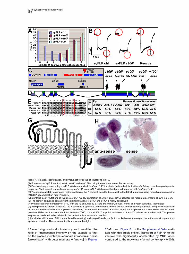
fusion efficiency downstream of SNARE-dependent tTvesicle priming.stResultsGrvha100-1 (v100) Is a Neuronal v-ATPasetV0 Subunit a1oIn a forward genetic screen for genes involved in syn-sapse formation and function we identified five EMS
(ethyl methane sulfonate)-induced alleles of a comple-mentation group named 3R3 (Mehta et al., 2005; Ver- D
Tstreken et al., 2003). We screened mosaic flies in whichchromosome arms carrying randomly induced muta- T
vtions were made homozygous in the visual systemusing the eyFLP system (Newsome et al., 2000; Stow- a
sers and Schwarz, 1999). Flies with eyes homozygousfor any of the five alleles of the 3R3 complementation w
vgroup consistently fail to walk toward light in a photo-taxis assay (Figure 1A; Benzer, 1967). Furthermore, all i
salleles display defects in electroretinograms (ERGs),extracellular recordings that measure the response of 2photoreceptor neurons to a light stimulus. Group 3R3eyFLP mutants respond with a normal depolarization to d
Tlight stimuli, but exhibit a complete loss of “on” and“off” transients, indicative of a failure to evoke a post- b
osynaptic response (Pak et al., 1969; Figure 1B), similarto the ERGs observed in neuronal synaptobrevin (n-syb) c
Fand synaptotagmin (syt) mutants (Stowers and Schwarz,1999; data not shown). v
rWe mapped the lethality associated with comple-mentation group 3R3 to a w50 kb interval at cytological
dlocation 98F12 using meiotic recombination mappingwith P elements (Figure 1C; Figure 2E in Zhai et al., c
m2003) and temperature-gradient capillary electrophore-sis (Figure 3 in Zhai et al., 2003). As shown in Figure l
i1D, all five alleles contain single-nucleotide changes:v1001 and v1004 exhibit altered splice acceptor sites w
opredicted to produce short, truncated proteins; v1002
and v1003 cause changes of conserved amino acids ptin the first transmembrane domain, and v1005 has a
premature stop codon (Figures 1D, 1E, and 1G). All mu- eqtations fail to complement each other, and all transhet-
erozygous allelic combinations, including any allele m
ver a deficiency (Df(3R)3450; 98E3-99A6) are late em-ryonic lethal. These data suggest that the five allelesre strong hypomorphic or null alleles. We obtained aDNA of the v100 transcript CG1709-RC (shown inreen in Figure 1D). Photoreceptor-specific expressionf this cDNA rescues the “on” and “off” transients inyFLP mutants of all alleles, indicating that the ERGhenotypes associated with the eyFLP v1001–5 mutantsre indeed caused by lesions in v100. The photorecep-or-specific rescue of the ERG further suggests a selec-ively presynaptic requirement of V100 (Figure 1B).
v100 is a highly conserved subunit of the V0 subcom-lex of the vesicular ATPase. Four similar genes (sub-nits a1–a4) exist in Drosophila, C. elegans, mouse, anduman. Drosophila v100 is more similar to that of theorm, mouse, and human subunit a1 genes (56%,8%, and 59%, respectively) than to the other subunits2–a4 (Figure 1F).To determine where v100 is expressed we performed
n situ hybridization. The gene is widely expressed inhe embryo and the larval nervous system (Figure 1H).o test if it is required in tissues other than the nervousystem, we expressed the full-length cDNA under con-rol of the neuron-specific elav-GAL4 driver using theAL4/UAS system (Brand and Perrimon, 1993). Neu-
on-specific expression of v100 in mutants rescues le-hality and results in viable and fertile adult flies with nobvious behavioral defects. Hence, v100 is a v-ATPaseubunit that is required in the nervous system.
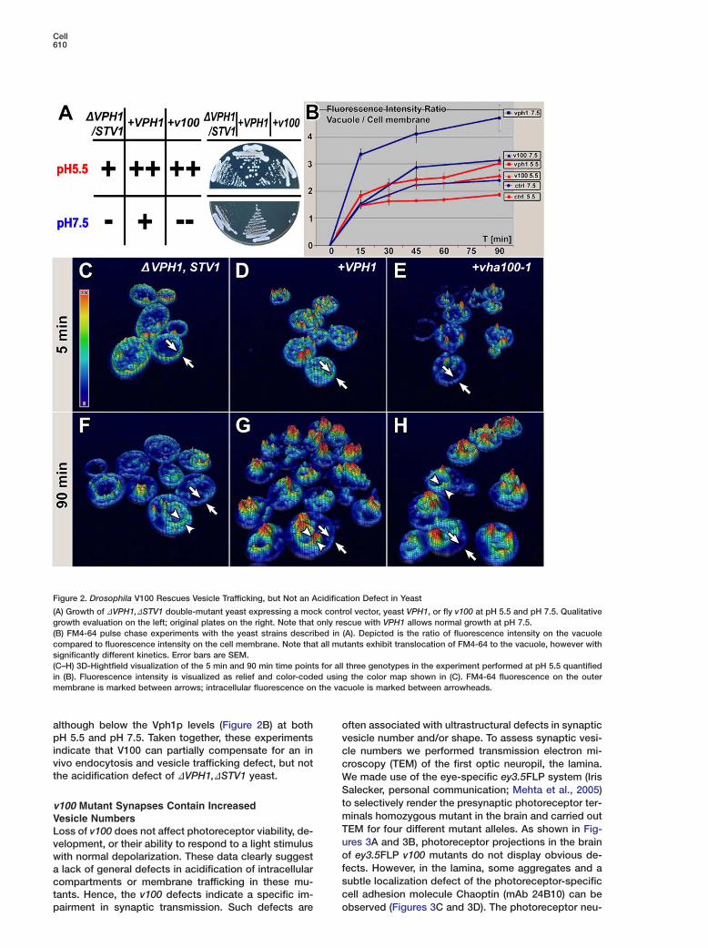
rosophila v100 Partially Rescues Vesiclerafficking but Not Acidification Defects in Yeasthe v-ATPase subunit a is the only component of the-ATPase encoded by two genes in S. cerevisiae, VPH1nd STV1. Vph1p is a vacuolar subunit and Stv1p re-ides within the Golgi/endosomal compartments (Ka-asaki-Nishi et al., 2001; Perzov et al., 2002). Whileacuolar fusion has been shown to require Vph1p, acid-
fication persists in the DVPH1 mutant due to compen-atory Stv1p function (Bayer et al., 2003; Perzov et al.,002).We tested the ability of Drosophila V100 to rescue
efects in a DVPH1,DSTV1 double-mutant yeast strain.he double mutant is nonviable on medium at pH 7.5,ut can grow at pH 5.5. We therefore tested the abilityf v100 to rescue the lethality at pH 7.5, using the res-ue with yeast VPH1 as a positive control. As shown inigure 2A, viability was not rescued with Drosophila100 at pH 7.5. This result indicates that v100 cannotescue the acidification defect in yeast.
In addition to the acidification defect, DVPH1,DSTV1ouble-mutant yeast are also defective in vesicle endo-ytosis and trafficking as shown in pulse chase experi-ents with FM4-64 (Perzov et al., 2002). FM4-64 is a
ipophilic dye commonly used to study vesicle traffick-ng. Wild-type yeast endocytose and transport the dyeithin minutes from the plasma membrane to the vacu-le (Perzov et al., 2002). We performed pulse chase ex-eriments at both pH 5.5 and pH 7.5 with double mu-
ants transformed with control vector, VPH1, or thexperimental vector carrying v100 (Figures 2B–2H). Touantify the translocation of the dye from the plasmaembrane to the vacuole, we imaged the cells every
V0 in Synaptic Vesicle Exocytosis609
Figure 1. Isolation, Identification, and Presynaptic Rescue of Mutations in v100
(A) Phototaxis of eyFLP control, v1001, v1002, and n-syb flies using the counter-current Benzer assay.(B) Electroretinogram recordings. eyFLP v100 mutants lack “on” and “off” transients (red circles), indicative of a failure to evoke a postsynapticresponse. Photoreceptor-specific expression of v100 in an eyFLP v100 mutant background restores both “on” and “off.”(C) Twenty-seven kilobyte genomic region containing the P element found to be closest to the lethal mutations using recombination mapping(KG6567, recombination rate 1/19,364).(D) Identified point mutations of five alleles. CG1709-RC annotation shown in blue; cDNA used for the rescue experiments shown in green.(E) The protein sequence containing the point mutations of v1002 and v1003 is highly conserved.(F) Protein sequence homology of V100 with the fly subunits a2–a4 and the human, mouse, worm, and yeast subunit a1 homologs.(G) V100 predicted protein structure. The N terminus is cytosolic and contains two coiled-coil domains (gray gradients). The protein has sevenor nine transmembrane domains (TMDs), depending on the transmembrane prediction algorithm. Depicted are seven TMDs; the two otherpossible TMDs are the loops depicted between TMD 2/3 and 4/5. The point mutations of the v100 alleles are marked 1–5. The proteinsequences predicted to be deleted in the mutant splice variants is marbled.(H) In situ hybridizations of third instar larval brains (top) and stage-15 embryos (bottom). Antisense staining on the left shows strong nervoussystem expression. The sense control is shown on the right.
15 min using confocal microscopy and quantified theratio of fluorescence intensity on the vacuole to thaton the plasma membrane (compare intracellular peaks[arrowheads] with outer membrane [arrows] in Figures
2C–2H and Figure S1 in the Supplemental Data avail-able with this article online). Transport of FM4-64 to thevacuole was significantly accelerated by V100 whencompared to the mock-transfected control (p < 0.005),
Cell610
Figure 2. Drosophila V100 Rescues Vesicle Trafficking, but Not an Acidification Defect in Yeast
(A) Growth of DVPH1,DSTV1 double-mutant yeast expressing a mock control vector, yeast VPH1, or fly v100 at pH 5.5 and pH 7.5. Qualitativegrowth evaluation on the left; original plates on the right. Note that only rescue with VPH1 allows normal growth at pH 7.5.(B) FM4-64 pulse chase experiments with the yeast strains described in (A). Depicted is the ratio of fluorescence intensity on the vacuolecompared to fluorescence intensity on the cell membrane. Note that all mutants exhibit translocation of FM4-64 to the vacuole, however withsignificantly different kinetics. Error bars are SEM.(C–H) 3D-Hightfield visualization of the 5 min and 90 min time points for all three genotypes in the experiment performed at pH 5.5 quantifiedin (B). Fluorescence intensity is visualized as relief and color-coded using the color map shown in (C). FM4-64 fluorescence on the outermembrane is marked between arrows; intracellular fluorescence on the vacuole is marked between arrowheads.
although below the Vph1p levels (Figure 2B) at both opH 5.5 and pH 7.5. Taken together, these experiments vindicate that V100 can partially compensate for an in cvivo endocytosis and vesicle trafficking defect, but not cthe acidification defect of DVPH1,DSTV1 yeast. W
Stv100 Mutant Synapses Contain IncreasedmVesicle NumbersTLoss of v100 does not affect photoreceptor viability, de-uvelopment, or their ability to respond to a light stimulusowith normal depolarization. These data clearly suggestfa lack of general defects in acidification of intracellularscompartments or membrane trafficking in these mu-ctants. Hence, the v100 defects indicate a specific im-
pairment in synaptic transmission. Such defects are
ften associated with ultrastructural defects in synapticesicle number and/or shape. To assess synaptic vesi-le numbers we performed transmission electron mi-roscopy (TEM) of the first optic neuropil, the lamina.e made use of the eye-specific ey3.5FLP system (Irisalecker, personal communication; Mehta et al., 2005)
o selectively render the presynaptic photoreceptor ter-inals homozygous mutant in the brain and carried outEM for four different mutant alleles. As shown in Fig-res 3A and 3B, photoreceptor projections in the brainf ey3.5FLP v100 mutants do not display obvious de-
ects. However, in the lamina, some aggregates and aubtle localization defect of the photoreceptor-specificell adhesion molecule Chaoptin (mAb 24B10) can be
observed (Figures 3C and 3D). The photoreceptor neu-
V0 in Synaptic Vesicle Exocytosis611
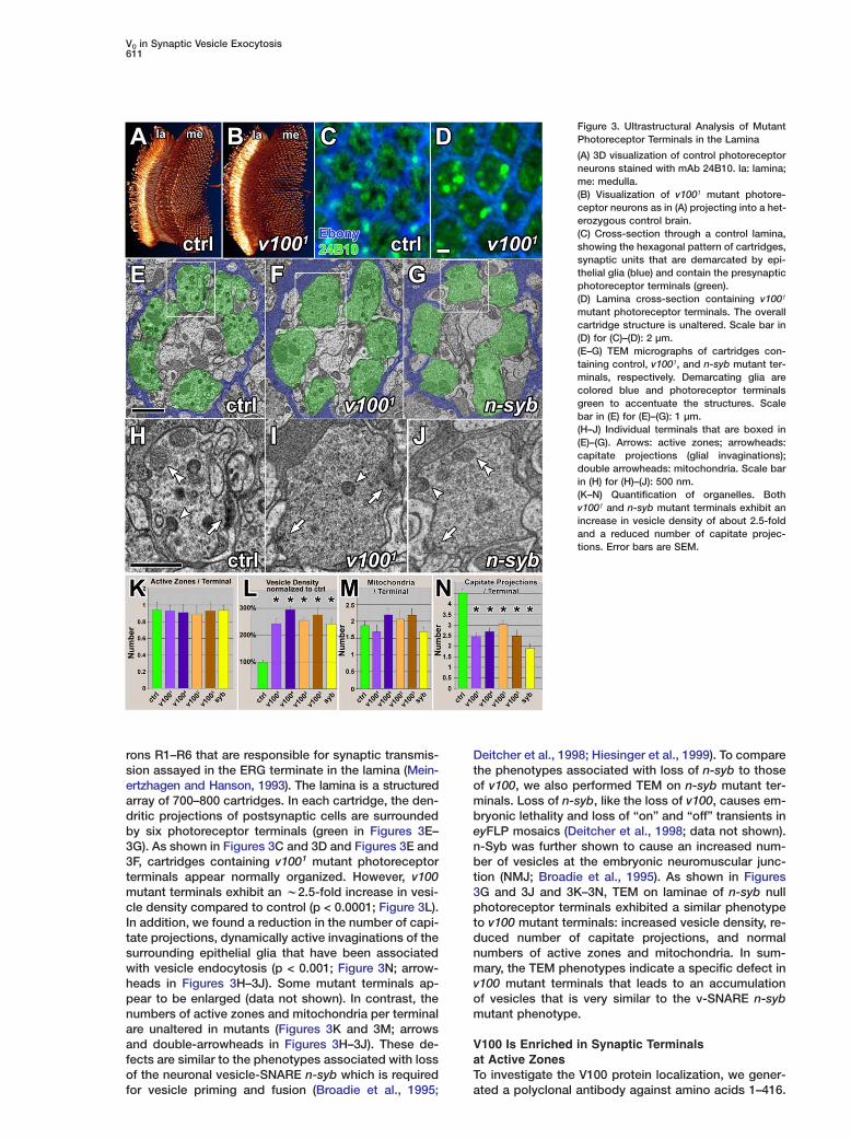
Figure 3. Ultrastructural Analysis of MutantPhotoreceptor Terminals in the Lamina
(A) 3D visualization of control photoreceptorneurons stained with mAb 24B10. la: lamina;me: medulla.(B) Visualization of v1001 mutant photore-ceptor neurons as in (A) projecting into a het-erozygous control brain.(C) Cross-section through a control lamina,showing the hexagonal pattern of cartridges,synaptic units that are demarcated by epi-thelial glia (blue) and contain the presynapticphotoreceptor terminals (green).(D) Lamina cross-section containing v1001
mutant photoreceptor terminals. The overallcartridge structure is unaltered. Scale bar in(D) for (C)–(D): 2 µm.(E–G) TEM micrographs of cartridges con-taining control, v1001, and n-syb mutant ter-minals, respectively. Demarcating glia arecolored blue and photoreceptor terminalsgreen to accentuate the structures. Scalebar in (E) for (E)–(G): 1 µm.(H–J) Individual terminals that are boxed in(E)–(G). Arrows: active zones; arrowheads:capitate projections (glial invaginations);double arrowheads: mitochondria. Scale barin (H) for (H)–(J): 500 nm.(K–N) Quantification of organelles. Bothv1001 and n-syb mutant terminals exhibit anincrease in vesicle density of about 2.5-foldand a reduced number of capitate projec-tions. Error bars are SEM.
rons R1–R6 that are responsible for synaptic transmis-sion assayed in the ERG terminate in the lamina (Mein-ertzhagen and Hanson, 1993). The lamina is a structuredarray of 700–800 cartridges. In each cartridge, the den-dritic projections of postsynaptic cells are surroundedby six photoreceptor terminals (green in Figures 3E–3G). As shown in Figures 3C and 3D and Figures 3E and3F, cartridges containing v1001 mutant photoreceptorterminals appear normally organized. However, v100mutant terminals exhibit an w2.5-fold increase in vesi-cle density compared to control (p < 0.0001; Figure 3L).In addition, we found a reduction in the number of capi-tate projections, dynamically active invaginations of thesurrounding epithelial glia that have been associatedwith vesicle endocytosis (p < 0.001; Figure 3N; arrow-heads in Figures 3H–3J). Some mutant terminals ap-pear to be enlarged (data not shown). In contrast, thenumbers of active zones and mitochondria per terminalare unaltered in mutants (Figures 3K and 3M; arrowsand double-arrowheads in Figures 3H–3J). These de-fects are similar to the phenotypes associated with lossof the neuronal vesicle-SNARE n-syb which is required
for vesicle priming and fusion (Broadie et al., 1995;Deitcher et al., 1998; Hiesinger et al., 1999). To comparethe phenotypes associated with loss of n-syb to thoseof v100, we also performed TEM on n-syb mutant ter-minals. Loss of n-syb, like the loss of v100, causes em-bryonic lethality and loss of “on” and “off” transients ineyFLP mosaics (Deitcher et al., 1998; data not shown).n-Syb was further shown to cause an increased num-ber of vesicles at the embryonic neuromuscular junc-tion (NMJ; Broadie et al., 1995). As shown in Figures3G and 3J and 3K–3N, TEM on laminae of n-syb nullphotoreceptor terminals exhibited a similar phenotypeto v100 mutant terminals: increased vesicle density, re-duced number of capitate projections, and normalnumbers of active zones and mitochondria. In sum-mary, the TEM phenotypes indicate a specific defect inv100 mutant terminals that leads to an accumulationof vesicles that is very similar to the v-SNARE n-sybmutant phenotype.
V100 Is Enriched in Synaptic Terminalsat Active ZonesTo investigate the V100 protein localization, we gener-
ated a polyclonal antibody against amino acids 1–416.
Cell612
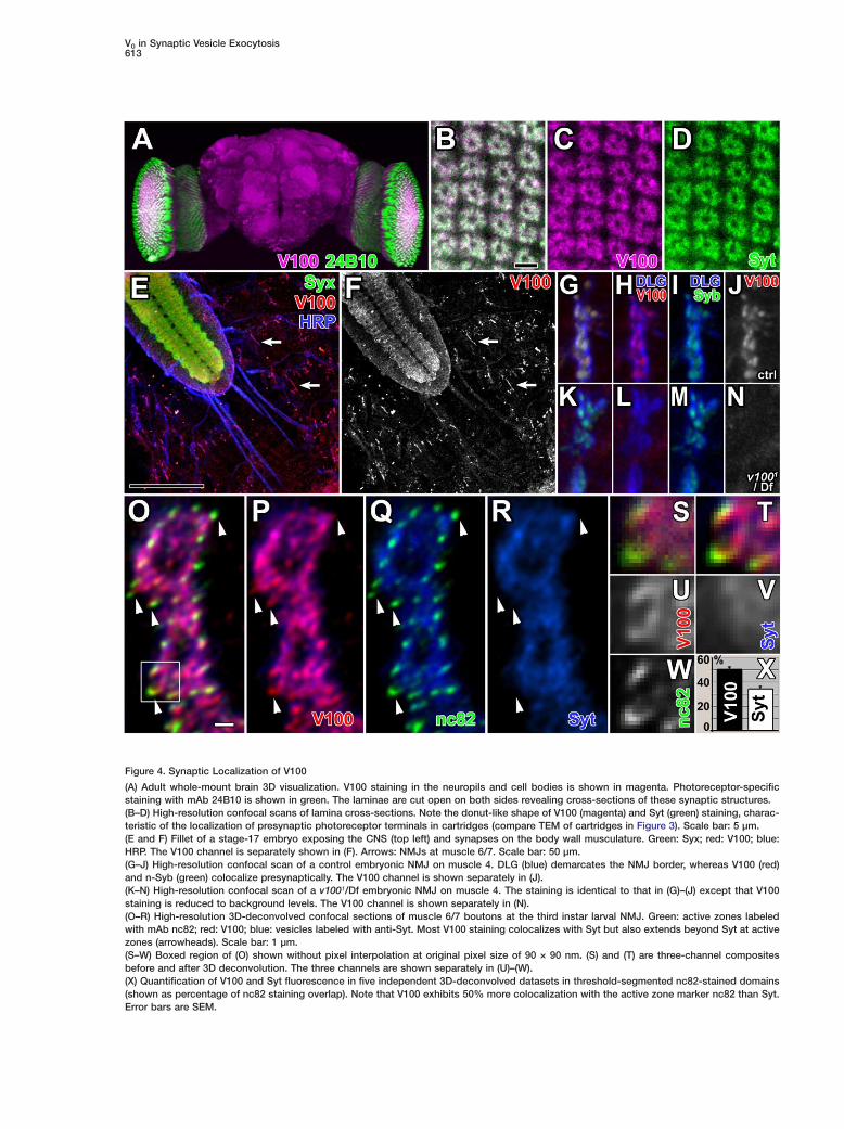
Anti-V100 immunoreactivity is abundant in the neuro- tpils of the adult brain but is also present in cell bodies c(Figure 4A). In the lamina, anti-V100 stains cartridges in ca donut-like pattern characteristic of the presynaptic (photoreceptor terminals, similar to Synaptotagmin c(Syt), a synaptic vesicle marker (Figures 4B–4D). As vmuch of the characterization of the v100 mutant pheno-types was carried out at the NMJ, we investigated the Vprotein localization in wild-type and mutant embryos V(22–24 hr). As shown in Figures 4E–4J, the V100 protein wis enriched at the embryonic NMJ and in the neuropil eof the central nervous system (CNS). Protein levels in av1001/Df and other mutant combinations in the CNS dneuropil as well as NMJ staining are strongly reduced wto a weak punctate staining that is found in all tissues, iincluding control animals (Figures 4K–4N and S2). This psuggests that there is either residual protein or some mminor nonspecific component that is recognized by the iantiserum. In summary, V100 is enriched at synaptic tterminals in the embryonic and adult CNS as well as at wthe NMJ. V
All v100 mutations develop as morphologically nor- rmal L1 larvae that fail to hatch. When dissected out of tthe egg case, they do not exhibit contraction waves nthat progress from anterior to posterior but rather show Suncoordinated twitching. The immobile late embryos/ fearly L1 larvae live for approximately 24–48 hr but can- tnot be kept alive longer using fluid yeast. To determine aif there are morphological defects at the NMJs or aber- Nrant localization of known synaptic markers, we stainedNMJs using antibodies against cell adhesion molecules d(HRP, Fas2), synaptic vesicle markers (Syt, Cysteine wString Protein, n-Syb), target SNAREs (Syntaxin [Syx], VSNAP-25), active zone markers (nc82, DPAK) and post- psynaptic receptors (GluRII). We did not observe any ob- tvious morphological defects or aberrant protein local-
bization of the analyzed markers (Figures 4K–4N, S2,
t7A–7F, S4I–S4N, and data not shown). Hence, v100 is
aeither not required for embryonic development or a ma-
pternal protein or RNA contribution to the egg is respon-isible for normal development. We therefore generatedtmaternal knockout (MKO) embryos for v1001 and v1002
m(Chou and Perrimon, 1992). These embryos display nocdevelopmental delay and present the same behaviortand morphology as all other mutant allelic combina-ttions (data not shown). In summary, these data indicateSthat embryonic and NMJ development does not de-
pend on the presence of v100.iTo define V100 localization inside synaptic boutonsrat high resolution, we analyzed the colocalization ofpV100 with vesicle and active zone markers at the L3alarval NMJ using 3D deconvolution of confocal imageestacks (see Supplemental Experimental Procedures).dThese boutons are typically 3–6 µm in diameter. V100Sstaining inside boutons colocalizes mostly with the ves-1icle marker Syt (Figures 4O and 4R). However, we con-esistently observed an increased V100 staining at activerzones labeled with mAb nc82 (Wucherpfennig et al.,p2003; arrowheads in Figures 4O–4R). A quantitative col-localization analysis revealed significantly more colocal-mization of V100 with nc82 than Syt with nc82, indicatingfan enrichment of V100 relative to Syt at active zonesp(Figures 4S–4X). We further analyzed V100 protein
content in a vesicle preparation versus membrane frac- d
ion (Schulze et al., 1995). V100 is enriched in the vesi-le fraction but is also present in the membrane fractionontaining both vesicles and presynaptic membranesFigure S3). These data indicate that V100 is a synapti-ally enriched protein that localizes to both synapticesicles and active zones.
100 Interacts with t-SNAREs100 homologs have previously been shown to interactith SNARE proteins in yeast and vertebrates (Bennettt al., 1992; Galli et al., 1996; Morel, 2003; Peters etl., 2001), and V100 contains two N-terminal coiled-coilomains (coil 1, 44–75; coil 2, 90–135) that may interactith SNARE proteins. We therefore investigated V100
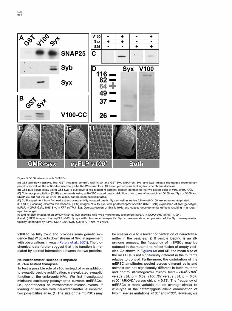
nteractions with SNAREs. First, we tested bacteriallyroduced V100 protein domains that lack the trans-embrane regions in GST pull-down assays. As shown
n Figure 5A, both GST-V100 and GST-Syx retrieve His-agged SNAP-25. Furthermore, GST-V100 also interactsith His-Syx. In contrast, only GST-Syx, but not GST-100, pulls down His-n-Syb, indicating that V100 di-
ectly interacts with Syx and SNAP-25, the SNAREs athe target membrane, but not with the vesicle-SNARE-Syb. We then reversed the experiment using GST-yntaxin to pull down a His-tagged V100 N-terminal
ragment of 133 amino acids (amino acids 10–143) con-aining the two coiled-coil domains (Figure 1G). Syxlso pulled down this V100 fragment, indicating an-terminal Syx interaction domain (Figure 5B).To confirm these interactions, we verified the pull-
own results with coimmunoprecipitation assays. Beadsere coated with purified IgGs extracted from anti-100 antiserum and tested for the ability to immuno-recipitate Syx or SNAP-25, either alone or in combina-ion with V100. Consistent with the pull-down results,oth Syx and SNAP-25 can be immunoprecipitated in
he presence of V100 (Figure 5C). Finally, we testednti-Syx-coated beads for their ability to coimmuno-recipitate native V100 from fly head extract. As shown
n Figure 5D, both Syx and V100 are coimmunoprecipi-ated from head extract (that was cleared of insolubleaterial and membraneous fragments) with anti-Syx
oated beads, indicating that they exist at least par-ially as a complex in vivo (Figure 5D). In summary,hese results indicate that V100 and the t-SNAREsNAP-25 and Syx directly interact.The in vivo significance of the V100—Syx interaction
s supported by the results of a suppressor screen car-ied out in parallel to our original eyFLP screen. Overex-ression of Syx driven by GMR-Gal4 in photoreceptorst low levels (in flies reared at 18°C) causes a roughye phenotype and high levels of Syx (28°C) cause celleath. We reasoned that loss of a protein required foryx action may suppress this toxic effect and screened81 mutants isolated on chromosome arm 3R in theyFLP screen (Mehta et al., 2005) for suppression of theough eye phenotype. Eight mutants displayed sup-ression, two of which are v1001 and v1004, severe
oss-of-function alleles (Figures 5E–5J). In contrast,utations causing single-amino acid changes in the
irst transmembrane domain, v1002 and v1003, dis-layed no suppression (data not shown). This result in-icates that overexpressed Syx requires the function of
V0 in Synaptic Vesicle Exocytosis613
Figure 4. Synaptic Localization of V100
(A) Adult whole-mount brain 3D visualization. V100 staining in the neuropils and cell bodies is shown in magenta. Photoreceptor-specificstaining with mAb 24B10 is shown in green. The laminae are cut open on both sides revealing cross-sections of these synaptic structures.(B–D) High-resolution confocal scans of lamina cross-sections. Note the donut-like shape of V100 (magenta) and Syt (green) staining, charac-teristic of the localization of presynaptic photoreceptor terminals in cartridges (compare TEM of cartridges in Figure 3). Scale bar: 5 µm.(E and F) Fillet of a stage-17 embryo exposing the CNS (top left) and synapses on the body wall musculature. Green: Syx; red: V100; blue:HRP. The V100 channel is separately shown in (F). Arrows: NMJs at muscle 6/7. Scale bar: 50 µm.(G–J) High-resolution confocal scan of a control embryonic NMJ on muscle 4. DLG (blue) demarcates the NMJ border, whereas V100 (red)and n-Syb (green) colocalize presynaptically. The V100 channel is shown separately in (J).(K–N) High-resolution confocal scan of a v1001/Df embryonic NMJ on muscle 4. The staining is identical to that in (G)–(J) except that V100staining is reduced to background levels. The V100 channel is shown separately in (N).(O–R) High-resolution 3D-deconvolved confocal sections of muscle 6/7 boutons at the third instar larval NMJ. Green: active zones labeledwith mAb nc82; red: V100; blue: vesicles labeled with anti-Syt. Most V100 staining colocalizes with Syt but also extends beyond Syt at activezones (arrowheads). Scale bar: 1 µm.(S–W) Boxed region of (O) shown without pixel interpolation at original pixel size of 90 × 90 nm. (S) and (T) are three-channel compositesbefore and after 3D deconvolution. The three channels are shown separately in (U)–(W).(X) Quantification of V100 and Syt fluorescence in five independent 3D-deconvolved datasets in threshold-segmented nc82-stained domains(shown as percentage of nc82 staining overlap). Note that V100 exhibits 50% more colocalization with the active zone marker nc82 than Syt.Error bars are SEM.
Cell614
Figure 5. V100 Interacts with SNAREs
(A) GST pull-down assays. Top: GST (negative control), GST-V100, and GST-Syx. SNAP-25, Syb, and Syx indicate His-tagged recombinantproteins as well as the antibodies used to probe the Western blots. All fusion proteins are lacking transmembrane domains.(B) GST pull-down assay using GST-Syx to pull down a His-tagged N-terminal domain containing the two coiled-coils of V100 (V100-CC).(C) Coimmuoprecipitation (CoIP) experiments using anti-V100 coated beads. Addition of mixtures of recombinant V100 and Syx or V100 andSNAP-25, but not Syx or SNAP-25 alone, can be immunoprecipitated.(D) CoIP experiment from fly head extract using anti-Syx-coated beads. Syx as well as native full-length V100 are immunoprecipitated.(E and F) Scanning electron microscopic (SEM) images of a fly eye with photoreceptor-specific (GMR-Gal4) expression of Syx (genotype:eyFLP/+; GMR-Gal4, UAS-Syx/+; FRT cl/TM3, Sb). Overexpression of Syx is toxic and causes developmental defects resulting in a rough-eye phenotype.(G and H) SEM images of an eyFLP v1001 fly eye showing wild-type morphology (genotype: eyFLP/+; +/CyO; FRT cl/FRT v1001).(I and J) SEM images of an eyFLP v1001 fly eye with photoreceptor-specific Syx expression show suppression of the Syx overexpressiontoxicity (genotype: eyFLP/+; GMR-Gal4, UAS-Syx/+; FRT cl/FRT v1001).
V100 to be fully toxic and provides some genetic evi- bdence that V100 acts downstream of Syx, in agreement mwith observations in yeast (Peters et al., 2001). The bio- ochemical data further suggest that this function is me- rdiated by a direct interaction between the two proteins. c
trNeurotransmitter Release Is Impairedmat v100 Mutant SynapsesaTo test a possible role of v100 instead of or in additionato synaptic vesicle acidification, we evaluated synapticvfunction at the embryonic NMJ. We first investigatedvminiature excitatory postsynaptic currents (mEPSCs),mi.e., spontaneous neurotransmitter release events. Ifwloading of vesicles with neurotransmitter is impaired
two possibilities arise. (1) The size of the mEPSCs may t
e smaller due to a lower concentration of neurotrans-itter in the vesicles. (2) If vesicle loading is an all-r-none process, the frequency of mEPSCs may beeduced in the mutants to reflect fusion of empty vesi-les. As shown in Figures 6A and 6B, the mean size ofhe mEPSCs is not significantly different in the mutantselative to control. Furthermore, the distribution of theEPSC amplitudes pooled across different cells and
nimals are not significantly different in both mutantsnd control (Kolmogorov-Smirnov tests—v1002/v1003
ersus ctrl, p = 0.59; v1001/Df versus ctrl, p = 0.67;1001 MKO/Df versus ctrl, p = 0.73). The frequency ofEPSCs is more variable but on average similar toild-type in the heterozygous allelic combination of
wo missense mutations, v1002 and v1003. However, we
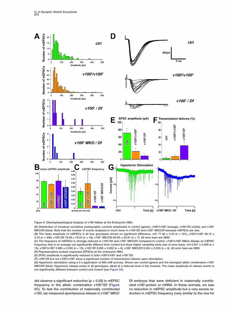
V0 in Synaptic Vesicle Exocytosis615
Figure 6. Electrophysiological Analysis of v100 Alleles at the Embryonic NMJ
(A) Distribution of miniature excitatory postsynaptic currents amplitudes in control (green), v1002/v1003 (orange), v1001/Df (violet), and v1001
MKO/Df (blue). Note that the number of events analyzed is much lower in v1001/Df and v1001 MKO/Df because mEPSCs are rare.(B) The mean amplitude of mEPSCs in all four genotypes shows no significant difference. ctrl: 77.48 ± 5.34 (n = 161), v1002/v1003 69.15 ±3.19 (n = 330), v1001/Df 79.29 ± 19.23 (n = 16), v1001 MKO/Df 60.93 ± 23.91 (n = 7). All error bars are SEM.(C) The frequency of mEPSCs is strongly reduced in v1001/Df and v1001 MKO/Df compared to control. v1002/v1003 NMJs display an mEPSCfrequency that is on average not significantly different from control but show higher variability (note size of error bars). ctrl 0.041 ± 0.009 (n =15), v1002/v1003 0.060 ± 0.035 (n = 13), v1001/Df 0.005 ± 0.002 (n = 6), v1001 MKO/Df 0.004 ± 0.003 (n = 6). All error bars are SEM.(D) Representative evoked responses (EPSCs) at the embryonic NMJ.(E) EPSC amplitude is significantly reduced in both v1002/v1003 and v1001/Df.(F) v1001/Df but not v1002/v1003 show a significant number of transmission failures upon stimulation.(G) Hypertonic stimulation using a 3 s application of 850 mM sucrose. Shown are control (green) and the strongest allelic combination v1001
MKO/Df (blue). Hypertonic release exists in all genotypes, albeit at a reduced level in the mutants. The mean amplitude of release events isnot significantly different between control and mutant (see Figure S4).
did observe a significant reduction (p < 0.05) in mEPSCfrequency in the allelic combination v1001/Df (Figure6C). To test the contribution of maternally contributedv100, we measured spontaneous release in v1001 MKO/
Df embryos that were deficient in maternally contrib-uted v100 protein or mRNA. In these animals, we sawno reduction in mEPSC amplitude but a very severe re-duction in mEPSC frequency (very similar to the one for
Cell616
v1001/Df) relative to control (p < 0.05). These results lsuggest that in late-stage embryos that have lost the vzygotic contribution, the maternal component has largely rdisappeared. In summary, our mEPSC analysis indi-cates that neurotransmitter content is normal in vesi-cles. However, it leaves open the question whether the Freduced mEPSC frequency is a result of the fusion of Mempty vesicles or an impairment of the fusion process Iitself. d
Since mutant synapses contain loaded vesicles that ccan be released spontaneously, the defect at the pho- ntoreceptor synapse as well as the lack of coordinated tmovements in the late embryo may be explained by Taberrant evoked neurotransmitter release. To test this dpossibility, we examined EPSCs at the neuromuscular 4junction evoked by stimulation of the motor nerve sup- tplying muscle 6. As shown in Figures 6D and 6E, we erecorded a significant reduction in the mean EPSC am- eplitudes (p < 0.01) from 712.7 ± 125 pA (SEM; n = 15) in icontrol to 218.3 ± 90 pA (n = 8) in v1002/v1003 mutant oembryos. v1001/Df mutants exhibit an even more se- uvere phenotype as the mean evoked amplitude is only c94.6 ± 36 pA (n = 9; p < 0.001). Furthermore, v1001/Df synapses display a marked increase in transmission ffailures: 31.1% ± 10.5% relative to 3.3% ± 3.3% (p < o0.005) in control animals (Figure 6F). There was no sig- anificant difference between the failure rate of v1002/ sv1003 and wild-type. These data indicate a strong im- bpairment of the synapse to release vesicles upon nerve estimulation. The normal mEPSC size and frequency in Sv1002/v1003 mutants combined with a significantly re- wduced evoked response in this allelic combination ar- mgue against a defect in vesicle acidification: if the re- aduction in the evoked response were due to the release tof empty vesicles (“shooting blanks”), these unloaded svesicles should be reflected in an equally reduced fre- tquency of spontaneous release events. Therefore, the sreduction in evoked release is likely to be due to a re- fduction in the number of vesicles released per stimulus. cThis effect may be due to a reduced presynaptic Ca2+
esignal, a reduction of the readily releasable pool of syn- saptic vesicles, or an impairment in the fusion process. dWe tested these possibilities using a Ca2+-independent cstimulation paradigm (Rosenmund and Stevens, 1996)
Tby pressure application of a 3 s pulse of hypertonic
tsucrose solution (850 mM) at the NMJ in the presence
wof 1 µM TTX. We detected postsynaptic currents in re-tsponse to hypertonic solution in wild-type and mutantw(v1002/v1003 and v1001/Df) animals (data not shown).sFurthermore, we tested maternal knockout v1001 MKO/cDf embryos (the most severe allelic combination) in the7presence of reduced Ca2+ (0.5 mM), and we also ob-tserved a response with hypertonic solution, indicatingathat loaded vesicles are docked and can be released inoa Ca2+-independent manner (Figure 6G). However, weeobserved fewer responses in mutants than in wild-type.tImportantly, the average amplitude of individual eventsiin control and maternal knockout v1001 MKO/Df em-dbryos is not significantly different (Kolmogorov-Smir-cnov test, p = 0.94; Figure S4). These data indicate thatKthe boutons contain vesicles that are loaded with neu-mrotransmitter and that vesicles can be released by hy-dpertonic stimulation. They also argue that the SNAREs
are functional and that vesicles can be primed for re- p
ease. In addition, our data do not suggest a defect inesicle acidification. We therefore argue that V100 isequired for synaptic vesicle fusion.
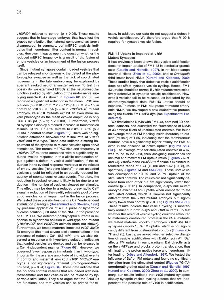
M1-43 Uptake Is Impaired at v100utant Synapses
t has previously been shown that vesicle acidificationoes not impair uptake of FM1-43 in cerebellar granuleells (Cousin and Nicholls, 1997), in rat hippocampaleuronal slices (Zhou et al., 2000), and at Drosophilahird instar larval NMJs (Kuromi and Kidokoro, 2000).hese studies imply that defective vesicle acidificationoes not affect synaptic vesicle cycling. Hence, FM1-3 uptake should be normal if v100 mutants were selec-ively defective in synaptic vesicle acidification. How-ver, if vesicles fail to be released, as indicated by thelectrophysiological data, FM1-43 uptake should be
mpaired. To measure FM1-43 uptake at mutant embry-nic NMJs, we developed a new quantitative protocolsing the fixable FM1-43FX dye (see Experimental Pro-edures).We first labeled NMJs with FM1-43, obtained 3D con-
ocal datasets, and quantified the fluorescence of NMJsf 33 embryo fillets of unstimulated controls. We foundn average ratio of FM labeling inside (boutons) to out-ide (muscle) of 1.55, indicating that the membranousoutons have a significantly higher affinity for FM1-43ven in the absence of active uptake (Figures S5C–5E). The average ratio for stimulated controls (n = 47)as found to be 2.23, thus assigning boundaries forinimal and maximal FM uptake ratios (Figures 7A–7C
nd 7J). v1001/Df and v1002/v1003 animals exhibited in-ermediate ratios of 1.74 (±0.08) and 1.62 (±0.08), re-pectively (Figures 7J and S5I–S5N). These uptake ra-ios correspond to 10.8% and 29.7% uptake of thetimulated controls. The values are not significantly dif-erent from each other but are significantly lower thanontrol (p < 0.001). In comparison, n-syb null mutantmbryos exhibit 44.5% uptake when compared to thetimulated control, which is likewise not significantlyifferent from the v100 mutant embryos but signifi-antly lower than control (p < 0.005; Figures S5F–S5H).hese results indicate that vesicle cycling is substan-ially reduced in both n-syb and v100 mutants. To testhether this residual vesicle cycling could be attributed
o maternally contributed protein in the v100 mutants,e tested maternal knockout embryos. v1001 MKO/Dfynapses display 1.8% FM uptake, which is not signifi-antly different from unstimulated controls (Figures 7D–F and 7J). Lastly, we asked whether selective disrup-ion of vesicle acidification with Bafilomycin A1 (Baf)ffects FM uptake in our paradigm. Baf directly actsn the v-ATPase and blocks proton translocation, thusliminating the proton motive force and neurotransmit-er loading (Dröse and Altendorf, 1997). We tested thenfluence of Baf on FM uptake and found no significanteviation from the stimulated control (Figures 7G–7J),orroborating other studies (Cousin and Nicholls, 1997;uromi and Kidokoro, 2000; Zhou et al., 2000). In sum-ary, our results indicate that v100 mutant synapsesisplay synaptic vesicle cycling defects that are inde-endent of a possible role of V100 in acidification.
V0 in Synaptic Vesicle Exocytosis617
Figure 7. FM1-43 Uptake at the Embry-onic NMJ
(A–C) FM1-43FX uptake experiment using a5 min 30 mM K+ stimulation paradigm.(D–F) FM1-43FX uptake using the samestimulation paradigm in a v1001 MKO/Dfmutant.(G–I) FM1-43FX uptake using the same stim-ulation paradigm after 15 min preincubationwith 1 µM Bafilomycin A1 (Baf) as well as 1µM Baf in the FM1-43 staining solution.(J) Quantification of FM1-43 uptake experi-ments in stimulated control (A–C), unstimu-lated control (Figures S4C–S4E), n-syb(Figures S4F–S4H), v1002/v1003 (Figures S4I–S4K), v1001/Df (Figures S4L–S4N), v1001
MKO/Df (D–F), and control with added Baf(G–I). Shown is the ratio of the mean fluores-cence intensity of the FM1-43 channel withinthe boundaries of HRP staining versus mus-cle background in 3D high-resolution confo-cal datasets. The yellow area marks theboundaries defined by unstimulated andstimulated control. Percentages of FM1-43uptake are given with respect to theseboundaries. The number of individual em-bryos analyzed is indicated inside bars. Errorbars are SEM.
Discussion
Here we report the identification of mutations in v100,the subunit a1 of the v-ATPase V0 complex in Drosoph-ila. We present an initial characterization of v100 mu-tant phenotypes that is inconsistent with a role of v100in acidification only. Our data indicate that V0 functionsin a late step in synaptic vesicle exocytosis.
The v-ATPase: Acidification and More?Vesicular or vacuolar ATPases are the most prominentintracellular proton pumps, consisting of at least 12subunits in two sectors (V0 and V1). Acidification is im-portant for many cellular functions, including receptor-ligand dissociation, degradative pathways, and thegeneration of intercompartment proton motive forcesthat are in turn utilized as driving forces for numeroussecondary transport processes (Nelson, 2003; Nishi
and Forgac, 2002). Here, we report the consequencesof the selective disruption of a V0 subunit a1 homologin neurons. The V0 subunit a is encoded by four homol-ogous genes in flies, worms, mouse, and human. Thedata from yeast and C. elegans indicate a crucial roleof V0 subunit a proteins for specific functions in distinctintracellular compartments and different cell types (Ka-wasaki-Nishi et al., 2001; Perzov et al., 2002; Oka et al.,2001; Pujol et al., 2001).
We isolated mutations in v100 based on the specificdefect of photoreceptor neurons to evoke a postsynap-tic response. Photoreceptors are an excellent “testtube” because they are not required for viability of thefly, and numerous assays can be used to assess mor-phology and function. Since loss of v100 does not af-fect photoreceptor specification, development, viabil-ity, and the ability to sense light, we surmise that mostintracellular vesicle trafficking and acidification pro-
Cell618
cesses are unaffected. Indeed, mutations that affect actwo key protein components of the V1 subcomplex
(subunits A and B) are cell lethal when removed in pho- aatoreceptors, and acidification as measured with Lyso-
Sensor in v100 mutant photoreceptor cell bodies is un- tlaffected (P.R.H. and H.J.B., unpublished data). Hence,
if acidification is the cause of the observed pheno- wttypes, it is likely to only affect synaptic vesicles.
Several methods can be used to directly or indirectly 1iassess the acidification of synaptic vesicles. The pH-
sensitive dye LysoSensor or genetically encoded pHlu- vsorin, a pH-sensitive GFP fusion protein that localizes
within vesicles, can be used in Drosophila neurons nrto directly assess synaptic vesicle acidification (Mie-
senböck et al., 1998). Unfortunately, the intensity differ- (iences at embryonic NMJs are too small to be observed
with our confocal system (P.R.H. and H.J.B., unpub- splished data). However, several lines of evidence allow
us to assess the contribution of a possible acidification fedefect to the observed phenotypes. We find that sev-
eral results are not consistent with a defect in synaptic spvesicle acidification alone. These include (1) the accu-
mulation of vesicles in mutant terminals, (2) the normalsmEPSC amplitude and frequency combined with a se-
verely reduced evoked response in a hypomorphic al- nclelic combination, and (3) the impairment of FM1-43 up-
take. In addition, several lines of evidence are not ttreadily explained with the function of V100 as part of
a proton pump: (1) selective partial rescue of vesicle sstrafficking but not acidification in yeast, (2) selective in-
teraction with t-SNAREs, and (3) localization at active przones. Taken together, our results indicate that V100
exerts additional or alternative functions to synaptic pvesicle acidification at Drosophila synapses.
E
SNAREs and V0: Fusion of “Proximity” and “Pore”? DThe hypothesis that SNAREs form the basic molecular yapparatus that forces lipid bilayers to fuse is widely s
ssupported (Jahn, 2004; Sørensen, 2004). However, thisgdoes not imply that SNAREs alone are required to in-sduce synaptic vesicle exocytosis. SNAREs alone ared
sufficient to induce fusion of liposomes, but the kineticsof these events does not mimic the kinetics that has Mbeen observed in synaptic vesicle fusion in vivo (Hu et U
Lal., 2003; Weber et al., 1998). The yeast vacuolar fusionoassay is the only system in which evidence for an addi-stional component downstream of SNARE function has1
been identified (Peters et al., 2001; Bayer et al., 2003). vInterestingly, Israël et al. (1986) reported the isolation of Aa proteolipid pore complex from synaptosomes from F
relectroplaques of Torpedo, named the “mediatophore.”oThis pore complex was later shown to contain a subunit
of the V0 complex and transfection of this component inT
some cells allowed quantal release of neurotransmitter I(Falk-Vairant et al., 1996; Morel, 2003). Subunit a1 was Srecently shown to localize to synaptic terminals and in-
Yteract with the v-SNARE n-Syb (Morel et al., 2003).SWhile the localization is in agreement with our findings
at Drosophila synapses, we found selective interac-Ptions of V100 with the t-SNAREs Syx and SNAP-25 inG
agreement with the observations made in yeast (Peters Bet al., 2001). d
gOur analyses reveal many similarities between v100
nd n-syb mutants: both die as late embryos withoutoordinated movement, accumulate vesicles at syn-pses, exhibit reduced spontaneous vesicle releasend FM1-43 uptake, contain vesicles with normalransmitter content that is poorly released upon stimu-ation, and interact with t-SNAREs. The only assay inhich n-syb mutants behaved differently from v100 is
he hypertonic stimulation (Rosenmund and Stevens,996). Vesicle release induced with hypertonic solution
n Drosophila SNARE mutants is largely abolished (Ara-amudan et al., 1999). In contrast, v100 mutants exhibitome responses with a reduced number of events butormal amplitude. This implies (1) the presence of neu-otransmitter-loaded vesicles in mutant terminals and2) that SNARE function required for hypertonic releases at least partially possible. V100 is not crucial for thistep, placing its role downstream of SNARE-dependentriming, congruous with the findings in yeast vacuolar
usion (Bayer et al., 2003; Peters et al., 2001; Thorngrent al., 2004). A function downstream of priming is alsoupported by the observation that Syx overexpressionhenotypes are suppressed by the loss of v100.Our results only show a late exocytic role for the V0
ubunit a1, while the implication of other V0 compo-ents remains to be tested. Hence, our data is formallyonsistent with a role of the V0 subunit a1 outside ofhe V0 complex in association with SNAREs. However,o our knowledge, no role outside the V0 complex haso far been shown for a subunit a in any system. Inummary, our data indicate a function for V100 andossibly the V0 proteolipid pore as a mediator of vesicle
elease efficiency downstream of SNARE-dependentriming.
xperimental Procedures
rosophila Strains, Mutagenesis, and Screenw;; P{ry+t7.2 = neo FRT} 82Bisogenized flies were used for mutagene-is and unmutagenized flies used as control animals. All further flytrains are described in detail below. Mutagenesis, electroretino-rams, and phototaxis assays were performed as described (Ver-treken et al., 2003). The eyFLP screen of chromosome arm 3R isescribed in Mehta et al. (2005).
olecular Biology and Antibody ProductionAS-v100 TransgeneD21248 containing a full-length cDNA of CG1709 in pOT2 wasbtained from the Drosophila Gene Collection (BDGP), and the in-ert was subcloned into the pUAST vector (Brand and Perrimon,993) at EcoRI and XhoI. Two independent insertions were reco-ered for each chromosome (X, 2 and 3).ntibody Productionor details see the Supplemental Experimental Procedures. Antise-um from guinea pig 11 was used at 1:2000 on tissue and 1:10,000n Western blots.
ransmission Electron Microscopy, Immunohistochemistry,mage Acquisition, and Processingee the Supplemental Experimental Procedures.
east Strains, Plasmids, and Methodsee the Supplemental Experimental Procedures.
rotein InteractionST Pull-Downsiochemical interactions of core complex proteins with V0 wereetermined by overnight incubation (4°C) of recombinant 0.25 µMlutathione S-transferase (GST)-tagged proteins (GST-Syx or GST-
V0 in Synaptic Vesicle Exocytosis619
V0, immobilized on glutathione-Sepharose beads) with 1.0 µM puri-fied recombinant Syx, n-Syb, or SNAP-25 in buffer A (50 mMHEPES [pH 7.4], 150 mM potassium acetate, 0.05% Tween 20).Samples were washed three times with buffer A + 1 mg/ml gelatinand two times with buffer A + 5% glycerol. Proteins were eluted in1× sample buffer (50 mM Tris [pH 6.8], 2.5% 2-mercaptoethanol,2% SDS, 5% glycerol).ImmunoprecipitationsImmunoprecipitations of core complex proteins with anti-Syx mAb8C3 or IgG purified guinea pig anti-V0 were performed using theSeize primary immunoprecipitation kit (Pierce Biotechnology, Rock-ford, Illinois). Per reaction, 0.5 µM Syx, n-Syb, or SNAP-25 or 500µg adult head extract was incubated with approximately 40 µgfixed antibody in IP buffer (25 mM Tris [pH 7.2], 150 mM NaCl,0.05% Tween 20). Samples were washed three times with IP buf-fer + 1 mg/ml gelatin and three times with IP buffer + 5% glycerol.Proteins were eluted in the kit’s provided elution buffer. Head ex-tract was prepared from Canton S flies using solubilization with1% Triton X-100 for 1 hr and subsequent ultracentrifugation at100,000 × g for 20 min to ensure the removal of membraneousfragments. The preparation of head extract is described in detail inthe Supplemental Experimental Procedures.Western BlotsSee the Supplemental Experimental Procedures.
ElectrophysiologyAll experiments were performed on late-stage embryos within 2–4hr before hatching. The embryos were selected under halocarbonoil (95, Halocarbon Products, River Edge, New Jersey) for the ab-sence of a GFP-marked balancer chromosome, and the chorionand the vitelline membrane were removed manually using sharpforceps. The embryo dissection was modified from Broadie et al.(1995) and was carried out in calcium-free HL3 saline: 70 mM NaCl,5 mM KCl, 11 mM MgCl2, 10 mM NaHCO3, 5 mM trehalose, 115mM sucrose, 5 mM HEPES. Briefly, the embryo was attached ante-riorly and posteriorly to a Sylgard-coated coverslip using cyanoac-rylate glue (Histoacryl B|Braun, Aesculap AG, Tuttlingen, Germany).The animal was cut open with a dorsal incision using a broken razorblade; the cut edges were attached to the coverslip with more glue,and the gut was removed to give a flat preparation with access tothe body wall muscles and the nerves supplying them. The muscleswere then treated with 100 U/ml of Collagenase Type 1A (Sigma-Aldrich, St. Louis, Missouri) for two minutes to remove the overly-ing sheath.
Whole-cell patch clamp recordings were made using an Axo-patch 200B amplifier from muscle 6 of abdominal segments A2–A4using thin-walled borosilicate pipettes fire-polished to a resistanceof 3–5 M�. Series resistance compensation was not used. Thepatch pipette was filled with a solution of the following composi-tion: 120 mM KCl, 20 mM KOH, 4 mM MgCl2, 0.25 mM CaCl2, 5 mMEGTA, 24 mM sucrose, 5 mM HEPES, and 4 mM Na2ATP. The mus-cle cells were clamped to −60 mV taking into account the cal-culated liquid junction potential of 6.7 mV. Conditions for the re-cording of spontaneous EPSCs, evoked EPSCs, and hypertonicstimulation are available in the Supplemental Experimental Pro-cedures.
FM UptakeIn brief, embryo fillets were exposed to 10 µM FM1-43FX with orwithout 30 mM K+ for a 5 min stimulation period (stimulated andunstimulated control). The samples were subsequently extensivelywashed with 0 Ca2+ saline for another 5 min using 0.5 mM EGTAand 1 mM Advasep-7 (Biotium, Hayward, California). Samples werethen fixed and stained for another 5 min with concentrated HRPantibody (1:10) in the absence of detergent. HRP recognizes acommon epitope of several neuron-specific surface molecules andthus allows us to demarcate the boundaries of the neuronal pro-cesses at the NMJ using threshold segmentation (Figure S4B). Co-labeling of HRP was similarly performed with highly concentratedCy5-conjugated secondary antibody. Standardized 3D confocalhigh-resolution datasets were obtained immediately afterwards.The surface of HRP-demarcated boutons was subsequently recon-structed and the average voxel intensity in the FM1-43 channel
calculated for the total volumes inside (boutons) and outside (mus-cle). Finally, inside/outside ratios were calculated and are pre-sented in Figure 7J.
Supplemental DataSupplemental Data include five figures and Supplemental Experi-mental Procedures and can be found with this article online athttp://www.cell.com/cgi/content/full/121/4/607/DC1/.
Acknowledgments
We would like to thank Alois Hofbauer, Sean Carroll, Morris Manol-son, Larry Zipursky, Peter Bryant, and the Bloomington Stock Cen-ter and the University of Iowa Developmental Studies HybridomaBank for reagents. We especially thank Iris Salecker for providingthe ey3.5FLP flies prior to publication. We are indebted to TimFergestad and Kendal Broadie for helping with embryonic FM up-take protocols as well as Tom Lloyd for help with the Syntaxin sup-pressor screen. Scanning EM was performed at the High Resolu-tion Microscopy Facility of the University of Texas MD AndersonCancer Center with the help of Kenneth Dunner, Jr. We thank Chris-tian Rosenmund and Erwin Neher for discussions and critical read-ing of the manuscript. P.R.H., A.F., R.G.Z., K.L.S., and H.J.B. aresupported by the HHMI. P.R.H. was further supported by an EMBOlong-term fellowship. This work was also supported by grantsGM068098 from the National Institutes of Health and Q-1536 fromthe Welch Foundation (both to J.K.). H.J.B. is an HHMI Investigator.
Received: December 22, 2004Revised: February 14, 2005Accepted: March 16, 2005Published: May 19, 2005
References
Amara, S.G., and Kuhar, M.J. (1993). Neurotransmitter transporters:recent progress. Annu. Rev. Neurosci. 16, 73–93.
Aravamudan, B., Fergestad, T., Davis, W.S., Rodesch, C.K., andBroadie, K. (1999). Drosophila UNC-13 is essential for synaptictransmission. Nat. Neurosci. 2, 965–971.
Bayer, M.J., Reese, C., Bühler, S., Peters, C., and Mayer, A. (2003).Vacuole membrane fusion: V0 functions after trans-SNARE pairingand is coupled to the Ca2+-releasing channel. J. Cell Biol. 162,211–222.
Bennett, M.K., Calakos, N., Kreiner, T., and Scheller, R.H. (1992).Synaptic vesicle membrane proteins interact to form a multimericcomplex. J. Cell Biol. 116, 761–775.
Benzer, S. (1967). Behavioral mutants of Drosophila isolated bycountercurrent distribution. Proc. Natl. Acad. Sci. USA 58, 1112–1119.
Brand, A.H., and Perrimon, N. (1993). Targeted gene expression asa means of altering cell fates and generating dominant phenotypes.Development 118, 401–415.
Broadie, K., Prokop, A., Bellen, H.J., O’Kane, C.J., Schulze, K.L.,and Sweeney, S.T. (1995). Syntaxin and synaptobrevin functiondownstream of vesicle docking in Drosophila. Neuron 15, 663–673.
Chen, Y.A., and Scheller, R.H. (2001). SNARE-mediated membranefusion. Nat. Rev. Mol. Cell Biol. 2, 98–106.
Chou, T.B., and Perrimon, N. (1992). Use of a yeast site-specificrecombinase to produce female germline chimeras in Drosophila.Genetics 131, 643–653.
Cousin, M.A., and Nicholls, D.G. (1997). Synaptic vesicle recyclingin cultured cerebellar granule cells: role of vesicular acidificationand refilling. J. Neurochem. 69, 1927–1935.
Deitcher, D.L., Ueda, A., Stewart, B.A., Burgess, R.W., Kidokoro,Y., and Schwarz, T.L. (1998). Distinct requirements for evoked andspontaneous release of neurotransmitter are revealed by mutationsin the Drosophila gene neuronal-synaptobrevin. J. Neurosci. 18,2028–2039.
Cell620
Dröse, S., and Altendorf, K. (1997). Bafilomycins and concanamyc- ACins as inhibitors of V-ATPases and P-ATPases. J. Exp. Biol. 200,
1–8. PmFalk-Vairant, J., Corrèges, P., Eder-Colli, L., Salem, N., Roulet, E.,
Bloc, A., Meunier, F., Lesbats, B., Loctin, F., Synguelakis, M., et Pal. (1996). Quantal acetylcholine release induced by mediatophore Ctransfection. Proc. Natl. Acad. Sci. USA 93, 5203–5207. SGalli, T., McPherson, P.S., and De Camilli, P. (1996). The V0 sector Pof the V-ATPase, synaptobrevin, and synaptophysin are associated Mon synaptic vesicles in a Triton X-100-resistant, freeze-thawing isensitive, complex. J. Biol. Chem. 271, 2193–2198. PHiesinger, P.R., Reiter, C., Schau, H., and Fischbach, K.F. (1999). DNeuropil pattern formation and regulation of cell adhesion mole- ncules in Drosophila optic lobe development depend on synapto- 1brevin. J. Neurosci. 19, 7548–7556. RHu, C., Ahmed, M., Melia, T.J., Söllner, T.H., Mayer, T., and Roth- cman, J.E. (2003). Fusion of cells by flipped SNAREs. Science 300, O1745–1749. RIsraël, M., Morel, N., Lesbats, B., Birman, S., and Manaranche, R. r(1986). Purification of a presynaptic membrane protein that medi- 1ates a calcium-dependent translocation of acetylcholine. Proc. SNatl. Acad. Sci. USA 83, 9226–9230. nJahn, R. (2004). Principles of exocytosis and membrane fusion. dAnn. N Y Acad. Sci. 1014, 170–178. sJahn, R., Lang, T., and Südhof, T.C. (2003). Membrane fusion. Cell S112, 519–533. r
3Jena, B.P. (2004). Discovery of the Porosome: revealing the molec-ular mechanism of secretion and membrane fusion in cells. J. Cell. SMol. Med. 8, 1–21. e
aKawasaki-Nishi, S., Nishi, T., and Forgac, M. (2001). Yeast V-ATPasecomplexes containing different isoforms of the 100-kDa a-subunit Sdiffer in coupling efficiency and in vivo dissociation. J. Biol. Chem. r276, 17941–17948. TKuromi, H., and Kidokoro, Y. (2000). Tetanic stimulation recruits Avesicles from reserve pool via a cAMP-mediated process in Dro- asophila synapses. Neuron 27, 133–143. VLindau, M., and Almers, W. (1995). Structure and function of fusion Zpores in exocytosis and ectoplasmic membrane fusion. Curr. Opin. SCell Biol. 7, 509–517. uMayer, A. (2002). Membrane fusion in eukaryotic cells. Annu. Rev. WCell Dev. Biol. 18, 289–314. M
mMehta, S.Q., Hiesinger, P.R., Beronja, S., Zhai, R.G., Schulze, K.L.,Verstreken, P., Cao, Y., Zhou, Y., Tepass, U., Crair, M.C., and Bellen, WH.J. (2005). Mutations in Drosophila sec15 reveal a specific func- ttion in neuronal targeting for a subset of exocyst components. Neu- Wron 46, 219–232. (Meinertzhagen, I.A., and Hanson, T.E. (1993). The development of tthe optic lobe. In The Development of Drosophila melanogaster, M. 6Bate and A. Martinez Arias, eds. (Plainview, NY: Cold Spring Harbor ZLaboratory Press), pp. 1363–1491. CMiesenböck, G., De Angelis, D.A., and Rothman, J.E. (1998). Visual- Mizing secretion and synaptic transmission with pH-sensitive green ifluorescent proteins. Nature 394, 192–195. ZMorel, N. (2003). Neurotransmitter release: the dark side of the vac- vuolar-H+ATPase. Biol. Cell. 95, 453–457. rMorel, N., Dedieu, J.C., and Philippe, J.M. (2003). Specific sortingof the a1 isoform of the V-H+ATPase a subunit to nerve terminalswhere it associates with both synaptic vesicles and the presynapticplasma membrane. J. Cell Sci. 116, 4751–4762.
Nelson, N. (2003). A journey from mammals to yeast with vacuolarH+-ATPase (V-ATPase). J. Bioenerg. Biomembr. 35, 281–289.
Newsome, T.P., Åsling, B., and Dickson, B.J. (2000). Analysis ofDrosophila photoreceptor axon guidance in eye-specific mosaics.Development 127, 851–860.
Nishi, T., and Forgac, M. (2002). The vacuolar (H+)-ATPases—nature’s most versatile proton pumps. Nat. Rev. Mol. Cell Biol. 3,94–103.
Oka, T., Toyomura, T., Honjo, K., Wada, Y., and Futai, M. (2001).Four subunit a isoforms of Caenorhabditis elegans vacuolar H+-
TPase. Cell-specific expression during development. J. Biol.hem. 276, 33079–33085.
ak, W.L., Grossfield, J., and White, N.V. (1969). Nonphototacticutants in a study of vision of Drosophila. Nature 222, 351–354.
erzov, N., Padler-Karavani, V., Nelson, H., and Nelson, N. (2002).haracterization of yeast V-ATPase mutants lacking Vph1p ortv1p and the effect on endocytosis. J. Exp. Biol. 205, 1209–1219.
eters, C., Bayer, M.J., Bühler, S., Andersen, J.S., Mann, M., andayer, A. (2001). Trans-complex formation by proteolipid channels
n the terminal phase of membrane fusion. Nature 409, 581–588.
ujol, N., Bonnerot, C., Ewbank, J.J., Kohara, Y., and Thierry-Mieg,. (2001). The Caenorhabditis elegans unc-32 gene encodes alter-ative forms of a vacuolar ATPase a subunit. J. Biol. Chem. 276,1913–11921.
ichmond, J.E., and Broadie, K.S. (2002). The synaptic vesicle cy-le: exocytosis and endocytosis in Drosophila and C. elegans. Curr.pin. Neurobiol. 12, 499–507.
osenmund, C., and Stevens, C.F. (1996). Definition of the readilyeleasable pool of vesicles at hippocampal synapses. Neuron 16,197–1207.
chulze, K.L., Broadie, K., Perin, M.S., and Bellen, H.J. (1995). Ge-etic and electrophysiological studies of Drosophila syntaxin-1Aemonstrate its role in nonneuronal secretion and neurotransmis-ion. Cell 80, 311–320.
ørensen, J.B. (2004). Formation, stabilisation and fusion of theeadily releasable pool of secretory vesicles. Pflugers Arch. 448,47–362.
towers, R.S., and Schwarz, T.L. (1999). A genetic method for gen-rating Drosophila eyes composed exclusively of mitotic clones ofsingle genotype. Genetics 152, 1631–1639.
üdhof, T.C. (2004). The synaptic vesicle cycle. Annu. Rev. Neu-osci. 27, 509–547.
horngren, N., Collins, K.M., Fratti, R.A., Wickner, W., and Merz,.J. (2004). A soluble SNARE drives rapid docking, bypassing ATPnd Sec17/18p for vacuole fusion. EMBO J. 23, 2765–2776.
erstreken, P., Koh, T.W., Schulze, K.L., Zhai, R.G., Hiesinger, P.R.,hou, Y., Mehta, S.Q., Cao, Y., Roos, J., and Bellen, H.J. (2003).ynaptojanin is recruited by endophilin to promote synaptic vesiclencoating. Neuron 40, 733–748.
eber, T., Zemelman, B.V., McNew, J.A., Westermann, B., Gmachl,., Parlati, F., Söllner, T.H., and Rothman, J.E. (1998). SNAREpins:inimal machinery for membrane fusion. Cell 92, 759–772.
eimer, R.M., and Jorgensen, E.M. (2003). Controversies in synap-ic vesicle exocytosis. J. Cell Sci. 116, 3661–3666.
ucherpfennig, T., Wilsch-Bräuninger, M., and González-Gaitán, M.2003). Role of Drosophila Rab5 during endosomal trafficking athe synapse and evoked neurotransmitter release. J. Cell Biol. 161,09–624.
hai, R.G., Hiesinger, P.R., Koh, T.W., Verstreken, P., Schulze, K.L.,ao, Y., Jafar-Nejad, H., Norga, K.K., Pan, H., Bayat, V., et al. (2003).apping Drosophila mutations with molecularly defined P element
nsertions. Proc. Natl. Acad. Sci. USA 100, 10860–10865.
hou, Q., Petersen, C.C., and Nicoll, R.A. (2000). Effects of reducedesicular filling on synaptic transmission in rat hippocampal neu-ones. J. Physiol. 525, 195–206.