Assessment of recovery in the hemiparkinson rat: Drug ...
11
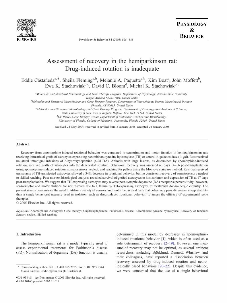
Assessment of recovery in the hemiparkinson rat: Drug-induced rotation is inadequate Eddie Castan ˜eda a, T , Sheila Fleming a,b , Melanie A. Paquette a,b , Kim Boat a , John Moffett b , Ewa K. Stachowiak b,c , David C. Bloom d , Michal K. Stachowiak b,c a Molecular and Structural Neurobiology and Gene Therapy Program, Department of Psychology, Arizona State University, Tempe, Arizona 85287-1104, United States b Molecular and Structural Neurobiology and Gene Therapy Program, Department of Neurobiology, Barrow Neurological Institute, Phoenix, AZ 85013, United States c Molecular and Structural Neurobiology and Gene Therapy Program, Department of Pathology and Anatomical Sciences, State University of New York at Buffalo, Buffalo, New York 14214, United States d UF Powell Gene Therapy Center, Department of Molecular Genetics and Microbiology, University of Florida, College of Medicine, Gainesville, Florida 32610, United States Received 24 May 2004; received in revised form 3 January 2005; accepted 24 January 2005 Abstract Recovery from apomorphine-induced rotational behavior was compared to sensorimotor and motor function in hemiparkinsonian rats receiving intrastriatal grafts of astrocytes expressing recombinant tyrosine hydroxylase (TH) or control h-galactosidase (h-gal). Rats received unilateral intranigral infusions of 6-hydroxydopamine (6-OHDA). Animals with large lesions, as determined by apomorphine-induced rotation, received grafts of astrocytes into the denervated striatum. Behavioral recovery was assessed on days 14–16 post-transplantation using apomorphine-induced rotation, somatosensory neglect, and reaching for pellets using the Montoya staircase method. Rats that received transplants of TH-transfected astrocytes showed a 34% decrease in rotational behavior, but no consistent recovery of somatosensory neglect or skilled reaching. Post-mortem histological analyses revealed survival of grafted astrocytes in host striatum and expression of TH at 17 days post-transplantation. We suggest that TH-expressing astrocytes may reverse post-synaptic dopamine (DA) receptor supersensitivity; however, sensorimotor and motor abilities are not restored due to a failure by TH-expressing astrocytes to reestablish dopaminergic circuitry. The present results demonstrate the need to utilize a variety of sensory and motor behavioral tests that cohesively provide greater interpretability than a single behavioral measure used in isolation, such as drug-induced rotational behavior, to assess the efficacy of experimental gene therapies. D 2005 Elsevier Inc. All rights reserved. Keywords: Apomorphine; Astrocytes; Gene therapy; 6-hydroxydopamine; Parkinson’s disease; Recombinant tyrosine hydroxylase; Recovery of function; Sensory neglect; Skilled reaching 1. Introduction The hemiparkinsonian rat is a model typically used to assess experimental treatments for Parkinson’s disease (PD). Normalization of dopamine (DA) function is usually determined in this model by decreases in apomorphine- induced rotational behavior [1], which is often used as a sole determinant of recovery [2–19]. However, one mea- sure of recovery may not be optimal, as several eminent researchers, including Bjfrklund, Dunnett, Whishaw, and their colleagues, have reported a dissociation between recovery assessed by drug-induced rotation and neuro- logically based behaviors [20–22]. Despite this evidence, we were concerned that the use of a single behavioral 0031-9384/$ - see front matter D 2005 Elsevier Inc. All rights reserved. doi:10.1016/j.physbeh.2005.01.019 T Corresponding author. Tel.: +1 480 965 2265; fax: 1 480 965 8544. E-mail address: [email protected] (E. Castan ˜eda). Physiology & Behavior 84 (2005) 525 – 535
Transcript of Assessment of recovery in the hemiparkinson rat: Drug ...
Physiology & Behavior
Assessment of recovery in the hemiparkinson rat:
Drug-induced rotation is inadequate
Eddie Castanedaa,T, Sheila Fleminga,b, Melanie A. Paquettea,b, Kim Boata, John Moffettb,
Ewa K. Stachowiakb,c, David C. Bloomd, Michal K. Stachowiakb,c
aMolecular and Structural Neurobiology and Gene Therapy Program, Department of Psychology, Arizona State University,
Tempe, Arizona 85287-1104, United StatesbMolecular and Structural Neurobiology and Gene Therapy Program, Department of Neurobiology, Barrow Neurological Institute,
Phoenix, AZ 85013, United StatescMolecular and Structural Neurobiology and Gene Therapy Program, Department of Pathology and Anatomical Sciences,
State University of New York at Buffalo, Buffalo, New York 14214, United StatesdUF Powell Gene Therapy Center, Department of Molecular Genetics and Microbiology,
University of Florida, College of Medicine, Gainesville, Florida 32610, United States
Received 24 May 2004; received in revised form 3 January 2005; accepted 24 January 2005
Abstract
Recovery from apomorphine-induced rotational behavior was compared to sensorimotor and motor function in hemiparkinsonian rats
receiving intrastriatal grafts of astrocytes expressing recombinant tyrosine hydroxylase (TH) or control h-galactosidase (h-gal). Rats receivedunilateral intranigral infusions of 6-hydroxydopamine (6-OHDA). Animals with large lesions, as determined by apomorphine-induced
rotation, received grafts of astrocytes into the denervated striatum. Behavioral recovery was assessed on days 14–16 post-transplantation
using apomorphine-induced rotation, somatosensory neglect, and reaching for pellets using the Montoya staircase method. Rats that received
transplants of TH-transfected astrocytes showed a 34% decrease in rotational behavior, but no consistent recovery of somatosensory neglect
or skilled reaching. Post-mortem histological analyses revealed survival of grafted astrocytes in host striatum and expression of TH at 17 days
post-transplantation. We suggest that TH-expressing astrocytes may reverse post-synaptic dopamine (DA) receptor supersensitivity; however,
sensorimotor and motor abilities are not restored due to a failure by TH-expressing astrocytes to reestablish dopaminergic circuitry. The
present results demonstrate the need to utilize a variety of sensory and motor behavioral tests that cohesively provide greater interpretability
than a single behavioral measure used in isolation, such as drug-induced rotational behavior, to assess the efficacy of experimental gene
therapies.
D 2005 Elsevier Inc. All rights reserved.
Keywords: Apomorphine; Astrocytes; Gene therapy; 6-hydroxydopamine; Parkinson’s disease; Recombinant tyrosine hydroxylase; Recovery of function;
Sensory neglect; Skilled reaching
1. Introduction
The hemiparkinsonian rat is a model typically used to
assess experimental treatments for Parkinson’s disease
(PD). Normalization of dopamine (DA) function is usually
0031-9384/$ - see front matter D 2005 Elsevier Inc. All rights reserved.
doi:10.1016/j.physbeh.2005.01.019
T Corresponding author. Tel.: +1 480 965 2265; fax: 1 480 965 8544.
E-mail address: [email protected] (E. Castaneda).
determined in this model by decreases in apomorphine-
induced rotational behavior [1], which is often used as a
sole determinant of recovery [2–19]. However, one mea-
sure of recovery may not be optimal, as several eminent
researchers, including Bjfrklund, Dunnett, Whishaw, and
their colleagues, have reported a dissociation between
recovery assessed by drug-induced rotation and neuro-
logically based behaviors [20–22]. Despite this evidence,
we were concerned that the use of a single behavioral
84 (2005) 525–535
E. Castaneda et al. / Physiology & Behavior 84 (2005) 525–535526
measure perseveres in assessment of the efficacy of
potential therapies for neurological disorders, and this
practice may mislead our advancement towards effective
treatments. We applied a more comprehensive behavioral
analysis to clarify the ameliorative effects of transgenic
astrocytes in the hemiparkinsonian rat. The present
investigation is significant because a large amount of
research involving transgenic astrocytes limits behavioral
assessment of recovery to only apomorphine-induced
rotational behavior.
The use of astrocytes as vectors for delivery of
potentially therapeutic genes has been a popular line of
investigation. Various approaches have expressed the TH
gene in vitro in rat [2,9,17] or human [4,15] astrocytes for
later transplantation into the host [2,4,6,9,15,17]. There are
a number of reasons to believe that astrocytes could be
successful vectors for gene therapy. Astrocytes are
endogenous to the striatum and thus should optimize
survival when transplanted back into this environment. For
this reason, astrocytes might serve as autologous trans-
plants. In addition, transfected astrocytes show little
proliferation, allowing transgenes to persist episomally.
Finally, astrocytes demonstrate a propensity to accept
transgenes via retroviral [2,9,17] and adenoviral vectors
[6,7], as well as direct injection of a DNA-liposomal
complex [13]. Unfortunately, none of the above studies
utilizing TH-expressing astrocytes have incorporated a
neurological approach to assess behavioral recovery; they
are limited by their sole reliance on reductions in drug-
induced rotational behavior as an index of recovery
[2,4,6,7,13,15–17]. A comprehensive behavioral approach
may better assess neurological recovery.
For the present study, we selected intrastriatal grafts of
astrocytes expressing recombinant tyrosine hydroxylase
(TH), the rate-limiting catecholamine biosynthetic
enzyme. We were particularly interested in determining
how recovery would be assessed by behavioral measures
in addition to the drug-based behavioral assay. To
accomplish this, we chose to compare apomorphine-
induced rotation to somatosensory neglect and skilled
reaching, two behavioral deficits that have been charac-
terized in the hemiparkinsonian rat and are analogous to
human PD [23–26], and thus are more likely to be
predictive of recovery in human PD patients [27].
Notably, these sensorimotor and motor behaviors are
dependent upon pre-synaptic events involving exocytosis,
whereas apomorphine-induced rotational behavior relies
solely upon activation of supersensitive post-synaptic
receptors by a direct agonist. Thus, different neuronal
mechanisms are involved in the expression of these
behaviors. We hypothesized that TH-expressing astro-
cytes would promote recovery in the hemiparkinsonian
rat on apomorphine-induced rotation, but not sensorimo-
tor and motor behaviors, likely due to a failure by
transgenic astrocytes to restore pre-synaptic factors of
DA neurotransmission.
2. Materials and methods
2.1. Subjects
Male Fischer 344 rats weighing 250–300 g were group-
housed and handled for at least two weeks prior to behavioral
testing and surgery. Animals were maintained at 90% free-
feeding weight during training and testing on the reaching
apparatus (Montoya staircase) and were food-deprived 24 h
prior to surgery. At all other times, food and water were
available ad libitum. Animals were maintained on a reverse
12:12 h light–dark cycle (lights on at 1800 hours) and were
tested during the dark portion. The protocol for this experi-
ment was approved by the Arizona State University Institu-
tional Animal Care and Use Committee in conformance with
the NIH Guide for the Care and Use of Laboratory Animals
(NIH Publications No. 80-23, revised 1996).
2.2. 6-OHDA lesion
Lesions were created as described previously [28,29].
The hemisphere contralateral to the forelimb dominant for
skilled reaching (see Fig. 4A) was selected a priori to
receive 6-OHDA [30]. Animals were anesthetized with
sodium pentobarbital (Sigma, 50 mg/kg, i.p.), and supple-
ments of chloral hydrate (Sigma, 80 mg/kg, i.p.) were
administered when necessary. Atropine (Henry Schein, 5
mg/kg, s.c.) was administered prophylactically. To protect
norepinephrine cells, animals were pretreated with desipr-
amine HCl (RBI, 15 mg/kg, i.p.), a noradrenergic reuptake
blocker, 30 min prior to 6-OHDA infusion. Using standard
stereotaxic procedures, a 28 ga injector needle (Plastics
One), connected to a 1 ml Hamilton gastight syringe by
polyethylene tubing (PE 20/Clay Adams, Intramedic), was
lowered to the level of the substantia nigra (SN; with
bregma and lambda horizontal; AP �5.8 mm, ML +2.5
mm, DV �8.0 mm [31]. One min later, 6-OHDA HBr
(RBI), dissolved in deoxygenated 0.9% saline containing
10% ascorbic acid, was infused (10 Ag/2.5 Al at 0.2 Al/min
over 12.5 min) via a syringe pump (Harvard Apparatus).
The needle remained lowered for 1 min after the infusion to
allow for diffusion from the tip. Immediately following
infusion of 6-OHDA, animals were returned to their home
cages for two weeks to allow a neurotoxin-induced state of
chronic DA depletion to develop, in accordance with the
traditional Ungerstedt paradigm [32–34].
2.3. In vitro procedures
2.3.1. Astrocyte cultures
Astrocytes were isolated from the caudate nucleus of 5–
6-week-old rats. The tissue was dissociated with collagenase
(0.4 mg/ml) and DNase (10 U/ml) for 5 min. Cells were then
plated in astrocyte media (Clonetics) in a 75 cm2 tissue
culture flask (Corning). Astrocytes were maintained in a
humidified incubator at 37 8C with 5% CO2. Cultures were
E. Castaneda et al. / Physiology & Behavior 84 (2005) 525–535 527
passaged 1:2 and transfected by electroporation at passage
three or four.
2.3.2. Plasmid construction
Two expression vectors based on the pBkCMV plasmids
(Stratagene) were constructed. The vector pBkCMV/TH
was created by inserting the gene encoding a mutated form
of the human TH cDNA into the Xba I site of pBkCMV.
The 2.2 kb TH cDNA (a gift from Dr. Menek Goldstein) has
a substitution of Leu 40 with Ser, resulting in over a 10-fold
increase in TH activity. The high activity of mutated TH is
phosphorylation-independent and has been shown to con-
tribute to the production of L-DOPA [35,36]. To construct
pBkCMV/h-gal, the Lac Z gene encoding h-gal was excisedfrom pCH110 (Pharmacia) with Bam HI and Hind III and
subcloned into the Hind III/Sma I sites of pBkCMV after
end-filling of the Bam HI site.
2.3.3. Transfection
The pCMV/h-gal and pCMV/TH were transfected into
astrocyte cultures using electroporation as described in
Stachowiak et al. [37]. Briefly, cells were trypsinized and
washed in astrocyte media and 1X Hebes buffer. Cells were
then placed into cuvettes (0.4 cm gap), 150 Ag of pCMV/TH
or pCMV/h-gal was added, and cells were electroporated at
170mV. Electroporated cells were allowed to recover for 48 h
in culture flasks before in vitro staining or transplantation.
2.3.4. Validation of transfection efficiency
Cells were plated on glass well slides (n=4) immediately
after electroporation, and staining for TH or h-gal was
performed 48 h later. For TH immunostaining, wells were
washed with phosphate buffered saline (PBS), pH 7.4, and
then fixed with PBS containing 2.5% paraformaldehyde for
15 min. Wells were washed with PBS, pH 8.4, and treated
with 1% Triton X-100 for 15 min, then 3% bovine serum
albumin (BSA) in PBS at 37 8C for 30 min. Cells were
then incubated overnight at 4 8C with polyclonal rabbit
anti-TH antibodies (Chemicon, 1:500) in 3% BSA followed
by anti-rabbit IgG conjugated with alkaline phosphatase
(Jackson, 1:500). The immune complexes were stained
using avidin–biotin-peroxidase (Elite ABC rabbit IgG Kit,
Vector) and 3,3-diaminobenzidine (DAB, Sigma), accord-
ing to manufacturer’s instructions. Transfection efficiency
was calculated from three wells while the fourth well,
lacking primary antibody, served as a control. To assess h-gal activity, the wells were washed with PBS, pH 7.4, and
fixed in a formaldehyde solution [2% formaldehyde (37%),
0.2% gluteraldehyde, 0.1% NaDOC, and 0.02% NP-40 in
PBS] for 15 min. Wells were washed and then incubated in
the X-gal solution [2 mM MgCl2, 5 mM K3Fe(CN)6, 5 mM
K4Fe(CN)6, and 1 mg/ml of 5V-bromo-4-chloro-3-indolyl-b-
d-galactoside (X-gal; Eastman Kodak Co.)] in the dark at 4
8C for up to 12 h. Transfection efficiency was calculated
from three wells while the fourth well, containing non-
transfected astrocytes, served as a control.
2.4. Transplantation of astrocytes
2.4.1. Cell preparation and labeling
Forty-eight hours after electroporation, when astrocytes
express transgenes in vitro (see Fig. 2), astrocytes were
detached by trypsinization, centrifuged at 1000 rpm for 5
min, then washed with PBS and resuspended in PBS at a
density of 1�105 cells/Al. At this point, to further validate
placement and diffusion of transgenic cells, both h-gal andTH-expressing astrocyte grafts were labeled with the
carbocyanine dye diI [1,1’-dioctadecyl-3,3,3’,3’-tetramethy-
lindocarbocyanine perchlorate; diI-C18-(3), Molecular
Probes] at a concentration of 0.1%.
2.4.2. Transplantation
Immediately after completing tests to affirm the presence
of behavioral deficits (post-lesion days 14–16), on post-
lesion day 17, animals received control h-gal or TH-
expressing astrocytes based on pseudo-random assignment,
counterbalanced for turning rate in the apomorphine-induced
rotation test. Astrocytes were infused into the striatum
ipsilateral to the lesion (experimental, n=8; control, n=7)
using the general stereotaxic surgery procedures for the 6-
OHDA lesions (see above) except that four infusions were
made (3 Al/site at 0.5 Al/min to deliver 3�105 cells/site).
Coordinates for the four sites with bregma and lambda
horizontal were: 1) AP +1.8, ML +2.5, DV �4.5; 2) AP
+1.2, ML +2.5, DV �6.0; 3) AP +0.7, ML +4.0, DV �6.4;
4) AP +0.2, ML +3.5, DV �7.0. Thus infusions were made
in a rostral–caudal direction, with rostral infusions targeting
the corpus of the striatum, while caudal infusions targeted the
ventrolateral striatum, which has been shown to be important
for forelimb use during food handling in rats [38]. The
cannula was left in place for 5 min following each infusion.
Animals were allowed two weeks post-operative recovery.
2.5. Behavioral tests
All animals (experimental, n=8; control, n=7) were
tested for somatosensory neglect, skilled reaching, and
apomorphine-induced rotation by an investigator blind to
the experimental treatment. Somatosensory neglect and
skilled reaching were assessed over three consecutive days
at three time points: prior to any surgery (days 1–3 pre-
lesion), two weeks after unilateral DA depletion (days 14–
16 post-lesion), and two weeks after transplantation (days
31–33 post-lesion). Apomorphine-induced rotation was
assessed on days 16 and 33 post-lesion, immediately after
the other two tests.
2.5.1. Somatosensory neglect
Small adhesive stickers (13 mm diameter) were placed
concurrently on the ulnar surface of both forelimbs of the
rats, as adapted from Schallert et al. [39]. Latencies in
seconds to contact and to remove the stickers were recorded
separately for the left and right forelimbs [26].
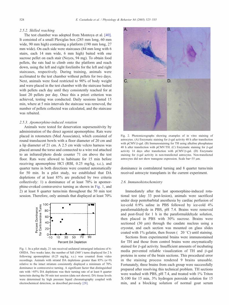
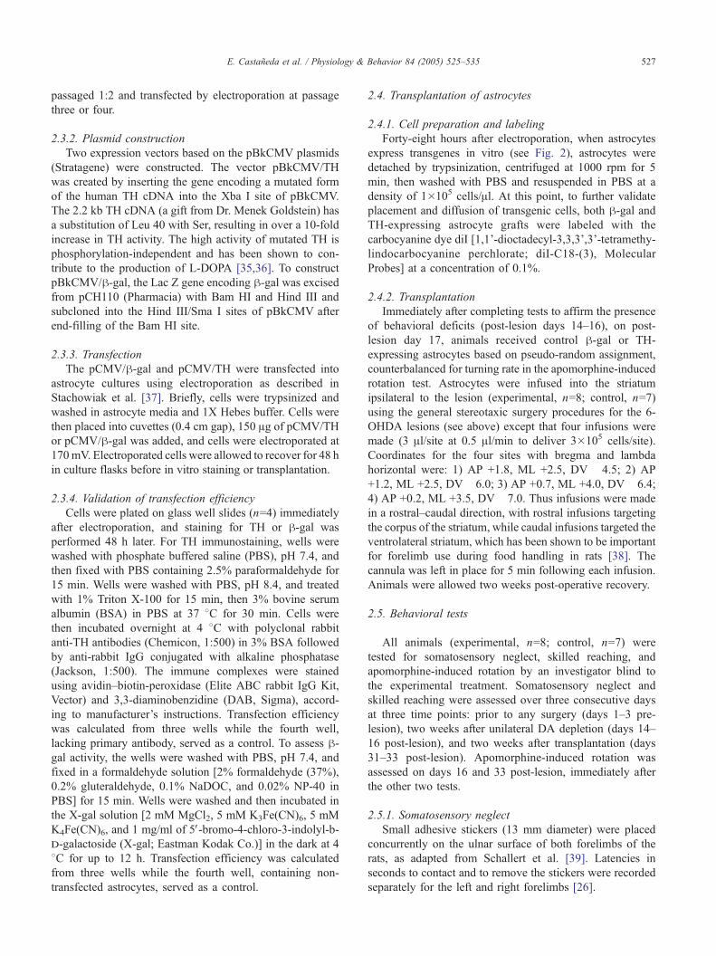
Fig. 2. Photomicrographs showing examples of in vitro staining of
astrocytes. (A) Enzymatic staining for h-gal activity 48 h after transfection
with pCMV/h-gal. (B) Immunostaining for TH using alkaline phosphatase
48 h after transfection with pCMV/TH. (C) Enzymatic staining for h-galactivity 14 days after transfection with pCMV/h-gal. (D) Enzymatic
staining for h-gal activity in non-transfected astrocytes. Non-transfected
astrocytes did not show transgene expression. Scale bar=35 Am.
E. Castaneda et al. / Physiology & Behavior 84 (2005) 525–535528
2.5.2. Skilled reaching
The test chamber was adopted from Montoya et al. [40].
It consisted of a small Plexiglas box (285 mm long, 60 mm
wide, 90 mm high) containing a platform (190 mm long, 27
mm wide). On each side were staircases (84 mm long with 6
stairs, each 14 mm wide, 6 mm high) baited with one
sucrose pellet on each stair (Noyes, 94 mg). To obtain food
pellets, the rats had to climb onto the platform and reach
down, using the left and right forelimbs for the left and right
staircases, respectively. During training, animals were
acclimated to the test chamber without pellets for two days.
Next, animals were food restricted to 90% of body weight
and were placed in the test chamber with the staircase baited
with pellets each day until they consistently reached for at
least 20 pellets per day. Once this a priori criterion was
achieved, testing was conducted. Daily sessions lasted 15
min, where at 5 min intervals the staircase was removed, the
number of pellets collected was calculated, and the staircase
was rebaited.
2.5.3. Apomorphine-induced rotation
Animals were tested for denervation supersensitivity by
administration of the direct agonist apomorphine. Rats were
placed in rotometers (Med Associates), which consisted of
round translucent bowls with a floor diameter of 20 cm and
a lip diameter of 21 cm. A 2.5 cm wide velcro harness was
placed around the torso and connected to a wire rod attached
to an infrared/photo diode counter 71 cm above the test
floor. Rats were allowed to habituate for 15 min before
receiving apomorphine HCl (RBI, 0.25 mg/kg, s.c.), and
quarter turns in both directions were counted automatically
for 50 min. In a pilot study, we established that DA
depletions of at least 85% are predicted by two criteria
collectively: 1) a dominance of at least 70% in apomor-
phine-evoked contraversive turning as shown in Fig. 1, and
2) at least 8 quarter turns/min throughout the 50 min test
session. Therefore, only animals that displayed at least 70%
Fig. 1. In a pilot study, 21 rats received unilateral intranigral infusions of 6-
OHDA. Two weeks later, the number of full (3608) turns displayed for 2 h
following apomorphine (0.25 mg/kg, s.c.) was counted from video
recordings. Animals with striatal DA depletions greater than 85% (n=9)
relative to the intact striatum consistently displayed a minimum of 70%
dominance in contraversive turning. A significant factor that distinguished
rats with N85% DA depletions was their turning rate of at least 8 quarter
turns/min during the 50 min test session (data not shown). DA tissue levels
were determined by high pressure liquid chromatography coupled with
electrochemical detection, as described previously [28].
dominance in contralateral turning and 8 quarter turns/min
received astrocyte transplants in the current experiment.
2.6. Immunohistochemistry
Immediately after the last apomorphine-induced rota-
tional test (day 33 post-lesion), animals were sacrificed
under deep pentobarbital anesthesia by cardiac perfusion of
ice-cold 0.9% saline in PBS followed by ice-cold 4%
paraformaldehyde in PBS, pH 7.4. Brains were removed
and post-fixed for 1 h in the paraformaldehyde solution,
then placed in PBS with 30% sucrose. Brains were
sectioned (30 Am) through the caudate nucleus using a
cryostat, and each section was mounted on glass slides
coated with 1% gelatin, then frozen (�20 8C) until staining.Sections from experimental brains were immunostained
for TH and those from control brains were enzymatically
stained for h-gal activity. Insufficient amounts of incubating
media prevented reliable visualization of TH and h-galproteins in some of the brain sections. This procedural error
in the staining process rendered 9 brains unusable.
Fortunately, three brains from each group were successfully
prepared after resolving this technical problem. TH sections
were washed with PBS, pH 7.4, and treated with 1% Triton
X-100 for 15 min, 3% hydrogen peroxide solution for 15
min, and a blocking solution of normal goat serum
E. Castaneda et al. / Physiology & Behavior 84 (2005) 525–535 529
(polyclonal) in PBS for 30 min at 37 8C. Sections were thenincubated with polyclonal rabbit anti-TH (Chemicon, 1:500)
in blocking solution overnight at 4 8C, followed by
biotinylated goat anti-rabbit TH, avidin–biotin-peroxidase,
and DAB. h-gal sections were washed with PBS, pH 7.4,
and fixed in a formaldehyde solution [2% formaldehyde
(37%), 0.2% gluteraldehyde, 0.1% NaDOC, and 0.02% NP-
40 in PBS] for 1 h at 4 8C. They were then washed and
treated with staining solution [0.87% NaCl, 0.1 M
Na2HPO4, 2 mM MgCl2, 0.01% NaDOC, 0.02% NP-40, 5
mM K3Fe(CN)6, 5 mM K4Fe(CN)6, and 1 mg/ml X-gal] in
the dark at 31 8C for up to 12 h, as described in Tabbaa et al.
[41]. For each brain, the section with the most staining,
presumably reflecting the locus of infusion, was selected for
analysis.
2.7. Statistical analyses
2.7.1. Somatosensory neglect
Latencies to contact and to remove stickers placed on the
contralateral and ipsilateral forelimbs were assessed. To
assess deficits following a unilateral DA depletion, an
ANOVA was performed on a 2�2 within subject design,
comparing limb (contralateral and ipsilateral) and time (pre-
surgery and post-lesion). To determine recovery following
transplantation, an ANOVAwas performed on a 2�2 mixed
design comparing group (TH and h-gal) and time (post-
lesion and post-transplant). A priori planned comparisons
were analyzed with Fisher’s LSD.
2.7.2. Skilled reaching
The number of pellets collected by the contralateral and
ipsilateral forelimbs was quantified. Statistical analyses
were conducted as has been reported previously [21,40]
and as described for somatosensory neglect.
2.7.3. Apomorphine-induced rotation
The number of contralateral quarter turns was quantified,
and a square root transformation was performed on the data
to achieve homogeneity of variance among groups. To
determine recovery following transplantation, statistical
analyses were conducted as described for somatosensory
neglect.
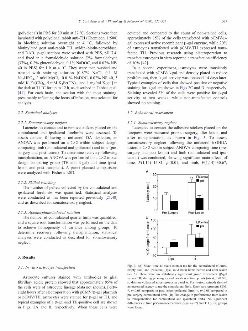
Fig. 3. (A) Mean time to make contact (s) for the contralateral (Contra,
empty bars) and ipsilateral (Ipsi, solid bars) limbs before and after lesion
(n=15). There were no statistically significant group differences (h-galversus TH) during pre-surgery and post-lesion time points (t-test, pN0.05),
so data are collapsed across groups in panel A. Post-lesion, animals showed
an increased latency to use the contralateral limb. Error bars represent SEM.
*, pb0.05 compared to post-lesion ipsilateral limb; +, pb0.05 compared to
pre-surgery contralateral limb. (B) The change in performance from lesion
to transplantation for contralateral and ipsilateral limbs. No significant
differences in limb performance between h-gal (n=7) and TH (n=8) groups
were found.
3. Results
3.1. In vitro astrocyte transfection
Astrocyte cultures stained with antibodies to glial
fibrillary acidic protein showed that approximately 95% of
the cells were of astrocytic lineage (data not shown). Forty-
eight hours after electroporation with pCMV/h-gal plasmids
or pCMV/TH, astrocytes were stained for h-gal or TH, andtypical examples of a h-gal-and TH-positive cell are shown
in Figs. 2A and B, respectively. When these cells were
counted and compared to the count of non-stained cells,
approximately 15% of the cells transfected with pCMV/h-gal expressed active recombinant h-gal enzyme, while 20%
of astrocytes transfected with pCMV/TH expressed trans-
fected TH. Previous research using electroporation to
transfect astrocytes in vitro reported a transfection efficiency
of 10% [42].
In a second experiment, astrocytes were transiently
transfected with pCMV/h-gal and densely plated to reduce
proliferation, then h-gal activity was assessed 14 days later.
Typical examples of cells that showed positive or negative
staining for h-gal are shown in Figs 2C and D, respectively.
Staining revealed 5% of the cells were positive for h-galactivity at two weeks, while non-transfected controls
showed no staining.
3.2. Behavioral assessment
3.2.1. Somatosensory neglect
Latencies to contact the adhesive stickers placed on the
forepaws were measured prior to surgery, after lesion, and
after transplantation, as shown in Fig. 3. To assess
somatosensory neglect following the unilateral 6-OHDA
lesion, a 2�2 within subject ANOVA comparing time (pre-
surgery and post-lesion) and limb (contralateral and ipsi-
lateral) was conducted, showing significant main effects of
time, F(1,14)=15.81, pb0.01, and limb, F(1,14)=30.67,
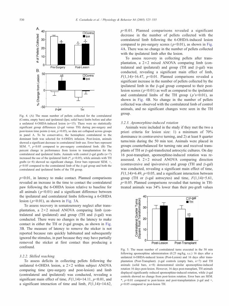
Fig. 4. (A) The mean number of pellets collected for the contralateral
(Contra, empty bars) and ipsilateral (Ipsi, solid bars) limbs before and after
a unilateral 6-OHDA-induced lesion (n=15). There were no statistically
significant group differences (h-gal versus TH) during pre-surgery and
post-lesion time points (t-test, pN0.05), so data are collapsed across groups
in panel A. To be conservative, the hemisphere contralateral to the
dominant limb was selected for 6-OHDA infusion. Post-lesion, animals
showed a significant decrease in contralateral limb use. Error bars represent
SEM. *, pb0.05 compared to pre-surgery contralateral limb. (B) The
percent change in performance from lesion to transplantation for the
contralateral and ipsilateral limbs. Animals with control h-gal grafts (n=7)increased the use of the ipsilateral limb (*, pb0.05), while animals with TH
grafts (n=8) showed no significant change. Error bars represent SEM. +,
pb0.05 compared to the contralateral limb of the h-gal group and both the
contralateral and ipsilateral limbs of the TH group.
Fig. 5. The mean number of contralateral turns executed in the 50 min
following apomorphine administration (0.25 mg/kg, s.c.) 16 days after a
unilateral 6-OHDA-induced lesion (Post-Lesion) and 16 days after trans-
plantation (Post-Transplant). h-gal controls (empty bars, n=7) and TH
animals (solid bars, n=8) demonstrated similar apomorphine-induced
rotation 16 days post-lesion. However, 16 days post-transplant, TH animals
displayed significantly reduced apomorphine-induced rotation, while h-galcontrols showed no change from post-lesion rotation. Error bars are SEM.
*, pb0.01 compared to post-lesion and post-transplantation h-gal and +,
pb0.05 compared to post-lesion TH.
E. Castaneda et al. / Physiology & Behavior 84 (2005) 525–535530
pb0.01, in latency to make contact. Planned comparisons
revealed an increase in the time to contact the contralateral
paw following the 6-OHDA lesion relative to baseline for
all animals ( pb0.01) and a significant difference between
the ipsilateral and contralateral limbs following a 6-OHDA
lesion ( pb0.01), as shown in Fig. 3A.
To assess recovery in somatosensory neglect after trans-
plantation, a 2�2 mixed ANOVA comparing limb (con-
tralateral and ipsilateral) and group (TH and h-gal) was
conducted. There were no changes in the latency to make
contact in either the TH or h-gal groups, as shown in Fig.
3B. The measure of latency to remove the sticker is not
reported because rats quickly habituated and subsequently
ignored the stimulus, in part because they may have partially
removed the sticker at first contact thus producing a
confound.
3.2.2. Skilled reaching
To assess deficits in collecting pellets following the
unilateral 6-OHDA lesion, a 2�2 within subject ANOVA
comparing time (pre-surgery and post-lesion) and limb
(contralateral and ipsilateral) was conducted, revealing a
significant main effect of time, F(1,14)=14.11, pb0.01, and
a significant interaction of time and limb, F(1,14)=14.62,
pb0.01. Planned comparisons revealed a significant
decrease in the number of pellets collected with the
contralateral limb following the 6-OHDA-induced lesion
compared to pre-surgery scores ( pb0.01), as shown in Fig.
4A. There was no change in the number of pellets collected
with the ipsilateral limb after the lesion.
To assess recovery in collecting pellets after trans-
plantation, a 2�2 mixed ANOVA comparing limb (con-
tralateral and ipsilateral) and group (TH and h-gal) was
conducted, revealing a significant main effect of limb,
F(1,14)=16.47, pb0.01. Planned comparisons revealed a
significant increase in the number of pellets collected by the
ipsilateral limb in the h-gal group compared to their post-
lesion scores ( pb0.01) as well as compared to the ipsilateral
and contralateral limbs of the TH group ( p’sb0.01), as
shown in Fig. 4B. No change in the number of pellets
collected was observed with the contralateral limb of control
animals, and no significant changes were seen in the TH
group.
3.2.3. Apomorphine-induced rotation
Animals were included in the study if they met the two a
priori criteria for lesion size: 1) a minimum of 70%
dominance in contraversive turning, and 2) at least 8 quarter
turns/min during the 50 min test. Animals were placed in
groups counterbalanced for turning rate and received trans-
plants of TH or h-gal-transfected astrocytic cultures. On day
16 post-transplant, apomorphine-induced rotation was re-
assessed. A 2�2 mixed ANOVA comparing direction
(contraversive and ipsiversive) and group (TH and h-gal)was conducted, revealing a significant main effect of time,
F(1,14)=6.49, pb0.05, and a significant interaction between
group (TH or h-gal astrocytes) and time, F(1,14)=5.61,
pb0.05. Planned comparisons revealed that turning in TH-
treated animals was 34% lower than their pre-graft values
E. Castaneda et al. / Physiology & Behavior 84 (2005) 525–535 531
( pb0.01) and also significantly reduced compared to h-galcontrol performance measured at post-lesion and post-
transplantation time points ( p’sb0.05), as shown in Fig. 5.
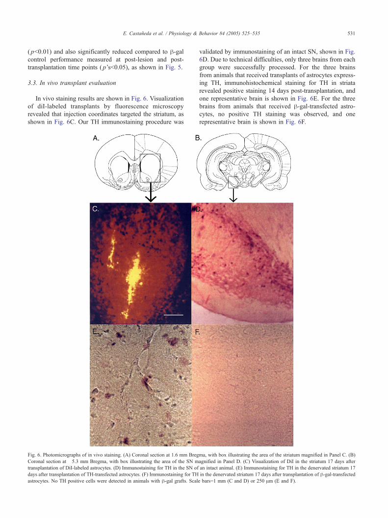
3.3. In vivo transplant evaluation
In vivo staining results are shown in Fig. 6. Visualization
of diI-labeled transplants by fluorescence microscopy
revealed that injection coordinates targeted the striatum, as
shown in Fig. 6C. Our TH immunostaining procedure was
Fig. 6. Photomicrographs of in vivo staining. (A) Coronal section at 1.6 mm Breg
Coronal section at �5.3 mm Bregma, with box illustrating the area of the SN m
transplantation of DiI-labeled astrocytes. (D) Immunostaining for TH in the SN of
days after transplantation of TH-transfected astrocytes. (F) Immunostaining for TH
astrocytes. No TH positive cells were detected in animals with h-gal grafts. Scal
validated by immunostaining of an intact SN, shown in Fig.
6D. Due to technical difficulties, only three brains from each
group were successfully processed. For the three brains
from animals that received transplants of astrocytes express-
ing TH, immunohistochemical staining for TH in striata
revealed positive staining 14 days post-transplantation, and
one representative brain is shown in Fig. 6E. For the three
brains from animals that received h-gal-transfected astro-
cytes, no positive TH staining was observed, and one
representative brain is shown in Fig. 6F.
ma, with box illustrating the area of the striatum magnified in Panel C. (B)
agnified in Panel D. (C) Visualization of DiI in the striatum 17 days after
an intact animal. (E) Immunostaining for TH in the denervated striatum 17
in the denervated striatum 17 days after transplantation of h-gal-transfectede bars=1 mm (C and D) or 250 Am (E and F).
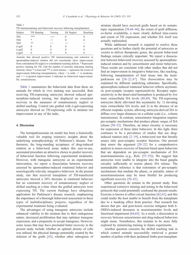
Table 1
TH immunostaining and behavioral recovery following transplantation
Subject TH Staining Turning SS Neglect Reaching
TH1 *** +++ 0 0
TH2 * ++ 0 0
TH3 ** ++ ++ +
h-gal1 0 0 0 0
h-gal2 0 0 0 0
h-gal3 0 0 0 0
Animals that showed positive TH immunostaining and reduction in
apomorphine-induced rotation did not consistently show improvement
from contralateral SS neglect or contralateral reaching deficits. * Represents
positive staining for TH, with the number of asterisks indicating staining
intensity where * is least and *** is most intense. + represents the extent of
improvement following transplantation, where + is mild, ++ is moderate,
and +++ is greatest improvement. 0 indicates no behavioral improvement.
SS, somatosensory.
E. Castaneda et al. / Physiology & Behavior 84 (2005) 525–535532
Table 1 summarizes the behavioral data from these six
animals for which in vivo staining was successful. Rats
receiving TH-expressing astrocytes showed reduced apo-
morphine-induced turning, but displayed no consistent
recovery in the measures of somatosensory neglect or
skilled reaching. Control rats grafted with h-gal-expressingastrocytes showed no TH-expressing cells in striatum nor
improvement in any of the tasks.
4. Discussion
The hemiparkinsonian rat model has been a historically
valuable tool for reaping extensive insights about the
underlying neurophysiology of Parkinson’s disease. Fur-
thermore, the long-standing acceptance of drug-induced
rotation as a behavioral assay makes this easy-to-use,
automated procedure an attractive choice to predict recovery
of behavioral function following experimental treatments.
However, with transgenic astrocytes as an experimental
intervention, we report a dissociation between recovery
assessed by apomorphine-induced rotational behavior and
neurologically-relevant, integrative behaviors. In the present
study, rats that received transplants of TH-transfected
astrocytes showed a 34% decrease in rotational behavior,
but no consistent recovery of somatosensory neglect or
skilled reaching at a time when the grafted astrocytes were
expressing TH. The current findings have ubiquitous
application for Parkinson’s disease research, emphasizing
the importance of a thorough behavioral assessment in these
types of multidisciplinary projects, regardless of the
experimental treatment being evaluated.
The advantages of using transgenic astrocytes include
enhanced viability in the striatum due to their endogenous
nature, decreased proliferation that may optimize transgene
expression, and a propensity to accept transgenes. However,
some issues that may have precluded better recovery in the
present study include whether an optimal density of cells
was infused, the physical damage potentially created by the
infusion of the grafts [43], whether other subregions of
striatum should have received grafts based on its somato-
topic organization [38,44–46], the extent of graft diffusion,
co-factor availability, a more clearly defined time-course
and extent of TH expression, and whether DA itself was
actually replenished.
While additional research is required to resolve these
questions and to further clarify the potential of astrocytes as
vectors to deliver therapeutic genes, the present behavioral
findings remain critically important. We report a dissocia-
tion between behavioral recovery assessed by apomorphine-
induced rotation and by sensorimotor and motor behaviors.
These results are consistent with other studies that did not
find an improvement in integrative behaviors, like reaching,
following transplantation of fetal tissue into the hemi-
parkinson rat [20–22,47]. This dissociation may be
explained by different underlying substrates. Specifically,
apomorphine-induced rotational behavior reflects asymmet-
ric post-synaptic receptor supersensitivity. Receptor super-
sensitivity in the denervated hemisphere is produced by low
tonic extracellular DA levels [48,49]. The TH-expressing
astrocytes likely alleviated this asymmetry by: 1) elevating
tonic extracellular DA levels, and 2) in the absence of an
efficient reuptake system, allowing astrocyte-derived DA to
diffuse over larger distances on a cellular scale (i.e., volume
transmission). In contrast, sensorimotor integration requires
pre-synaptic mechanisms that produce phasic surges of DA
release [50–52]. Therefore, an intact circuitry is necessary
for expression of these latter behaviors. In this light, there
continues to be a prevalence of studies that use drug-
induced rotation only [2–19], a behavioral test that literally
assays only post-synaptic receptor function. Our present
data renew the argument [20–22] for a comprehensive
analysis to assess recovery of function based upon behaviors
that are dependent on pre-synaptic mechanisms of DA
neurotransmission (e.g., Refs. [53–55]). We suggest that
astrocytes were unable to integrate into the basal ganglia
circuitry sufficiently to restore phasic DA release. The
unmistakable inference is that restoration of pre-synaptic
mechanisms that mediate the phasic, or pulsatile, nature of
neurotransmission may be most fruitful for producing
significant recovery [56–61].
Other questions do remain in the present study. Rats
experienced extensive training and testing in the behavioral
protocols that could potentially confound the present results.
Exercise is known to affect circuit function [62,63], and this
could render the host unable to benefit from the transplants
due to a masking effect from practice. Past research has
shown that pre- and post-lesion exercise mitigates both 6-
OHDA-induced decreases in neurotransmitter levels and
functional impairment [64,65]. As a result, a dissociation in
recovery between sensorimotor and drug-induced behaviors
might ensue. Nonetheless, this remains to be more fully
explored by minimizing training in future experiments.
Another question concerns the skilled reaching task in
which control animals successfully retrieved a greater
number of pellets with their ipsilateral limbs post-transplant.
E. Castaneda et al. / Physiology & Behavior 84 (2005) 525–535 533
Past research has shown such behavioral compensation after
unilateral 6-OHDA, namely that rats show a preference to
use the ipsilateral limb [24,65,66]. In contrast, the group
receiving TH-expressing astrocytes did not develop this
pattern of reaching. One might speculate that the TH-
expressing astrocytes produced recovery in the contralateral
limb, abolishing the need for behavioral compensation by
the ipsilateral limb. It is too early to be confident about this
interpretation. It is just as likely that the control group was
reaching more but with less success, possibly due to the
additional damage produced by the transplant procedure. An
index of efficiency to retrieve pellets (number of pellets
collected as a function of the number of attempts) could
have resolved this issue.
Finally, another question is posed by the possibility for
additional damage produced by the transplant procedure.
Damage from the infusion volume that destroys post-
synaptic receptors on neurons intrinsic to the striatum
would decrease apomorphine-induced rotation, as well as
prevent recovery in motor and sensorimotor abilities. If this
is true, then there is no dissociation, and our results reflect
post-synaptic damage. Given the limited number of infu-
sions (4 injections, 3 Al/site) into the relatively large
striatum, we maintain that the explanation of dissociation
is more viable. Indeed, the possibility for volume trans-
mission produced by the transplants means that a healthy
area beyond the locus of damage could be affected.
However, only further investigation will resolve this issue,
for example by minimizing infusion volumes using opti-
mized cell densities.
In summary, transplants of astrocytes expressing TH
were demonstrated to reduce apomorphine-induced rotation
but did not alleviate deficits in somatosensory neglect or
skilled reaching despite survival of grafts and expression of
TH. We surmise that transfected astrocytes functioned as
tonic pumps, with no impact upon pre-synaptic mecha-
nisms. The overarching significance of these results is in the
message that changes in drug-induced rotation alone are not
sufficient to demonstrate recovery of function by any
putatively therapeutic approach. A thorough assessment
across a wide profile of abilities such as sensory, motor, self-
regulatory, and motivated behaviors would provide stronger
evidence for the potential of experimental treatments to
alleviate PD and other neurodegenerative disorders [66].
Moreover, such a bneuropsychologicalQ approach in tandem
with apomorphine-induced rotation is a powerful strategy
for dissecting molecular changes, such as pre-synaptic
versus post-synaptic function.
Acknowledgements
This research was supported by NINDS/NIH to MKS,
Arizona Disease Control Research Commission Contract
#9834, the Office of the Vice Provost for Research at ASU,
and the ASU Hispanic Research Center. We thank Victor
Luevano for technical assistance and Dr. Cheryl Conrad as
well as the anonymous reviewers for constructive feedback
that significantly improved the quality of this manuscript.
References
[1] Schwarting RK, Huston JP. The unilateral 6-hydroxydopamine lesion
model in behavioral brain research. Analysis of functional deficits,
recovery and treatments. Prog Neurobiol 1996;50:275–331.
[2] Cortez N, Trejo F, Vergara P, Segovia J. Primary astrocytes retrovirally
transduced with a tyrosine hydroxylase transgene driven by a glial-
specific promoter elicit behavioral recovery in experimental parkin-
sonism. J Neurosci Res 2000;59:39–46.
[3] Fisher LJ, Jinnah HA, Kale LC, Higgins GA, Gage FH. Survival and
function of intrastriatally grafted primary fibroblasts genetically
modified to produce l-dopa. Neuron 1991;6:371–80.
[4] Fitoussi N, Sotnik-Barkai I, Tornatore C, Herzberg U, Yadid G.
Dopamine turnover and metabolism in the striatum of parkinsonian
rats grafted with genetically-modified human astrocytes. Neuroscience
1998;85:405–13.
[5] Harvey BK, Mark A, Chou J, Chen GJ, Hoffer BJ, Wang Y.
Neurotrophic effects of bone morphogenetic protein-7 in a rat model
of Parkinson’s disease. Brain Res 2004;1022:88–95.
[6] Hida H, Hashimoto M, Fujimoto I, Nakajima K, Shimano Y, Nagatsu
T, et al. Dopa-producing astrocytes generated by adenoviral trans-
duction of human tyrosine hydroxylase gene: in vitro study and
transplantation to hemiparkinsonian model rats. Neurosci Res
1999;35:101–12.
[7] Horellou P, Vigne E, Castel MN, Barneoud P, Colin P, Perricaudet M,
et al. Direct intracerebral gene transfer of an adenoviral vector
expressing tyrosine hydroxylase in a rat model of Parkinson’s disease.
Neuroreport 1994;6:49–53.
[8] Kemmerer ES, Desmond TJ, Albin RL, Kilbourn MR, Frey KA.
Treatment effects on nigrostriatal projection integrity in partial 6-
OHDA lesions: comparison of L-DOPA and pramipexole. Exp Neurol
2003;183:81–6.
[9] Lundberg C, Horellou P, Mallet J, Bjfrklund A. Generation of DOPA-
producing astrocytes by retroviral transduction of the human tyrosine
hydroxylase gene: in vitro characterization and in vivo effects in the
rat Parkinson model. Exp Neurol 1996;139:39–53.
[10] Maesawa S, Kaneoke Y, Kajita Y, Usui N, Misawa N, Nakayama A,
et al. Long-term stimulation of the subthalamic nucleus in hemi-
parkinsonian rats: neuroprotection of dopaminergic neurons. J Neuro-
surg 2004;100:679–87.
[11] Olds ME, Jacques DB, Kopyov O. Behavioral and subthalamic
effects of combining a fetal ventral mesencephalic transplant in
striatum with an electrolytic lesion of the entopeduncular nucleus in
the rat with a unilateral 6-OHDA lesion of substantia nigra. Synapse
2003;48:90–9.
[12] Olds ME, Jacques DB, Kopyov O. Behavioral/neurophysiological
investigation of effects of combining a quinolinic acid entopeduncular
lesion with a fetal mesencephalic tissue transplant in striatum of the 6-
OHDA hemilesioned rat. Synapse 2003;49:1–11.
[13] Segovia J, Verara P, Brenner M. Astrocyte-specific expression of
tyrosine hydroxylase after intracerebral gene transfer induces behav-
ioral recovery in experimental parkinsonism. Gene Ther 1998;
5:1650–5.
[14] Shen Y, Muramatsu SI, Ikeguchi K, Fujimoto KI, Fan DS, Ogawa M,
et al. Triple transduction with adeno-associated virus vectors express-
ing tyrosine hydroxylase, aromatic-l-amino-acid decarboxylase, and
GTP cyclohydrolase I for gene therapy of Parkinson’s disease. Hum
Gene Ther 2000;11:1509–19.
[15] Tornatore C, Baker-Cairns B, Yadid G, Hamilton R, Meyers K,
Atwood W, et al. Expression of tyrosine hydroxylase in an
immortalized human fetal astrocyte cell line: in vitro characterization
E. Castaneda et al. / Physiology & Behavior 84 (2005) 525–535534
and engraftment into the rodent striatum. Cell Transplant 1996;
5:145–63.
[16] Wang Y, Chang CF, Morales M, Chiang YH, Harvey BK, Su TP, et al.
Diadenosine tetraphosphate protects against injuries induced by
ischemia and 6-hydroxydopamine in rat brain. J Neurosci 2003;23:
7958–65.
[17] Wang ZH, Ji Y, Shan W, Zeng B, Raksadawan N, Pastores GM, et al.
Therapeutic effects of astrocytes expressing both tyrosine hydroxylase
and brain-derived neurotrophic factor on a rat model of Parkinson’s
disease. Neuroscience 2002;113:629–40.
[18] Wolff JA, Fisher LJ, Xu L, Jinnah HA, Langlais PJ, Iuvone PM, et al.
Grafting fibroblasts genetically modified to produce l-dopa in a rat
model of Parkinson’s disease. Proc Natl Acad Sci 1989;86:9011–4.
[19] Zhou M, Xu DH, Cao L, Xu LF, Yu FR, Zheng ZC, et al. Long term
gene therapy of Parkinson’s disease using immortalized rat glial cell
line with tyrosine hydroxylase gene. Sheng Wu Hua Xue Yu Sheng
Wu Wu Li Xue Bao 2003;35:1066–71.
[20] Abrous DN, Shaltot AR, Torres EM, Dunnett SB. Dopamine-rich
grafts in the neostriatum and/or nucleus accumbens: effects on drug-
induced behaviours and skilled paw-reaching. Neuroscience 1993;53:
187–97.
[21] Dunnett SB, Whishaw IQ, Rogers DC, Jones GH. Dopamine-rich
grafts ameliorate whole body motor asymmetry and sensory neglect
but not independent limb use in rats with 6-hydroxydopamine lesions.
Brain Res 1987;415:63–78.
[22] Olsson M, Nikkhah G, Bentlage C, Bjfrklund A. Forelimb akinesia in
the rat Parkinson model: differential effects of dopamine agonists and
nigral transplants as assessed by a new stepping test. J Neurosci
1995;15:3863–75.
[23] Bernheimer H, Birkmayer W, Hornykiewicz O, Jellinger K, Seitel-
berger F. Brain dopamine and the syndromes of Parkinson and
Huntington. J Neurol Sci 1973;20:415–55.
[24] Miklyaeva EI, Castaneda E, Whishaw IQ. Skilled reaching deficits in
unilateral dopamine-depleted rats: impairments in movement and
posture and compensatory adjustments. J Neurosci 1994;14:7148–58.
[25] Miklyaeva EI, Martens DJ, Whishaw IQ. Impairments and compen-
satory adjustments in spontaneous movement after unilateral dop-
amine depletion in rats. Brain Res 1995;681:23–40.
[26] Schallert T, Upchurch M, Lobaugh N, Farrar R, Spirduso W, Gilliam
P, et al. Tactile extinction: distinguishing between sensorimotor and
motor asymmetries in rats with unilateral nigrostriatal damage.
Pharmacol Biochem Behav 1982;16:455–62.
[27] Metz GA, Whishaw IQ. Drug-induced rotation intensity in unilateral
dopamine-depleted rats is not correlated with end point or qualitative
measure of forelimb or hindlimb motor performance. Neuroscience
2002;111:325–36.
[28] Castaneda E, Whishaw IQ, Robinson TE. Changes in striatal
dopamine neurotransmission assessed with microdialysis following
recovery from a bilateral 6-OHDA lesion: variation as a function of
lesion size. J Neurosci 1990;10:1847–54.
[29] Zhang WQ, Tilson HA, Nanry KP, Hudson PM, Hong JS, Stachowiak
MK. Increased dopamine release from striata of rats after unilateral
nigrostriatal bundle damage. Brain Res 1988;461:335–42.
[30] Metz GA, Gonzalez CL, Piecharka DM, Whishaw IQ. Acute alcohol
administration improves skilled reaching success in intact but not 6-
OHDA dopamine depleted rats: a subsystems analysis of the motoric
and anxiolytic effects of alcohol. Behav Brain Res 2003;142(1-2):
167–74.
[31] Paxinos G, Watson C. The Rat Brain in Stereotaxic Coordinates. San
Diego7 Academic Press, Inc.; 1986.
[32] Hokfelt T, Ungerstedt U. Specificity of 6-hydroxydopamine induced
degeneration of central monoamine neurones: an electron and
fluorescence microscopic study with special reference to intracerebral
injection on the nigro-striatal dopamine system. Brain Res 1973;60:
269–97.
[33] Ungerstedt U. 6-hydroxydopamine induced degeneration of central
monoamine neurons. Eur J Pharmacol 1968;5:107–10.
[34] Ungerstedt U, Ljungberg T, Steg G. Behavioral, physiological, and
neurochemical changes after 6-hydroxydopamine-induced degener-
ation of the nigro-striatal dopamine neurons. Adv Neurol 1974;5:
421–6.
[35] Harada K, Wu J, Haycock JW, Goldstein M. Regulation of l-DOPA
biosynthesis by site-specific phosphorylation of tyrosine hydroxylase
in AtT-20 cells expressing wild-type and serine 40-substituted
enzyme. J Neurochem 1996;67:629–35.
[36] Wu J, Filer D, Friedhoff AJ, Goldstein M. Site-directed mutagenesis
of tyrosine hydroxylase. J Biol Chem 1992;267:25754–8.
[37] Stachowiak MK, Goc A, Hong JS, Poisner A, Jiang HK, Stachowiak
EK. Regulation of tyrosine hydroxylase gene expression in depolar-
ized non-transformed bovine adrenal medullary cells: second mes-
senger systems and promoter mechanisms. Mol Brain Res 1994;22:
309–19.
[38] Salamone JD, Mahan K, Rogers S. Ventrolateral striatal dopamine
depletions impair feeding and food handling in rats. Pharmacol
Biochem Behav 1993;44:605–10.
[39] Schallert T, Upchurch M, Wilcox RE, Vaughn DM. Posture
independent sensorimotor analysis of inter-hemispheric receptor
asymmetries in the neostriatum. Pharm Biochem Behav 1983;
18:753–9.
[40] Montoya CP, Campbell-Hope LJ, Pemberton KD, Dunnett SB. The
bstair-case testQ: a measure of independent forelimb reaching and
grasping abilities in rats. J Neurosci Methods 1991;36:219–28.
[41] Tabbaa S, Goulah C, Tran RK, Lis A, Korody R, Stachowski B, et al.
Gene transfer into the central nervous system using Herpes Simplex
Virus-1 vectors. Folia Morphol 2000;59:221–32.
[42] Mertz KD, Weisheit G, Schilling K, Luers GH. Electroporation of
primary neural cultures: a simple method for directed gene transfer in
vitro. Histochem Cell Biol 2002;118(6):501–6.
[43] Martin-Iverson MT, Altar CA. Spontaneous behaviours of rats
are differentially affected by substantia nigra infusions of brain-
derived neurotrophic factor and neurotrophin-3. Eur J Neurosci
1996;8:1696–706.
[44] Alexander GE, Crutcher MD, DeLong MR. Basal ganglia–thalamo-
cortical circuits: parallel substrates for motor, oculomotor, bprefrontalQand blimbicQ functions. Prog Brain Res 1990;85:119–46.
[45] Pisa M. Motor functions of the striatum in the rat: critical role of the
lateral region in tongue and forelimb reaching. Neuroscience
1988;24:453–63.
[46] Pisa M, Schranz JA. Dissociable motor roles of the rat’s striatum
conform to a somatotopic model. Behav Neurosci 1988;102:429–40.
[47] Montoya CP, Astell S, Dunnett SB. Effects of nigral and striatal grafts
on skilled forelimb use in the rat. Prog Brain Res 1990;82:459–66.
[48] Bunney BS, Walters JR, Roth RH, Aghajanian GK. Dopaminergic
neurons: effect of antipsychotic drugs and amphetamine on single cell
activity. J Pharmacol Exp Ther 1973;185:560–71.
[49] Grace AA, Bunney BS. The control of firing pattern in nigral
dopamine neurons: single spike firing. J Neurosci 1984;4:2866–76.
[50] Grace AA. Phasic versus tonic dopamine release and the modulation
of dopamine system responsivity: a hypothesis for the etiology of
schizophrenia. Neuroscience 1991;41:1–24.
[51] Grace AA. The tonic/phasic model of dopamine system regulation: its
relevance for understanding how stimulant abuse can alter basal
ganglia function. Drug Alcohol Depend 1995;37:111–29.
[52] Grace AA. The tonic/phasic model of dopamine system regulation and
its implications for understanding alcohol and psychostimulant
craving. Addiction 2000;95:S119–28.
[53] Kirik D, Georgievska B, Rosenblad C, Bjfrklund A. Delayed
infusion of GDNF promotes recovery of motor function in the
partial lesion model of Parkinson’s disease. Eur J Neurosci 2001;13:
1589–99.
[54] Kirik D, Rosenblad C, Bjfrklund A. Characterization of behavioral
and neurodegenerative changes following partial lesions of the
nigrostriatal dopamine system induced by intrastriatal 6-hydroxydop-
amine in the rat. Exp Neurol 1998;152:259–77.
E. Castaneda et al. / Physiology & Behavior 84 (2005) 525–535 535
[55] Kirik D, Winkler C, Bjfrklund A. Growth and functional efficacy of
intrastriatal nigral transplants depend on the extent of nigrostriatal
degeneration. J Neurosci 2001;21:2889–96.
[56] Chiodo LA, Caggiula AR, Antelman SM, Lineberry CG. Sensory
stimuli alter the discharge rate of dopamine (DA) neurons: evidence
for two functional types of DA cells in the substantia nigra. Brain Res
1980;189:544–9.
[57] Keller RW, Stricker EM, Zigmond MJ. Environmental stimuli but not
homeostatic challenges produce apparent increases in dopaminergic
activity in the striatum: an analysis by in vivo voltammetry. Brain Res
1983;279:159–70.
[58] Mirenowicz J, Schultz W. Preferential activation of midbrain
dopamine neurons by appetitive rather than aversive stimuli. Nature
1996;379:449–51.
[59] Salamone JD, Keller RW, Zigmond MJ, Stricker EM. Behavioral
activation in rats increases striatal dopamine metabolism measured by
dialysis perfusion. Brain Res 1989;487:215–24.
[60] Schultz W. Responses of midbrain dopamine neurons to behavioral
trigger stimuli in the monkey. J Neurophysiol 1986;56:1439–61.
[61] Schultz W, Romo R. Dopamine neurons of the monkey midbrain:
contingencies of responses to stimuli eliciting immediate behavioral
reactions. J Neurophysiol 1990;63:607–24.
[62] Kolb B, Brown R, Witt-Lajeunesse A, Gibb R. Neural compensations
after lesion of the cerebral cortex. Neural Plast 2001;8:1–16.
[63] Schallert T, Leasure JL, Kolb B. Experience-associated structural
events, subependymal cellular proliferative activity, and functional
recovery after injury to the central nervous system. J Cereb Blood
Flow Metab 2000;20:1513–28.
[64] Cohen AD, Tillerson JL, Smith AD, Schallert T, Zigmond MJ.
Neuroprotective effects of prior limb use in 6-hydroxydopamine-
treated rats: possible role of GDNF. J Neurochem 2003;85:299–305.
[65] Tillerson JL, Cohen AD, Philhower J, Miller GW, Zigmond MJ,
Schallert T. Forced limb-use effects on the behavioral and
neurochemical effects of 6-hydroxydopamine. J Neurosci 2001;21:
4427–35.
[66] Whishaw IQ, Haun F, Kolb B. Analysis of behavior in laboratory
rodents. In: Windhorst U, Johansson H, editors. Modern Techniques in
Neuroscience. Berlin7 Springer-Verlag; 1999. p. 1243–75.